0019-9567/02/$04.00⫹0 DOI: 10.1128/IAI.70.2.672–678.2002
Copyright © 2002, American Society for Microbiology. All Rights Reserved.
Failure of the
Mycobacterium bovis
BCG Vaccine: Some Species of
Environmental Mycobacteria Block Multiplication of BCG and
Induction of Protective Immunity to Tuberculosis
Lise Brandt,
1† Joana Feino Cunha,
2Anja Weinreich Olsen,
1Ben Chilima,
3,4Penny Hirsch,
4Rui Appelberg,
2and Peter Andersen
1*
Department of TB Immunology, Statens Serum Institut, Copenhagen, Denmark1; Laboratory of Microbiology and Immunology
of Infection, Institute of Molecular and Cell Biology, University of Porto, Portugal2; andDepartment of Infectious
and Tropical Diseases, London School of Hygiene and Tropical Medicine, London,3and Department of
Soil Science, Institute of Arable Crops Research-Rothamsted, Hertfordshire,4United Kingdom Received 15 August 2001/Returned for modification 5 October 2001/Accepted 7 November 2001
The efficacy ofMycobacterium bovisbacillus Calmette-Guérin (BCG) vaccine against pulmonary tuberculosis (TB) varies enormously in different populations. The prevailing hypothesis attributes this variation to inter-actions between the vaccine and mycobacteria common in the environment, but the precise mechanism has so far not been clarified. Our study demonstrates that prior exposure to live environmental mycobacteria can result in a broad immune response that is recalled rapidly after BCG vaccination and controls the multipli-cation of the vaccine. In these sensitized mice, BCG elicits only a transient immune response with a low frequency of mycobacterium-specific cells and no protective immunity against TB. In contrast, the efficacy of TB subunit vaccines was unaffected by prior exposure to environmental mycobacteria. Six different isolates from soil and sputum samples from Karonga district in Northern Malawi (a region in which BCG vaccination has no effect against pulmonary TB) were investigated in the mouse model, and two strains of the Mycobac-terium aviumcomplex were found to block BCG activity completely.
Tuberculosis (TB) is one of the most prevalent causes of death from infectious diseases in the world. As is the case for many intracellular pathogens, cell-mediated immunity plays an important role in host protection against TB (25, 29). In par-ticular, gamma interferon (IFN-␥)-secreting T cells have been shown to be important for the protective immune response (17). The only vaccine currently available against TB is the attenuated Mycobacterium bovis strain bacillus Calmette-Guérin (BCG). The efficacy of this vaccine varies from 0 to 80% in different populations, with a consistently low efficacy in many tropical regions of the world where the vaccine is most needed (15, 16, 35, 38). The reason for the failure of BCG in some populations has been a subject of debate since the 1950s, and many different hypotheses have been suggested to explain the observed variation. Some investigators have suggested that differences in the strain of BCG (23), the age at vaccination (40), or methodological differences are important factors for the variation reported (8). The most widely accepted hypoth-esis relates the efficacy of BCG to geographic location, with low to nondetectable levels of protection against pulmonary TB seen in tropical regions such as Africa and India, where exposure to nontuberculous mycobacteria is common (15). One exception from this general rule is the consistent high efficacy when BCG is used to vaccinate newborns. Neonatal vaccination with BCG imparts protection against the child-hood manifestations of TB (in particular, meningitis) (1, 9, 24),
but the efficacy wanes over a period of 10 to 15 years, and therefore it does not prevent against the later breakdown with pulmonary TB in the adult population in the third world (37). There is convincing evidence that exposure of laboratory animals to environmental mycobacteria can provide some pro-tection against infection withM. tuberculosis(7, 14, 20, 30, 33). The influence of such cross protection on the efficacy of sub-sequent BCG vaccination is not yet clarified, but based on animal experiments, it has been suggested that the protection provided by environmental mycobacteria may partly mask the effect of a subsequent BCG vaccination (33, 42) or that envi-ronmental mycobacteria have a direct antagonistic influence on subsequent BCG vaccination (34, 36). Our study demon-strates that prior sensitization with environmental mycobacte-ria can inhibit BCG multiplication and thereby prevent the induction of an efficient BCG-mediated immune response and protection against TB challenge. Interestingly, different species isolated from soil and sputum in Karonga, Malawi, an area in which BCG has been shown to provide no protection against TB (22), differed in their ability to inhibit BCG multiplication. In contrast, a TB subunit vaccine had the same protective effect in naive and sensitized animals.
MATERIALS AND METHODS
Animals.These studies were performed with pathogen-free 6- to 12-week-old CBA/J and C57BL/6J female mice, purchased from Bomholtegaard, Ry, Den-mark, or, in some of the experiments, purchased from Harlan UK, Ltd., Belton, England, or Harlan Interfauna Ibérica, Barcelona, Spain.
Bacteria.Mycobacterium avium (ATCC 15769),Mycobacterium scrofulaceum
(ATCC 19275), andMycobacterium vaccae(ATCC 15483) were grown in 7H9 broth until the mid-log phase of the bacterial growth.Mycobacterium tuberculosis(Edman) was grown at 37°C on Löwenstein-Jensen medium or in suspension in modified Sauton medium enriched with 0.5% sodium pyruvate and 0.5% glucose. In
prepa-* Corresponding author. Mailing address: Statens Serum Institut, Department of TB Immunology, Artillerivej 5, 2300 Copenhagen S, Denmark. Phone: 45 32683480. Fax: 45 32683035. E-mail: [email protected]. † Present address: Department of Microbiology, Colorado State University, Fort Collins, CO 80523..
ration for immunization of mice, frozen aliquots of the bacterial strains were thawed and sonicated for 5 min with a Branson 2210 ultrasonifier, and the viability of each strain was enumerated on 7H11 plates.Mycobacterium fortuitum(S78/2) andM. fortuitum(S160/5) are soil isolates from the north and south of Karonga, Malawi, respectively. We used standard decontamination of samples with 4% sodium hy-droxide and culture at 37°C on nutrient agar-based medium to isolate the organisms from soil.M. fortuitum(Sp2001),Mycobacterium chelonae(Sp2015), and two strains of theM. aviumcomplex (Sp1891) and (Sp2011) were sputum isolates from donors with suspected TB in the Karonga district. The organisms were isolated with acidi-fied Löwenstein-Jensen medium in Malawi, and their identity was confirmed at the Mycobacterium Reference Unit, Dulwich, United Kingdom, by standard biochem-ical identification tests for mycobacteria. Frozen aliquots of these strains were pre-pared for animal inoculation as described above.
Sensitization with environmental mycobacteria.Mice were immunized subcu-taneously (s.c.) in the back three times at 2-week intervals with 2⫻106CFU of each of three ATCC strains of environmental mycobacteria (M. avium,M. scrofulaceum,
andM. vaccae). To clear the remaining mycobacteria, sensitization was followed, 3 weeks after the last inoculation, by 1 month of treatment with rifampin (Sigma; 100 mg/liter), ethambutol (Sigma; 200 mg/liter), and clarithromycin (Abbott Laborato-ries, Solna, Sweden; 200 mg/liter) added to the drinking water.
To assess virulence of the strains isolated from Karonga, Malawi, mice were infected with 105CFU of each environmental mycobacterial strain in a volume of 0.2 ml of phosphate-buffered saline by intravenous (i.v.) injection via a lateral tail vein. At the appropriate time points, mice were killed (four in each group), and the organs were removed for bacterial enumeration. Whole organs were homogenized in a 0.04% Tween 80 (Sigma) solution in distilled water, serial 10-fold dilutions were plated on Middlebrook 7H10 medium at 37°C, and the numbers of CFU were determined.
Vaccinations.A single dose of BCG Danish 1331 (5⫻104CFU) was injected s.c. at the base of the tail. There were no significant differences in the protection obtained with doses ranging from 5⫻104to 107BCG bacteria (results not shown). In one experiment, an i.v dose of 5⫻106CFU of BCG was used to determine growth of BCG in naive versus sensitized mice. For subunit vaccina-tion, the mice were immunized s.c. three times at 2-week intervals with 10g (per dose) of either ESAT-6 or the Ag85B–ESAT-6 fusion protein emulsified in dioctadecylammonium bromide (DDA; 250g/dose; Eastman Kodak, Inc., Rochester, N.Y.) plus 25g of monophosphoryl lipid A (MPL; Corixa, Hamil-ton, Mont.) as described recently (5).
M. tuberculosisinfections.Animals were infected with approximately 100 CFU ofM. tuberculosis(Edman) per lung by the aerosol route in a Glas-Col inhalation exposure system. The mice were sacrificed 6 weeks after infection, and bacterial numbers in the lung and spleen were determined as described before (5).
The protective effect of BCG or subunit vaccination was expressed as the log10 reduction of the bacterial counts compared to that in the unvaccinated control mice. All results are based on five or six animals per group.
Mycobacterial antigens.A crude BCG antigen preparation (BCG Ag) was produced as an ammonium sulfate-precipitated culture filtrate from cultures at week 6 as described in reference 2. In one of the experiments (see Fig. 4), the BCG responses to an ammonium sulfate-precipitated extract of the cell wall were measured as described elsewhere (31). These two preparations were found to give similar responses in vitro.
Protein concentration was quantified by the Micro bicinchoninic acid method (Pierce, Rockford, Ill.).
Recombinant ESAT-6 was produced as described previously (18). The LPS content was below 0.3 ng/g of protein and had no influence on cellular activity. The fusion protein Ag85B–ESAT-6 was produced as described recently (26). The proteins were kept at⫺80°C until use.
Lymphocyte cultures.Lymphocytes from spleens and blood were isolated and cultured as described previously (5). Briefly, cells from individual mice were cultured in microtiter wells (96 well; Nunc, Roskilde, Denmark) containing 2⫻ 105cells in a volume of 200l of RPMI 1640 supplemented with 5⫻10⫺5M 2-mercaptoethanol, penicillin-streptomycin, 1 mM glutamine, and 5% (vol/vol) fetal calf serum. Based on previous dose-response investigations, BCG Ag and ESAT-6 were each used at 5g/ml in the cultures. Phytohemagglutinin at a concentration of 1g/ml was used in all experiments as a positive control for cell viability. IFN-␥, interleukin 4 (IL-4), and IL-5 were detected in 72-h culture supernatants by duplicate enzyme-linked immunosorbent assay (ELISA).
Enzyme-linked immunospot (ELISPOT) analyses were conducted with cells from individual mice or, when blood was analyzed, with cells pooled from groups of mice, as described in reference 6. The detection level was 10 spots.
Statistical methods.Because all of the data show a normal distribution, the assessment of experiments was carried out by analysis of variance. Differences between means were assessed by Dunnett’s test (Tables 1 and 2) or Student’st
test (see Fig. 2 and 4). APvalue of⬍0.05 was considered significant.
RESULTS
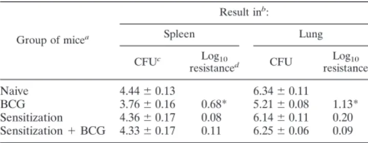
The multiplication of BCG is inhibited in mice sensitized with certain environmental mycobacteria. We inoculated CBA/J mice s.c. three times at 2-week intervals with a mixture TABLE 1. Sensitization with environmental mycobacteria
blocks the protective effect of BCG
Group of micea
Result inb:
Spleen Lung
CFUc Log10
resistanced CFU resistanceLog10
Naive 4.44⫾0.13 6.34⫾0.11 BCG 3.76⫾0.16 0.68* 5.21⫾0.08 1.13* Sensitization 4.36⫾0.17 0.08 6.14⫾0.11 0.20 Sensitization⫹BCG 4.33⫾0.17 0.11 6.25⫾0.06 0.09
aNaive or sensitized mice were BCG vaccinated (5
⫻104CFU) followed by aerosol challenge with virulentM. tuberculosis.
bThe experiment was repeated twice with similar results.
cBacterial numbers determined by growth of individual whole-organ
homog-enates 6 weeks postinfection.
dProtective effect expressed as the log
10reduction in bacterial loads compared to those of naive mice. Bacterial numbers significantly different (P⬍0.05) from those seen in naive mice are indicated by an asterisk.
TABLE 2. Bacterial numbers in organs of naive and sensitized mice after vaccination and aerosol challenge
with virulentM. tuberculosis
Vaccine groupa
Result in:
Lung Spleen
Log10
CFUb resistanceLog10 c LogCFU10 resistanceLog10
Expt 1 Naive
Control 6.36⫾0.08 4.71⫾0.05
BCG 5.83⫾0.06 0.53* 4.12⫾0.12 0.59* DDA-MPL 6.34⫾0.09 4.94⫾0.12
ESAT-6 5.76⫾0.09 0.60* 4.39⫾0.09 0.32* Sensitized
Control 6.18⫾0.08 4.82⫾0.16 BCG 6.27⫾0.07 ⬍0.05 4.79⫾0.05 ⬍0.05 DDA-MPL 6.39⫾0.05 4.73⫾0.11 ESAT-6 5.74⫾0.16 0.44* 4.43⫾0.05 0.39 Expt 2
Naive
Control 6.88⫾0.12 5.10⫾0.18 DDA-MPL 7.19⫾0.05 5.48⫾0.11
Ag85B–ESAT-6 6.03⫾0.12 0.85* 4.40⫾0.08 0.70* Sensitized
Control 6.30⫾0.08 4.39⫾0.09 DDA-MPL 6.49⫾0.05 4.27⫾0.08
Ag85B–ESAT-6 5.37⫾0.14 0.93* 3.89⫾0.07 0.50* aNaive or sensitized mice were immunized s.c. with BCG or injected three
times with a subunit vaccine emulsified in DDA-MPL.
bBacterial numbers are given as log
10CFU ofM. tuberculosisisolated from the lung and spleen 6 weeks after aerosol challenge with virulentM. tuberculosis.
cProtective effects of the two vaccines are expressed as log
of the mycobacterial strainsM. avium, M. scrofulaceum, andM. vaccae. These species have repeatedly been isolated from soil and water samples in tropical regions (21). Three weeks posti-noculation, a low but significant mycobacterium-specific recall response was measured in the spleen, with detectable levels of IFN-␥release in response to BCG Ag. (1.26 ⫾ 0.01 ng/ml) (data not shown). The BCG Ag preparation gave no IFN-␥ release (⬍0.05 ng/ml) from splenocytes isolated from naive mice. No IL-4 or IL-5 was detected in any of the supernatants. Three weeks after the last inoculation with environmental my-cobacteria, we subjected the mice to 4 weeks of chemotherapy to clear remaining live mycobacteria. After the end of chemo-therapy treatment, no environmental mycobacteria were de-tected in any of the target organs (liver, spleen, and lymph nodes).
We inoculated groups of sensitized and age-matched naive CBA/J mice i.v. 1 week after the end of chemotherapy treat-ment with 5 ⫻ 106 BCG and monitored the growth in the spleen and liver over time. Sensitization with environmental mycobacteria resulted in inhibition of the initial multiplication of BCG in the spleen and liver (Fig. 1A). In naive mice, the initial multiplication of BCG resulted in 10- to 30-fold more bacteria in the spleen postinoculation than in sensitized mice. A difference was also seen after a conventional s.c. vaccination, although the bacterial numbers were at lower levels (data not shown). Similar data were obtained with C57BL/6J mice, which are more susceptible to BCG (11). In this strain, larger differ-ences in BCG numbers were found between sensitized and nonsensitized mice (Fig. 1B).
Immune responses induced by BCG vaccination in sensi-tized and naive mice.We continued by investigating the im-mune response induced by BCG in sensitized and age-matched naive control CBA/J mice. ELISPOT was used to monitor
frequencies of BCG-specific T cells before and 3, 5, 8, and 11 weeks after the s.c. vaccination with BCG (Fig. 2). Before BCG vaccination, no mycobacterium-specific IFN-␥-producing T cells were detected in any of the mice. Three weeks after BCG inoculation, the number of BCG-specific IFN-␥-producing cells in the draining lymph nodes had increased and reached the same level in sensitized and in naive mice (Fig. 2A). The response in sensitized mice was, however, transient, and from 5 weeks after BCG inoculation and onwards, a higher fre-quency of mycobacterium-specific cells was found in naive vac-cinated mice. At the termination of the experiment (week 11), a 10-times-higher frequency of BCG-specific T cells was found in the naive vaccinated group than in the sensitized vaccinated group (P⫽0.032). A similar dynamic development of responses was found in the blood, although it was delayed so
FIG. 1. BCG multiplication is inhibited in mice previously sensi-tized with environmental mycobacteria. (A) CBA/J mice. (B) C57BL/6J mice. The growth of BCG was compared in naive mice (open symbols) and in sensitized mice (solid symbols). The data shown are the means of BCG CFU⫾standard errors. For both groups, five animals were sacrificed for each time point. The experiment was repeated twice with similar results.
that higher frequencies of specific T cells were found from week 8 onwards in naive vaccinated mice (Fig. 2B). At no time point after vaccination was IL-4 or IL-5 detected in the super-natants of the stimulated cultures (results not shown).
Sensitization with environmental mycobacteria blocks the protective effect of BCG, but not a TB subunit vaccine. We continued by vaccinating sensitized and naive age-matched control CBA/J mice 4 to 5 weeks after the end of chemother-apy-treatment, followed 2 months later by an aerosol challenge withM. tuberculosis. The mice were killed 6 weeks post-TB infection, andM. tuberculosis CFU were enumerated in the lungs and spleens. The BCG vaccine imparted appreciable protection to naive mice against the TB challenge, with signif-icantly reduced bacterial numbers in the organs (0.68 to 1.13 log10 reduction; Table 1). Sensitization with environmental mycobacteria on its own, or followed by BCG vaccination, failed to induce a statistically significant level of protection against TB (Table 1).
We also asked if a previous sensitization with environmental mycobacteria would influence protection induced by a subunit vaccine. Groups of naive and sensitized CBA/J mice were vaccinated with BCG or injected (three times at 2-week inter-vals) with recently developed TB subunit vaccines based on the immunodominant antigens ESAT-6 and Ag85B mixed with a DDA-MPL adjuvant emulsion (5, 26). ESAT-6-vaccinated
an-imals mounted a very strong recall immune response (5 to 7 ng of IFN-␥/ml) to the homologous preparation 1 week postvac-cination in the blood (data not shown). The protection ob-tained by BCG in control mice was log 0.53, and as in the previous experiment, BCG did not protect presensitized mice (Table 2, experiment 1) The ESAT-6 subunit vaccine, in con-trast, induced a similar degree of protection in both naive and sensitized mice. A subunit vaccine based on a fusion protein of Ag85B and ESAT-6 has recently been demonstrated to induce levels of protection similar to those of BCG in the mouse model (26), and this vaccine also protected against TB chal-lenge at the same level in naive and sensitized mice (Table 2, experiment 2).
Mycobacterial species isolated in Karonga, Malawi, differ in their ability to block BCG activity.We investigated six differ-ent isolates from soil and sputum samples from Karonga Dis-trict in Northern Malawi in the mouse model. Three of these isolates were typed as M. fortuitum, one was a strain of M. chelonae, and two were classified as belonging to theM. avium
complex (Fig. 3). The growth of these isolates in spleen, liver, and lung was investigated with C57BL/6J mice over a period of 30 days. Most of the isolates were rapidly cleared to below the level of detection, but the strains from theM. aviumcomplex multiplied and reached bacterial numbers 3 logs above those of
were treated with chemotherapy, followed by an injection of BCG according to our standard protocol. BCG counts in the spleen of these mice were quantified at week 2 postinoculation (Fig. 4A). M. fortuitumand M. chelonaedid not inhibit the growth of BCG, whereas bacteria from theM. aviumcomplex reduced BCG numbers by 1 to 1.5 log (P⬍0.01). This differ-ence correlated with the immune responses induced by the BCG vaccine. There was no influence on the level of IFN-␥ responses to BCG Ag by sensitization withM. chelonaeorM. fortuitum, whereas the previous inoculation with bacteria from the M. aviumcomplex completely ablated BCG immune re-sponses (Fig. 4B). All strains, on the other hand, induced low and variable responses to antigens extracted from the homol-ogous strain of environmental mycobacteria (results not shown).
DISCUSSION
This study demonstrates that animals exposed to certain environmental mycobacteria raise an immune response that controls the multiplication of BCG, thereby curtailing the vac-cine-induced immune response before it is fully developed. The finding is important for the long-held discussion on the
failure of BCG vaccination against TB in some parts of the world (15, 16, 38). One hypothesis to explain the failure of BCG was presented in 1966 by Palmer and Long, based on large-scale guinea pig experiments. They argued that because contact with nontuberculous bacteria offers some level of pro-tective immunity to TB, the propro-tective effect of a superimposed BCG vaccine would be masked (33). The present study con-firms the classical observation that priming with environmental mycobacteria promotes some levels of protective immunity to other mycobacteria (7, 10, 14, 33), in this case to BCG. How-ever, this effect was not sufficient to significantly reduce the growth ofM. tuberculosis, which multiplied at an almost un-changed rate in these sensitized animals. The difference from the partial protection imparted by environmental mycobacteria in the guinea pig model (14, 33) may be related to the fact that the earlier studies made no effort to clear the environmental mycobacteria by chemotherapy before challenge withM. tuber-culosis, as well as the different genetic makeup and suscepti-bility of mice versus guinea pigs. The differences in these mod-els and their relevance to human disease are the subject of an ongoing study.
That prior sensitization to environmental mycobacteria in-terferes in a similar way with human BCG vaccination is FIG. 4. (A) Growth of BCG in sensitized and naive animals. Mice were infected s.c. with 2⫻106CFU of eitherM. fortuitum,M. chelonae, or M. aviumor were left untreated. After chemotherapy, mice were infected with BCG Pasteur. BCG CFU in the spleen are shown as means with standard errors (n⫽4). Statistically significant differences between sensitized (solid bars) and naive (open bars) mice are indicated:ⴱ,P⬍0.05;
ⴱⴱ,P⬍0.01, according to Student’sttest. ND, not done. (B) Effects of sensitization on the IFN-␥response to BCG. Spleen cells were pooled from four individual mice sensitized with the environmental strains (shaded bars), naive mice inoculated with BCG (open bars), or sensitized mice inoculated with BCG (solid bars). IFN-␥production was assessed in the supernatants of spleen cell cultures stimulated in vitro with BCG Ag and are given as means with standard errors. Statistically significant effects of sensitization on the response to BCG infection are labeled:ⴱ,P⬍0.05;
strongly suggested by a number of classical epidemiological observations: (i) the finding of strong efficacy of BCG in trials in which tuberculin skin test-positive (and therefore sensitized) donors have been vigorously excluded (19); (ii) the consistent success with BCG in neonates vaccinated before any significant sensitization from environmental mycobacteria occurs (1, 9, 24); and, (iii) finally, the observation of a lower rate of skin test conversion, much smaller average diameter, and rapidly wan-ing responses after BCG vaccination in areas with environmen-tal sensitization (India and Egypt), compared with those in areas with minimal environmental exposure (Denmark) (4, 32). This observation was recently confirmed and extended by the observation of only minimal in vitro IFN-␥responses to purified protein derivative (PPD) induced by BCG vaccination in donors from Karonga, Malawi, compared to those from the United Kingdom (P. E. Fine and H. Dockrell, personal com-munication). Taken together, these findings are in agreement with the low and transient immune response in the group of animals sensitized with environmental mycobacteria before vaccination, whereas the naive animals developed strong and sustained responses (Fig. 3). Our experimental model is there-fore relevant to the many tropical regions where BCG is not protective against pulmonary TB and where the high incidence of TB indicates that any partial protection provided by expo-sure to environmental mycobacteria is insufficient for the pre-vention of TB.
Our main conclusion is that BCG, as a live vaccine, is par-ticularly sensitive to the influence of preexisting immune re-sponses to antigens shared with certain environmental strains. In this regard, a recent study has demonstrated the cross-recognition of a large number of antigens shared betweenM. aviumand BCG (T. Pais and R. Appelberg, unpublished re-sults). Multiplication is a precondition for the induction of immunity by BCG and killing of BCG by chemotherapy after administration has been demonstrated to abrogate subsequent immunity completely (13, 39). In the present study, this block-ing is achieved by immunological control instead of chemo-therapy, but the outcome in both cases is interference with the protective immune response, which would normally develop in response to the growing BCG. The requirement for BCG mul-tiplication can be explained as a simple consequence of dosage, but more likely is due to the fact that only live BCG secretes many antigens of importance for the induction of a protective immune response (3, 28). Interestingly, our data from the animal model also suggest that only environmental strains, which are capable of an initial multiplication in the host, block the activity of BCG. A detailed evaluation of a large number of different soil isolates from Karonga, Malawi, and of their in-teractions with BCG is ongoing. In the future, information on the geographical distribution of such strains would be a valu-able resource when trying to understand the huge variation in BCG efficacy in human trials.
This inhibitory effect of the environmental mycobacteria on the growth and activity of BCG provides an important argu-ment in the ongoing discussion of live attenuated vaccines versus nonviable subunit vaccines against TB (12, 27, 44). In comparison with live attenuated vaccines, the present study suggests that subunit vaccines may be much less influenced by prior contact with environmental mycobacteria. As mentioned above, neonatal BCG vaccination consistently imparts
protec-tion against the childhood manifestaprotec-tions of TB (mostly ex-trapulmonary disease), but as its efficacy wanes over a period of 10 to 15 years (37), the adult pulmonary manifestations of TB are prevented neither by neonatal vaccination, by vaccina-tion in adolescence after exposure to environmental mycobac-teria (41), nor by a BCG revaccination strategy (22, 43). A TB subunit vaccine could therefore fulfill the criterion of having consistently high efficacy in different populations and may have a particularly important use for revaccination of third world children in adolescence.
ACKNOWLEDGMENTS
This study has been supported by the Danish Research Council and The European Commission (contract no. 18CT970254). Lise Brandt is supported by the Faculty of Health Science, University of Copenha-gen.
Environmental mycobacteria from Malawi were isolated within the context of the Karonga Prevention Study (KPS) with the assistance of H. Phiri, S. Chagulkuka, and G. Black and were classified by M. Yates at the U.K. Mycobacterium Reference laboratory in Dulwich. The KPS is coordinated by Paul Fine and supported by The Wellcome Trust.
Paul Fine is thanked for valuable discussion, advice, and helpful comments on the manuscript. We thank Vita Skov, Lene Rasmussen, and Tina Lerche for excellent technical assistance.
REFERENCES
1.Al-Kassimi, F. A., M. S. Al-Hajjaj, I. O. Al-Orainey, and E. A. Bamgboye.
1995. Does the protective effect of neonatal BCG correlate with vaccine-induced tuberculin reaction? Am. J. Respir. Crit. Care Med.152:1575–1578. 2.Andersen, A. B., Z.-L. Yuan, K. Hasløv, B. Vergmann, and J. Bennedsen.
1986. Interspecies reactivity of five monoclonal antibodies toMycobacterium tuberculosisas examined by immunoblotting and enzyme-linked immunosor-bent assay. J. Clin. Microbiol.23:446–451.
3.Andersen, P.1997. Host responses and antigens involved in protective im-munity toMycobacterium tuberculosis. Scand. J. Immunol.45:115–131. 4.Baily, G. V.1980. Tuberculosis prevention trial, Madras. Indian J. Med. Res.
72:1–74.
5.Brandt, L., M. Elhay, I. Rosenkrands, E. B. Lindblad, and P. Andersen.
2000. ESAT-6 subunit vaccination againstMycobacterium tuberculosis. Infect. Immun.68:791–795.
6.Brandt, L., T. Oettinger, A. Holm, and P. Andersen.1996. Key epitopes on the ESAT-6 antigen recognized in mice during the recall of protective im-munity toMycobacterium tuberculosis. J. Immunol.157:3527–3533. 7.Brown, C. A., I. N. Brown, and S. Swinburne.1985. The effect of oral
Mycobacterium vaccaeon subsequent responses of mice to BCG sensitiza-tion. Tubercle66:251–260.
8.Clemens, J. D., J. J. Chuong, and A. R. Feinstein.1983. The BCG contro-versy. A methodological and statistical reappraisal. JAMA249:2362–2369. 9.Colditz, G. A., C. S. Berkey, F. Mosteller, T. F. Brewer, M. E. Wilson, E.
Burdick, and H. V. Fineberg.1995. The efficacy of bacillus Calmette-Guerin vaccination of newborns and infants in the prevention of tuberculosis: meta-analyses of the published literature. Pediatrics96:29–35.
10.Collins, F. M.1971. Immunogenicity of various mycobacteria and the cor-responding levels of cross-protection developed between species. Infect. Immun.4:688–696.
11.Denis, M., A. Forget, M. Pelletier, R. Turcotte, and E. Skamene.1986. Control of the Bcg gene of early resistance in mice to infections with BCG substrains and atypical mycobacteria. Clin. Exp. Immunol.63:517–525. 12.Doherty, T. M., and P. Andersen.2000. Tuberculosis vaccines:
developmen-tal work and the future. Curr. Opin. Pulm. Med.6:203–208.
13.Dworski, M.1973. Efficacy of bacillus Calmette-Guérin and isoniazid-resis-tant bacillus Calmette-Guérin with and without isoniazid chemoprophylaxis from day of vaccination. Am. Rev. Respir. Dis.108:294–300.
14.Edwards, M. L., J. M. Goodrich, D. Muller, A. Pollack, J. E. Ziegler, and D. W. Smith.1982. Infection withMycobacterium avium-intracellulareand the protective effects of Bacille Calmette-Guerin. J. Infect. Dis.145:733–741. 15.Fine, P. E.1989. The BCG story: lessons from the past and implications for
the future. Rev. Infect. Dis.11(Suppl. 2):S353–S359.
16.Fine, P. E.1995. Variation in protection by BCG: implications of and for heterologous immunity. Lancet346:1339–1345.
17.Flynn, J. L., J. Chan, K. J. Triebold, D. K. Dalton, T. A. Stewart, and B. R. Bloom.1993. An essential role for interferon gamma in resistance to Myco-bacterium tuberculosisinfection. J. Exp. Med.178:2249–2254.
of the ESAT-6 protein ofMycobacterium tuberculosis. Infect. Immun.66:
717–723.
19.Hart, P. D., and I. Sutherland.1977. BCG and vole bacillus vaccines in the prevention of tuberculosis in adolescence and early adult life. Br. Med. J.
2:293–295.
20.Kamala, T., C. N. Paramasivan, D. Herbert, P. Venkatesan, and R. Prab-hakar.1996. Immune response & modulation of immune response induced in the guinea-pigs byMycobacterium aviumcomplex (MAC) &M. fortuitum
complex isolates from different sources in the South Indian BCG trial area. Indian J. Med. Res.103:201–211.
21.Kamala, T., C. N. Paramasivan, D. Herbert, P. Venkatesan, and R. Prab-hakar.1994. Isolation and identification of environmental mycobacteria in the Mycobacterium bovisBCG trial area of South India. Appl. Environ. Microbiol.60:2180–2183.
22.Karonga Prevention Trial Group. 1996. Randomised controlled trial of single BCG, repeated BCG, or combined BCG and killedMycobacterium lepraevaccine for prevention of leprosy and tuberculosis in Malawi. Lancet
348:17–24.
23.Lagranderie, M. R., A. M. Balazuc, E. Deriaud, C. D. Leclerc, and M. Gheorghiu.1996. Comparison of immune responses of mice immunized with five differentMycobacterium bovisBCG vaccine strains. Infect. Immun.64:
1–9.
24.Miceli, I., I. N. de Kantor, D. Colaiacovo, G. Peluffo, I. Cutillo, R. Gorra, R. Botta, S. Hom, and H. G. ten Dam.1988. Evaluation of the effectiveness of BCG vaccination using the case-control method in Buenos Aires, Argentina. Int. J. Epidemiol.17:629–634.
25.North, R. J.1973. Importance of thymus-derived lymphocytes in cell-medi-ated immunity to infection. Cell Immunol.7:166–176.
26.Olsen, A. W., L. A. H. van Pinxteren, L. M. Okkels, P. B. Rasmussen, and P. Andersen.2001. Protection of mice with a tuberculosis subunit vaccine based on a fusion protein of antigen 85B and ESAT-6. Infect. Immun.69:2773– 2778.
27.Orme, I. M.1997. Progress in the development of new vaccines against tuberculosis. Int. J. Tuberc. Lung Dis.1:95–100.
28.Orme, I. M., P. Andersen, and W. H. Bloom. 1993. T cell response to
Mycobacterium tuberculosis. J. Infect. Dis.167:1481–1497.
29.Orme, I. M., E. S. Miller, A. D. Roberts, S. K. Furney, J. P. Griffin, K. M. Dobos, D. Chi, B. Rivoire, and P. J. Brennan.1992. T lymphocytes mediating protection and cellular cytolysis during the course ofMycobacterium tuber-culosisinfection. Evidence for different kinetics and recognition of a wide spectrum of protein antigens. J. Immunol.148:189–196.
30.Orme, I. M., A. R. Roberts, and F. M. Collins.1986. Lack of evidence for a reduction in the efficacy of subcutaneous BCG vaccination in mice infected with nontuberculous mycobacteria. Tubercle67:41–46.
31.Pais, T. F., R. A. Silva, B. Smedegaard, R. Appelberg, and P. Andersen.1998. Analysis of T cells recruited during delayed-type hypersensitivity to purified protein derivative (PPD) versus challenge with tuberculosis infection. Im-munology95:69–75.
32.Palmer, C. E.1952. BCG vaccination and tuberculin allergy. LancetMay 10:935–941.
33.Palmer, C. E., and M. W. Long.1966. Effects of infection with atypical mycobacteria on BCG vaccination and tuberculosis. Am. Rev. Respir. Dis.
94:553–568.
34.Rook, G. A., G. M. Bahr, and J. L. Stanford.1981. The effect of two distinct forms of cell-mediated response to mycobacteria on the protective efficacy of BCG. Tubercle62:63–68.
35.Smith, D. W., E. H. Wiegeshaus, and M. L. Edwards.1988. The protective effects of BCG vaccination against tuberculosis, p. 341–370.InM. Bendinelli and H. Friedman (ed.),Mycobacterium tuberculosis. Plenum Publishing Cor-poration, New York, N.Y.
36.Stanford, J. L., M. J. Shield, and G. A. Rook.1981. How environmental mycobacteria may predetermine the protective efficacy of BCG. Tubercle
62:55–62.
37.Sterne, J. A., L. C. Rodrigues, and I. N. Guedes.1998. Does the efficacy of BCG decline with time since vaccination? Int. J. Tuberc. Lung Dis.2:200– 207.
38.ten-Dam, H. G.1984. Research on BCG vaccination. Adv. Tuberc. Res.
21:79–106.
39.Toyohara, M., S. Kudoh, and Y. Obayashi.1959. Studies on the effect of isoniazid upon the antituberculous immunity induced by BCG vaccination. Tubercle40:184–191.
40.Tripathy, S. P.1983. The case for BCG. Ann. N. Y. Acad. Med. Sci.19:11– 21.
41.Tuberculosis Research Centre (ICMR).1999. Fifteen year follow up of trial of BCG vaccines in south India for tuberculosis prevention, Chennai. Indian J. Med. Res.110:56–69.
42.Weiszfeiler, J. C., and V. Karasseva.1981. Mixed mycobacterial infections. Rev. Infect. Dis.3:1081–1083.
43.World Health Organization.1995. W.H.O. news and activities. W.H.O. Bull. OMS73:805–807.
44.Young, D. B.2000. Current tuberculosis vaccine development. Clin. Infect. Dis.30:S254–S256.