Braz. J. of Develop., Curitiba, v. 6, n. 5, p. 30294-30305 may. 2020. ISSN 2525-8761
Potential risk of intracranial infection due to multiple odontogenic sinusitis
Potencial risco de infecção intracraniana devido a sinusite odontogênica
múltipla
DOI:10.34117/bjdv6n5-473
Recebimento dos originais: 20/04/2020 Aceitação para publicação: 22/05/2020
Marcelie Priscila de Oliveira Rosso
Doctoral Student (PhD) of Department of Biological Sciences (Anatomy), Bauru School of Dentistry, University of São Paulo (USP).
Institution: Bauru School of Dentistry, University of São Paulo (USP).
Address: Alameda Dr. Octávio Pinheiro Brisolla, 9-75, Vila Universitaria – Bauru, SP, 17012-901, Brazil.
E-mail: [email protected]
Letícia Gabriela Menezes
Dentistry Surgeon, Dentistry School, University of Itaúna. Institution: Dentistry School, University of Itaúna.
Address: Highway MG 431, Km 45, Itaúna, MG, 35.680-142, Brazil. E-mail: [email protected]
Amanda Silveira de Araújo
Medicine Student, Medical School, University of Itaúna. Institution: Medical School, University of Itaúna.
Address: Highway MG 431, Km 45, Itaúna, MG, 35.680-142, Brazil. E-mail: [email protected]
Beethoven Estevão Costa
Master's Degree student, Hospital for Rehabilitation of Craniofacial Anomalies (HRAC). Institution: Hospital for Rehabilitation of Craniofacial Anomalies (HRAC).
Address: Silvio Marchione Street, 3-20, Vila Nova Cidade Universitaria – Bauru, SP, 17012-900, Brazil.
E-mail: [email protected]
Daniela Vieira Buchaim
PhD by Bauru School of Dentistry, University of São Paulo (USP). Professor of the Postgraduate Program in Structural and Functional Interactions in Rehabilitation, University of Marilia
(UNIMAR) and in Medical School, University Center of Adamantina (UniFAI).
Institutions: University of Marilia (UNIMAR) and University Center of Adamantina (UniFAI). Address: Avenue Hygino Muzzy Filho, 1001, Marília, SP, 17525–902, Brazil.
Address: Nove de Julho Street, 730 - Centro, Adamantina, SP, 17800-000, Brazil. E-mail: [email protected]
Karina Torres Pomini
Doctoral student (PhD) of Department of Biological Sciences (Anatomy), Bauru School of Dentistry, University of São Paulo (USP).
Braz. J. of Develop., Curitiba, v. 6, n. 5, p. 30294-30305 may. 2020. ISSN 2525-8761 Institution: Bauru School of Dentistry, University of São Paulo (USP).
Address: Alameda Dr. Octávio Pinheiro Brisolla, 9-75, Vila Universitaria – Bauru, SP, 17012-901, Brazil.
E-mail: [email protected]
Carlos Henrique Bertoni Reis
Master's student, Postgraduate Program in Structural and Functional Interactions in Rehabilitation, University of Marilia (UNIMAR).
Institution: University of Marilia (UNIMAR).
Address: Avenue Hygino Muzzy Filho, 1001, Marília, SP, 17525–902, Brazil. E-mail: [email protected]
João Paulo Galletti Pilon
Master's student, Postgraduate Program in Structural and Functional Interactions in Rehabilitation, University of Marilia (UNIMAR).
Institution: University of Marilia (UNIMAR),
Address: Avenue Hygino Muzzy Filho, 1001, Marília, SP, 17525–902, Brazil. E-mail: [email protected]
Rogério Leone Buchaim*
Associate Professor, Bauru School of Dentistry, University of São Paulo. Coordinator of the Postgraduate Program in Structural and Functional Interactions in Rehabilitation, University of
Marilia (UNIMAR).
Institutions: Bauru School of Dentistry, University of São Paulo (USP) and University of Marilia (UNIMAR).
Address: Alameda Dr. Octávio Pinheiro Brisolla, 9-75, Vila Universitaria – Bauru, SP, 17012-901, Brazil
Address: Avenue Hygino Muzzy Filho, 1001, Marília, SP, 17525–902, Brazil. E-mail: [email protected]
* Correspondence: [email protected]; Tel.: +55-3235-8226
ABSTRACT
We report a clinical case that demonstrates the importance of multiprofessional action in the prevention of intracranial infection in a case of multiple sinusitis originating in the maxillary sinus after to dental procedure. Fifteen days after the extraction of the right upper third molar (tooth 18), a 19-year-old patient presented with symptoms of pulsatile pain at the surgical site, accompanied by oppressive headache, nasal obstruction, fetid mucopurulent rhinorrhea and posterior nasal discharge treated with antibiotics. There was persistence of intermittent headache and sinusitis remained chronic, with tomographic examination compatible with frontal, ethmoidal, sphenoidal and maxillary sinusopathy on the right side, with obliteration of the ipsilateral osteomeatal unit, small calcific foci at the right maxillary floor through areas of soft tissue density, evoking material of dental origin. A sinusotomy and new antibiotic therapy were performed, with a diagnosis of moderate/severe chronic sinusitis, evolving to absence of symptoms. In the presence of symptoms compatible with sinusitis and previous history of dental approach, it is widely necessary to have the attention of both dental surgeon and otolaryngologist to allow an assertive and early diagnosis of odontogenic sinusitis.
Braz. J. of Develop., Curitiba, v. 6, n. 5, p. 30294-30305 may. 2020. ISSN 2525-8761 RESUMO
Relatamos um caso clínico que demonstra a importância da ação multiprofissional na prevenção de infecção intracraniana em um caso de sinusite múltipla originada no seio maxilar após procedimento odontológico. Quinze dias após a extração do terceiro molar superior direito (dente 18), uma paciente de 19 anos apresentou sintomas de dor pulsátil no local da cirurgia, acompanhada de dor de cabeça opressiva, obstrução nasal, rinorréia fétida mucopurulenta e secreção nasal posterior, recebeu tratamento com antibióticos. Houve persistência de cefaleia intermitente e o quadro de sinusite permaneceu crônico, com exame tomográfico compatível com sinusopatia frontal, etmoidal, esfenoidal e maxilar do lado direito, obliteração da unidade osteomeatal ipsilateral, pequenos focos calcificados no assoalho maxilar direito em permeio às áreas de tecido mole, evocando possível material de origem dentária. Foi realizada sinusotomia e nova antibioticoterapia, com diagnóstico de sinusite crônica moderada / grave, evoluindo para ausência de sintomas. Na presença de sintomas compatíveis com sinusite e história prévia de abordagem odontológica, é amplamente necessário ter a atenção do cirurgião-dentista e do otorrinolaringologista para permitir um diagnóstico assertivo e precoce da sinusite odontogênica.
Palavras-chave: Doença crônica, Sinusite maxilar, Sinusite, Cirurgia oral.
1 INTRODUCTION
Maxillary sinusitis (MS) is defined as an inflammation of the sinus membrane that covers the paranasal cavity. It presents a multifactorial etiology, including inflammation by viruses, bacteria, fungi (ZHANG et al., 2019) and dental infection, with odontogenic sinusitis (OS) corresponding to 10% to 15% of diagnosed cases (AKHLAGHI; ESMAEELINEJAD; SAFAI, 2015; SAIBENE et al., 2019).
The molar roots usually project to the maxillary sinus and odontogenic infections can drain into the sinus causing reactive responses (BACHELET; LANDIS; SCOLOZZI, 2019) and such infections can spread to the other paranasal sinuses (MARTINES et al., 2014). When the diagnosis and treatment of OS is late, it can lead to serious complications such as orbital cellulitis and brain abscesses (MEHRA; MURAD, 2004; LONGHINI; FERGUSON, 2012; MOAZZAM et al., 2015).
Treatment of OS requires combating the source of infection with antibiotic therapy and/or treatment of the causative tooth. However, the otolaryngologists prefer the surgical technique associated with drug treatment, removing the foreign body from the maxillary sinus by endoscopy (TOMOMATSU et al., 2014; AKIYAMA et al., 2018).
Thus, the aim of this study was to report a clinical case demonstrating the importance of multiprofessional action in the prevention of intracranial infection in an unusual case of multiple sinusitis originating in the maxillary sinus due to dental procedure.
Braz. J. of Develop., Curitiba, v. 6, n. 5, p. 30294-30305 may. 2020. ISSN 2525-8761 2 CLINICAL REPORT
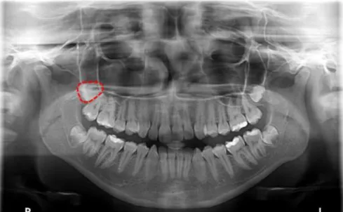
Patient LGM, 19 years old, female, attended the dental surgeon for extraction of tooth 18. He was classified according to the American Society of Anesthesiologists (ASA) (ASA HOUSE OF DELEGATES, 2014) for surgical risk assessment as ASA 1: No physiological, biochemical or psychiatric disorders. Then, a panoramic X-ray was requested (Figure 1), which showed an erupted tooth in distoangular position with incomplete root formation.
Figure 1: Preoperative panoramic radiography. Highlighted in red: unerupted tooth 18 in distoangular position with incomplete root formation.
The surgery was then performed on January 2016, lasting approximately one hour and twenty minutes. After 15 days of the procedure, the patient started to feel pulsatile pain at the surgical site, accompanied by oppressive headache, nasal obstruction, fetid mucopurulent rhinorrhea and posterior nasal discharge. The patient attended the dental surgeon who conducted the procedure, who performed drainage of the site with irrigation using 0.9% saline solution and prescribed antibiotic therapy. Symptoms were partially improved, but intermittent headache was maintained.
In March 2016, there was recurrence with high fever (38.5ºC), which was not associated with the dental procedure by the patient, who then decided to attend an otolaryngologist. A nasosinusal videoendoscopy was performed, which revealed purulent secretion in the right middle meatus, without other alterations. The doctor signed the diagnosis of sinusitis and prescribed antibiotics and anti-inflammatories. Treatment was refractory; the symptomatology was attenuated, but the condition persisted subacute for months.
On 09/15/2017 the patient attended another otolaryngologist, already with the eradication of pain, because the process entered a chronic state. Laboratory and imaging exams were requested:
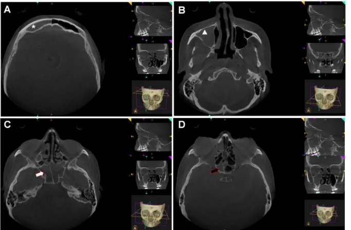
Braz. J. of Develop., Curitiba, v. 6, n. 5, p. 30294-30305 may. 2020. ISSN 2525-8761 hemogram, C-reactive protein (CRP), erythrocyte sedimentation rate (ESR) and computed tomography. The exams showed leukocytosis of 11.190 cells/mm³, CRP and increased ESR. The tomography (Figures 2 and 3) revealed frontal, ethmoidal, sphenoidal and maxillary sinusopathy on the right side, with obliteration of the ipsilateral osteomeatal unit, with enlarged aspect; small calcific foci at the right maxillary floor through material of soft-tissue density, evoking possible material of dental origin.
Figure 2: Computed tomography of the face, transversal section: asterisk - right frontal sinus (RFS); white triangle - right maxillary sinus (RMS); white arrow - right sphenoidal sinus (RSS); black arrow - right ethmoidal sinus (RES). A), B), C) and D) shows total opacification of RFS, RMS, RSS and RES, respectively.
Braz. J. of Develop., Curitiba, v. 6, n. 5, p. 30294-30305 may. 2020. ISSN 2525-8761 Figure 3: Computed tomography of the face: white triangle - right maxillary sinus (RMS) in A) Sagittal section and B) Coronal section showing calcific foci in the presence of soft tissue densities near the floor of the RMS evokes possible dental material (dashed circle and yellow arrow).
Due to the unfavorable clinical and tomographic evolution, the physician indicated a surgical approach. Sinusotomy surgery was performed, whose histopathology evaluation was compatible with chronic / moderate chronic sinusitis.
After clearing and cleaning the paranasal sinuses (Figure 4), the patient presented favorable clinical evolution and did not present relapse of any symptoms.
Braz. J. of Develop., Curitiba, v. 6, n. 5, p. 30294-30305 may. 2020. ISSN 2525-8761 Figure 4: Radiographic image after 1 year of sinusotomy surgery. Red highlights: RFS with thickening characteristics of the mucosal lining, and / or mucous fluid. Highlighted in yellow: radiolucency is observed within radiographic normality for the maxillary sinuses.
3 DISCUSSION
OS is a complication observed in the clinical routine of dental surgeons, which should be analyzed with a judicious approach to quickly diagnose and treat patients more appropriately. This case report shows the OS after extraction of tooth 18 which, after medical treatments guided by the otolaryngologist, presented chronicity and evolved reaching the frontal, ethmoidal, sphenoid sinus on the right side and obliteration of the ipsilateral osteomeatal unit, requiring surgical intervention (sinusotomy) 20 months after extraction, and the biopsy confirmed the presence of material of odontogenic origin.
Scientific studies reveal that 10 to 40% of cases of chronic MS are due to OS (HONG; SHIM; KWON, 2017; SAIBENE et al., 2019), leading to OS being misdiagnosed as common sinusitis (HONG; SHIM; KWON, 2017), among which only 1/3 are evaluated by a dental surgeon, since the otolaryngologic diagnosis focuses more on osteomeatal complex obstruction as the etiology of the disease (CARTWRIGHT; HOPKINS, 2016). Odontogenic origin was reported as responsible for 33% of cases of intracranial infections (EUFINGER; MACHTENS, 2001), emphasizing that the neglect of an adequate evaluation may delay the correct diagnosis, thus
Braz. J. of Develop., Curitiba, v. 6, n. 5, p. 30294-30305 may. 2020. ISSN 2525-8761 highlighting the importance of knowledge on the etiology, diagnosis and treatment of this pathology.
The signs and symptoms of OS are similar to MS, such as congestion or nasal obstruction, pressure, facial pain, and headache, but it has a unilateral origin that draws attention to the dental cause. They are associated with fetid secretion, ocular pain and postnasal drip (CARTWRIGHT; HOPKINS, 2016; BACHELET; LANDIS; SCOLOZZI, 2019), and their differentiation is important for treatment with antibiotics, since in OS there is greater presence of anaerobic bacteria (BOMELI; BRANSTETTER; FERGUSON, 2009). Surgery is indicated in 48% to 81% (HOSKISON et al., 2012; MATTOS; FERGUSON; LEE, 2016), but the ideal time of treatments is not yet clear (CRAIG et al., 2019).
The OS cause of this case refers to the presence of material of odontogenic origin in the maxillary sinus resulting from extraction performed 20 months earlier. Regarding the etiology of chronic OS, the literature reports several cases of iatrogenic origin, for example, placement of implants in 65.7%, complications after extraction in 29.6%, the upper teeth had greater relation with OS, with 17.4% for the third molar (LECHIEN et al., 2014). Due to the anatomical proximity of structures, it is clear that complications resulting from these dental procedures may progress to intracranial infections (MARTINES et al., 2014; GERMAN et al., 2015; AKASHI et al., 2017).
Regarding the symptoms presented by the patient, the professional initially indicated allopathic treatment with antibiotics and, after relapse, the otolaryngologist followed with more medications and surgery. When the cause of OS was identified, the treatment chosen was related to long-term antibiotic therapy and/or treatment of the causative tooth (tooth extraction or root canal treatment), with an efficacy rate of 59.5% (TOMOMATSU et al., 2014).
A review on major OS treatments concluded on the elimination of infection, which can sometimes be effective, yet the surgical approach by endoscopy of the sinuses may be indicated to remove inflammation of the mucosa and foreign bodies present as fragments of teeth, maintaining the physiology of the sinus for complete resolution of the case, however the appropriate time for a surgical approach has not yet been fully elucidated (CARTWRIGHT; HOPKINS, 2016). The chronicity of the case is closely related to the choice of endoscopic surgical option, being chosen as gold standard, with few complications in cases of chronic sinusitis (HOSKISON et al., 2012; AKIYAMA et al., 2018). Clinical professionals should follow cases of chronic MS with high degree of suspicion of an odontogenic cause in cases with previous dentoalveolar surgery (LECHIEN et al., 2014).
Anatomically, the average distance between the maxillary molar and maxillary sinus is 1.97 mm (MEHRA; MURAD, 2004; BUCHAIM et al., 2014). The proximity between the
Braz. J. of Develop., Curitiba, v. 6, n. 5, p. 30294-30305 may. 2020. ISSN 2525-8761 maxillary sinus ostium (natural drainage site) and the anterior ethmoid sinus facilitates the spread of inflammation of the maxillary sinus to the anterior ethmoid cells, causing anterior ethmoiditis (CROVETTO-MARTÍNEZ et al., 2014). With worsening of the condition due to late diagnosis, OS can culminate in serious complications such as orbital cellulitis and brain abscesses (BROOK, 2006). In the case presented, MS had already extended to frontal, ethmoid and sphenoid sinusitis, with intermittent headache even after treatment, and systemic alterations with elevated CRP and ESR increasing the risk of intracranial infection, since hematogenous dissemination is the main factor for the occurrence of a CNS infection (MOAZZAM et al., 2015) due to the difficulty in anticipating the assertive diagnosis.
4 CONCLUSION
In conclusion, in the present case there was difficulty in the early diagnosis of OS, leading to chronic sinusitis and need for surgery 20 months after extraction. The symptomatology of the patient after extraction should prompt immediate attention alerting the occurrence of OS, for early diagnosis and treatment to prevent progression of the condition and risk of intracranial infections.
CONFLICTS OF INTEREST
The authors declares that there is no conflict of interest regarding the publication of this paper.
REFERENCES
AKASHI, M. et al. Brain Abscess Potentially Resulting from Odontogenic Focus: Report of Three Cases and a Literature Review. Journal of Maxillofacial and Oral Surgery, v. 16, n. 1, p. 58–64, 2017.
AKHLAGHI, F.; ESMAEELINEJAD, M.; SAFAI, P. Etiologies and Treatments of Odontogenic Maxillary Sinusitis: A Systematic Review. Iranian Red Crescent Medical Journal, v. 17, n. 12, 2015. Disponível em: <http://www.ircmj.com/?page=article&article_id=25536>.
AKIYAMA, K. et al. Assessment of Simultaneous Surgery for Odontogenic Sinusitis. Journal of
Craniofacial Surgery, v. 30, n. 1, p. 239–243, 2018.
ASA HOUSE OF DELEGATES. ASA Physical Status Classification SystemAmerican Society
Braz. J. of Develop., Curitiba, v. 6, n. 5, p. 30294-30305 may. 2020. ISSN 2525-8761 physical-status-classification-system>.
BACHELET, J.-T.; LANDIS, B. N.; SCOLOZZI, P. Recurrent Maxillary Sinusitis and Periorbital Cellulitis Revealing an Unnoticed Medial Wall Orbital Fracture. Journal of Craniofacial Surgery, v. 30, n. 7, p. 2251–2252, out. 2019. Disponível em: <http://journals.lww.com/00001665-201910000-00082>.
BOMELI, S.; BRANSTETTER, B.; FERGUSON, B. Frequency of a dental source for acute maxillary sinusitis. Laryngoscope, v. 119, n. 3, p. 580–584, 2009.
BROOK, I. Sinusitis of odontogenic origin. Otolaryngol Head Neck Surg., v. 133, p. 349–55, 2006.
BUCHAIM, R. et al. Multidisciplinary Approach in the Teaching of Dental Sculpture and Anatomy. Int. J. Morphol., v. 32, n. 2, p. 399–403, jun. 2014. Disponível em:
<http://www.scielo.cl/scielo.php?script=sci_arttext&pid=S0717-95022014000200002&lng=en&nrm=iso&tlng=en>.
CARTWRIGHT, S.; HOPKINS, C. Odontogenic Sinusitis an underappreciated diagnosis: Our experience. Clinical Otolaryngology, v. 41, n. 3, p. 284–285, 2016.
CRAIG, J. et al. Optimal timing of endoscopic sinus surgery for odontogenic sinusitis.
Laryngoscope, p. 1–8, 2019.
CROVETTO-MARTÍNEZ, R. et al. Frequency of the odontogenic maxillary sinusitis extended to the anterior ethmoid sinus and response to surgical treatment. Medicina Oral, Patologia Oral y
Cirugia Bucal, v. 19, n. 4, 2014.
EUFINGER, H.; MACHTENS, E. Purulent pansinusitis, orbital cellulitis and rhinogenic intracranial complications. Journal of Cranio-Maxillofacial Surgery, v. 29, n. 2, p. 111–117, 2001.
GERMAN, I. J. S. et al. Identification of the bony canal of the posterior superior alveolar nerve and artery in the maxillary sinus: Tomographic, radiographic, and macroscopic analyses. Scientific
Braz. J. of Develop., Curitiba, v. 6, n. 5, p. 30294-30305 may. 2020. ISSN 2525-8761 World Journal, v. 2015, 2015.
HONG, S.; SHIM, G.; KWON, Y. Novel approach to the maxillary sinusitis after sinus graft.
Maxillofac Plast Reconstr Surg, v. 39, p. 18, 2017.
HOSKISON, E. et al. Evidence of an increase in the incidence of odontogenic sinusitis over the last decade in the UK. J Laryngol Otol., v. 126, n. 1, p. 43–6, 2012.
LECHIEN, J. et al. Chronic Maxillary Rhinosinusitis of Dental Origin: A Systematic Review of 674 Patient Cases. Int J Otolaryngol., v. 2014, p. 1–9, 2014.
LONGHINI, A. B.; FERGUSON, B. J. Clinical aspects of odontogenic maxillary sinusitis: a case series. Int. Forum Allergy Rhinol, v. 1, p. 409–415, 2012.
MARTINES, F. et al. Parietal subdural empyema as complication of acute odontogenic sinusitis: A case report. Journal of Medical Case Reports, v. 8, n. 1, p. 1–7, 2014.
MATTOS, J.; FERGUSON, B.; LEE, S. Predictive factors in patients undergoing endoscopic sinus surgery for odontogenic sinusitis. Int Forum Allergy Rhinol, v. 6, p. 697–700, 2016.
MEHRA, P.; MURAD, H. Maxillary sinus disease of odontogenic origin. Otolaryngol Clin North
Am, v. 37, p. 347–64, 2004.
MOAZZAM, A. et al. Intracranial bacterial infections of oral origin. Journal of Clinical
Neuroscience, v. 22, n. 5, p. 800–806, 2015. Disponível em:
<http://dx.doi.org/10.1016/j.jocn.2014.11.015>.
SAIBENE, A. M. et al. Odontogenic rhinosinusitis and sinonasal complications of dental disease or treatment: prospective validation of a classification and treatment protocol. European Archives of
Oto-Rhino-Laryngology, v. 276, n. 2, p. 401–406, 27 fev. 2019. Disponível em:
<http://dx.doi.org/10.1007/s00405-018-5220-0>.
TOMOMATSU, N. et al. Aperture width of the osteomeatal complex as a predictor of successful treatment of odontogenic maxillary sinusitis. International Journal of Oral and Maxillofacial
Braz. J. of Develop., Curitiba, v. 6, n. 5, p. 30294-30305 may. 2020. ISSN 2525-8761 <http://dx.doi.org/10.1016/j.ijom.2014.06.007>.
ZHANG, Y. et al. Formation of papillary mucosa folds and enhancement of epithelial barrier in odontogenic sinusitis. International Forum of Allergy & Rhinology, v. 9, n. 11, p. 1281–1288, 8 nov. 2019. Disponível em: <https://onlinelibrary.wiley.com/doi/abs/10.1002/alr.22277>.