Evaluation of the Prevalence of Maxillary Sinuses
Abnormalities through Spiral Computed
Tomography (CT)
João Paulo Nunes Drumond
1Bruna Bianca Allegro
2Neil Ferreira Novo
3Sérgio Luís de Miranda
4Wilson Roberto Sendyk
11Department of Oral Implantology, Universidade de Santo Amaro
Ringgold Standard Institution, São Paulo, SP, Brazil
2Department of Radiology and Diagnostic Imaging, Hospital
Heliopolis Ringgold Standard Institution, São Paulo, São Paulo, Brazil
3Department of Statistics and Health Sciences, Universidade de Santo
Amaro Ringgold Standard Institution, São Paulo, São Paulo, Brazil
4Department of Otolaryngology Head and Neck Surgery, Universidade
Federal de Sao Paulo Ringgold Standard Institution, São Paulo, São Paulo, Brazil
Int Arch Otorhinolaryngol 2017;21:126–133.
Address for correspondence João Paulo Nunes Drumond, DMD, MDSc, Department of Oral Implantology, Universidade de Santo Amaro, Medical School, Street Prof. Enéas de Siqueira Neto, 340 Jardim das Imbuias, São Paulo, SP 04829-300, Brazil (e-mail: [email protected]).
Keywords
►
maxillary sinus
►
spiral computed
tomography
►
diagnostic imaging
►
otolaryngology
►
epidemiology
Abstract
Introduction
Maxillary sinus disease is common and numerous disorders can affect
this anatomical area. Abnormalities can be classi
fi
ed as: non-neoplastic, neoplastic
benign, and neoplastic malignant.
Objective
Evaluate through CT the prevalence of diseases in maxillary sinuses, using
the Radiology Department
’
s database of a hospital in São Paulo city.
Methods
The sample consisted of 762 facial CT scans that we divided into
three groups: Group A (12
–
19 years old); Group B (20
–
49 years old); Group C
(above 50 years old); and male or female. We considered the following pathological
processes: I - Mucoperiosteal Thickening; II - Chronic Sinusitis; III - Chronic
Odontogenic Sinusitis; IV Rhinosinusitis; V Polypoid Lesions; VI Bone Lesions; VII
-Neoplasms; VIII - Antrolith; IX - Foreign Bodies; X - Oroantral Fistula.
Results
Our study found that 305 exams (40.02%) were normal and 457 exams
(59.97%) were abnormal. We found the following disease frequencies: focal
mucoper-iosteal thickening (21.25%); polypoid lesions (10.76%); chronic sinusitis (7.48%); chronic
odontogenic sinusitis (2.29%); neoplasms (2.03%); rhinosinusitis (1.77%); bone lesions,
foreign bodies and oroantral
fi
stula in 0.65%; 0.13% and 0.06% respectively. There was
no signi
fi
cant difference between male and female, and Groups A, B, or C when relating
the frequencies of abnormalities found. There was no signi
fi
cant difference between
male and female and the age group for the side of the altered maxillary sinus.
Conclusion
We observed a high prevalence of sinus maxillary diseases.
Mucoperios-teal thickening; acute, chronic, and odontogenic sinusitis; polypoid lesions and
neo-plasms have high prevalence in maxillary sinuses. Thus, facial CT exam was effective for
the evaluation of diseases in maxillary sinuses.
received May 19, 2016 accepted August 30, 2016 published online December 27, 2016
DOI http://dx.doi.org/ 10.1055/s-0036-1593834. ISSN 1809-9777.
Copyright © 2017 by Thieme-Revinter Publicações Ltda, Rio de Janeiro, Brazil Original Research
Introduction
The facial region is formed in general, by oral cavity, jaw bones, nose and paranasal sinuses. Due to the wide anatomic prox-imity between the facial structures, diseases in one component can easily affect others.1The maxillary sinuses are the largest of all paranasal sinuses and are located bilaterally within the maxilla bone, assuming a pyramidal shape.2
The maxillary sinus disease is common and numerous disorders can affect this anatomical area. Abnormalities can be grouped as: non-neoplastic, neoplastic benign, and neo-plastic malignant. Inflammatory processes, infections, cysts, polyps, and mucoceles are examples of non-neoplastic lesions. Papilloma,fibro-osseous, and mesenchymal tumors are benign neoplasms. The Squamous Cell Carcinoma, adeno-cystic carcinoma, adenocarcinoma, and sarcomas are some types of malignant tumors that affect the maxillary sinus.3
Maxillary sinus diseases can be classified according to their nature as: congenital (aplasia and hypoplasia); neoplas-tic (benign or malignant); dental conditions (benign dental tumor, odontogenic cyst or periapical inflammatory lesion); bone injury (ossifyingfibroma, Fibrous Dysplasia, andPaget’s
Disease); traumatic bone injury; iatrogenic (related to previ-ous surgical procedures); inflammatory (mucosal thickening, opacification, polyp, mucous retention cyst, antrolith); sys-temic; and silent sinus syndrome.4,5
Mucosal thickening is an inflammatory reaction with hyperplasia of the mucous lining of the maxillary sinus.2 This condition may result from harmful actions caused by trauma, infections, chemical agents, foreign body reaction, neoplasm, or airway conditions such as allergies, rhinitis, or asthma.6 The prevalence of allergic rhinitis in the world population is around 10.25%7. Continuous exposure to inhaled allergens produces a chronic inflammation in the nasal-sinus mucosa, identified as mucosal thickening on Computed Tomography (CT).8
Sinusitis is the most common disease of the paranasal sinuses. Forms of acute or chronic presentation when hassle, do not require diagnostic imaging; but when the symptoms are recurrent or refractory, research with imaging is needed for a better diagnosis.9There is one distinction between acute and chronic sinusitis. In acute sinusitis, liquid orfluid levels inside the maxillary sinus are isolated; but in chronic sinusi-tis, there is a thickening of the bone sinus wall.10 The odontogenic maxillary sinusitis differs from rhinogenic for its pathophysiology, microbiology, and treatment. The odon-togenic maxillary sinusitis is 10% to 40% of all maxillary sinusitis, and its incidence may be increasing.11
Mucosal cysts are a common incidentalfinding on imaging studies, with an incidence between 12.4 and 35.6%.12They are typically spherical opacities on CT scanning, and are not associated with symptoms of chronic rhinosinusitis.3 Muco-cele are pseudocysts expansive formations of the paranasal sinuses, whose wall consists of a modified sinus mucosa and the presence of cystic aseptic liquid inside, generally thick and viscous, and may be infected and became a mucopyocele.13 The accumulation of fluid increases intrasinusal pressure, resulting in expansion and bone destruction. Nasal polyps
develop from the thickening of chronically inflamed mucosa, causing irregular mucosal folds. The polyposis can develop singly or in multiple forms within the maxillary sinus.6
The maxillary sinus carcinoma is uncommon, representing 3% of head and neck carcinoma and 80% of tumors of the paranasal sinuses. Most tumors that affect the maxillary sinus have epithelial origin, and Squamous Cell Carcinomas account for85% of all sinus malignancies, followed by adenocystic carcinoma, with 5–15% frequency. These tumors, when small, are misdiagnosed as chronic sinusitis or polyps. In 40–60% of cases we observed facial asymmetry, bulging of the oral cavity, and invasion into the nasal cavity. In a more advanced stage, maxillary sinus carcinomas reach the pterygopalatine fossa, pterygoid muscles, orbital fissure, cavernous sinus, skull base structures, and central nervous system. This type of injury has very poor prognosis due to late detection and difficult complete surgical resection, with 5-year survival rate ranging from 20–40%.14
In our study, we observed the prevalence of pathological findings in maxillary sinuses in a sample of 1029 Cone Beam CT (CBCT) exams. Results from 450 patients (43.7%) showed normal images. We noted mucosal thickening in 392 patients (38.1%); partial sinus opacification was present in 123 patients (11.9%); total opacification was present in 73 patients (6.9%); and 67 cases (6.4%) had polypoid formations.15
We studied a sample of 513 CBCT scans obtained for ortho-dontic diagnosis and treatment planning. We observed mucosal thickening in 258 sinuses (25.1%), detected pseudocysts in 59 sinuses (5.75%), and 26 (5.1%) sinuses presented severe opaci-fication. Gender was a significant predictor of pseudocysts, with male subjects showing a 196.3% higher relative risk of this pathology. Age was a significant predictor of mucosal thickening, with subjects aged 41 - 60 years showing a 401% higher odds ratio than those aged 12 - 18 years.16
Through CBCT, we evaluated 1406 maxillary sinus and identified abnormalities. We diagnosed diseases in 68.2% of cases. Mucosal thickening was the most prevalent abnor-mality (66%), followed by polypoid lesions (10.1%), opaci-fication (7.8%), antrolith (3.2%), oroantral communication (2.2%), bone lesions (1.8%), neoplasms, and benign dental tumors in 1.3%.There was a significant difference between the genders, showing a greater occurrence of sinus abnor-malities in males (p<0.001). We observed no difference in the occurrence of abnormalities with regards to the age groups (p>0.05).17
formations, 8.9% partial opacification, and 6.5% complete opacification.19
In another study, two observers assessed 402 maxillary sinuses via CBCT searching incidental pathologicalfindings. The overall prevalence of diseases was 59.7%, and mucosal thickening was present in 35.1% of the maxillary sinuses, followed by opacification (16.6%), polypoid formations (7.2%), and otherfindings in 0.7%.20
A retrospective cohort study based on CT scans analyzed 504 patients who were being treated in an oral surgery clinic. The authors evaluated the relationship between etiological dental factors with imaging changes in maxillary sinuses, specifically mucosal thickening, air-fluid levels or opacifi ca-tion. They reported that 32.40% presented maxillary sinus without pathologic changes and without any etiological factor associated; 29.00% showed presence of factors and imaging changes; 20.60% had only imaging changes in the maxillary sinus; and 18.00% had only etiological factors and normal maxillary sinuses.21
This study evaluated the prevalence of all maxillary si-nuses disease groups, through images obtained by Facial CT scans in a public hospital database, common interest injuries among dental specialties, otolaryngology, and head and neck surgery. Due to the clinical significance of abnormalities in maxillary sinus and its high frequency, knowledge of the prevalence and characterization by imaging in a given popu-lation can be very useful for a more accurate diagnosis and subsequent indication for treatment with greater safety and efficacy.
Method
In this cross-sectional study, we conducted a quantitative assessment, with images analysis from the database of the Department of Radiology and Diagnostic Imaging of a public referral hospital - in São Paulo city, Brazil. The Data Transfer Agreement and Co-Participant Declaration were indispensable for the development of this research. The Research Ethics Committee previously approved the research project attached to the documents above, under CAE 42728815.5.0000.5449 and number 1113368 of 05/07/2015.
Selection of patients:the sample was composed of 762 Facial CT scans, performed in a hospital setting from Febru-ary 2012 to FebruFebru-ary 2014, requested by doctors and dentists. The sample included patients of both sexes, aged over 12 years and having performed Facial CT scan for any indication. The sample excluded: patients younger than 12 years, due to their incomplete formation of the maxillary sinuses; patients with a history of surgery in maxillary sinus; exams whose patient identification data were incomplete, or when the maxillary sinuses were not completely displayed on the exam.
To assess the prevalence of pathological processes varying according to age and sex, we divided the examinations into 3 groups according to the following age groups: Group A (aged 12–19 years); Group B (aged 20 to 49 years); Group C (age above 50 years); and male or female.
Acquisition of images:we obtained the images in a unit of Spiral Computed Tomography, modelSiemens Somatom
Emo-tion(Siemens AG, Forchheim, Germany). The images were processed and evaluated inEPACS WORKSTATION(EPEOPLE Technology Solutions, São Paulo, Brazil) version 5.13.10.17.
Image interpretation:the images were dynamically ma-nipulated in axial and coronal planes. A resident doctor in Radiology/Diagnostic Imaging and an Oral & Maxillofacial surgeon performed the analyses simultaneously; respectively calibrated andfixed throughout the study. The Radiological Reports would undergo consultation for verifying purposes in case of disagreement among examiners.
Data collection:we prepared a specific Data Collection Form. The evaluation process was done in two stages. In the first stage, we noted general demographic data, such as age and gender, or the presence or absence of sinus disease, and later marked the Data Collection Form as (1) Abnormal and (2) Normal. If abnormal, we also noted the affected side as (D) Right, (E) Left, or (DE) Bilateral. In the second stage, we only selected the exams classified as (1) Abnormal by at least one examiner, and the disease was classified according to the criteria established.
Rating:we considered the following pathological process-es: 1- Mucosal Thickening (soft thickening, mild, or focal thickness greater than 2 mm); 2 - Chronic Sinusitis (partial or complete opacification; or round mucosal thickening); 3 -Odontogenic Sinusitis; 4 - Acute Rhinosinusitis; 5 - Polypoid Lesions (mucous retention cyst, polyp, or mucocele); 6 - Bone Injury (bone cysts, Fibrous Dysplasia, fractures,Paget’s Disease); 7 Neoplasms (benign or malignant); 8 Antrolith; 9 -Foreign Bodies; 10 - Oroantral Fistula.
According to location within the maxillary sinus, we classified the sinus abnormalities as follows: (A) Anterior, (P) Posterior, (I) Inferior, (S) Superior, (M) Medial, (L) Lateral and, with combined locations: (C) Combined.
Verification of data and statistics:we determined data through manual counting, organized in tabs, and presented in the form of tables and graphs for demonstration of their frequency and prevalence of results, thus fulfilling the objec-tives of the study. The results underwent statistical analysis byChi-Square2. The objective of the statistical analysis was to correlate the age and gender of patients with the frequency of sinusfindings and its unilateral or bilateral occurrence. We applied the same test to analyze comparatively the frequen-cies of the three most prevalent alterations in this study with results observed in studies with similar methodology over the lastfive years. In all tests, the level of significance was set at 5% (p<0.05).
Result
We found the following diseases frequencies: focal mucosal thickening in 21.25%; polypoid lesions in 10.76%; chronic sinusi-tis in 7.48%; 2.29% of maxillary sinuses had chronic odontogenic sinusitis; neoplasms were present in 2.03%; acute rhinosinusitis in 1.77%; bone injury and foreign bodies in 0.65% and 0.13% respectively; and oroantral fistula in 0.06% (►Table 1). The
frequency of the respective anatomical locations inside the maxillary sinus is shown in►Table 2.►Figs. 1–8show some examples of the diseases detected.
TheChi-Square Test2was applied to verify the proposed relations. Regardless of gender and age group, we observed that the frequency of abnormalities was 59.97% (2¼1.82; p¼0.3942; IL¼56.50% and SL¼63.44%) (►Table 3). There
was no significant difference between male and female (2¼7.32 andp¼0.3961) (►Table 4), and Groups A, B, or
C (2¼16.08;p¼0.3088) (►Table 5); when comparing the
frequencies of abnormalities found. There was no significant difference between male and female and the age group for the side of the altered maxillary sinus (2¼1.65 andp¼0.4381;
2¼4.63 andp¼0.3280) (►Tables 6and7).
Discussion
In this study, we found an overall prevalence of pathological findings in 457 patients, which prevalence was 59.97%. Observing the overall of maxillary sinuses, 647 showed these abnormalities, representing a prevalence of 42.45%. Other results were observed in previous studies, whose authors reported the following total prevalence of maxil-lary sinuses diseases: 56.3%, 50.3%, 68.2%, 45.1%, 59.7%, and 49.6%.15–17,19–21
Importantly, the data obtained in this study are correlated to a population already suspected of having an abnormality in the maxillary sinuses. Patients to undergo the facial diagnostic imaging were under medical or dental care, or at least had symptoms that could justify the examination in hospital.
Some authors cite the mucosal thickening as the most frequentfinding in maxillary sinus, with prevalence ranging from 38.1%; 25.1%, 66%, 48.52%; 56.5%, and 35.1%.15–20In our
study, the occurrence of mucosal thickening in 324 of 1524 maxillary sinuses evaluated, representing a prevalence of 21.25%. Although the values we found were lower than those of the authors mentioned above, this condition was also the most frequent in our study. It is then possible that the epidemiological importance of this inflammatoryfinding is closely related to its variable etiology - traumatic, infectious, chemical, or allergic. Among these possible etiologies, there is allergic cause, taking into account the high incidence of allergic rhinitis in the general population as well as the fact that mucosal thickening is more common in individuals with allergic rhinitis.
Images classified as chronic sinusitis show round mucosal thickening, partial opacification, or complete opacification of the maxillary sinus. As for the images classified as acute rhinosinusitis, there are prominent fluid or liquid levels, indicative of the acute phase of this disease. Authors reported the following rates for acute or chronic sinusitis: 19.04%, 5.1%, 7.8%, 15.4%, 2.94%, and 16.6%.15–20In our study, we found 7.48% prevalence of chronic sinusitis and 1.77% for acute sinusitis, a total of 141 of the 1524 maxillary sinuses evaluat-ed, accumulating an overall prevalence of 9.25% for chronic and acute sinusitis, excluding odontogenic diseases.
We observed the occurrence of chronic odontogenic dis-eases in 35 of 1524 maxillary sinuses evaluated, resulting in a prevalence of 2.29%. However, if we just consider the overall maxillary sinuses with suggestion of sinusitis (176), the amount occupied by dental origin corresponds to a value of 19.88%; coinciding with the range of the aforementioned values.11 In a specific study to evaluate the presence of inflammatory processes related to dental factors, 29% of patients assessed had confirmed odontogenic sinusitis, also converging with the above values.21
The polypoid lesions described are represented by mucous cysts, pseudocysts, or mucoceles, sinus polyps, and antro-choanal polyps. These lesions are worth noting for epidemio-logical purposes in this study and other literature consulted. We observed the presence of polypoid lesions in 164 of the 1524 maxillary sinuses evaluated, indicating an overall prev-alence of 10.76%. Other authors cite the following rates: 5.9%, 5.75%, 10.1%, 28.2%, and 7.2%.15–17,19,20
Maxillary sinuses may be affected by benign or malignant neoplasms worthy of great clinical relevance not by the high Table 2 Frequency of anatomical location
Sinus Surface N (%) Abnormal Maxillary Sinuses
Anterior 9 1.40
Posterior 8 1.24
Inferior 157 24.26
Superior 24 3.71
Medial 41 6.33
Lateral 37 5.72
Combined 371 57.34 Table 1 Absolute prevalence of maxillary sinusfindings
Rating N (%) All Maxillary
Sinuses
Mucosal Thickening 324 21.25
Chronic Sinusitis 114 7.48
Odontogenic Sinusitis 35 2.29
Acute Rhinosinusitis 27 1.77
Polypoid Lesions 164 10.76
Bone Injury 10 0.65
Neoplasms 31 2.03
Foreign Bodies 2 0.13
Antrolith 0 0.00
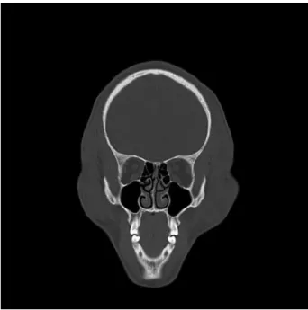
Fig. 1 Normal maxillary sinuses.
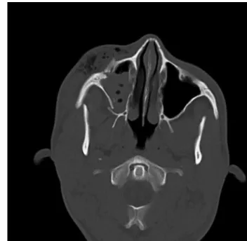
Fig. 2 Mucosal thickening.
Fig. 3 Chronic sinusitis.
Fig. 5 Acute rhinosinusitis.
Fig. 6 Polypoid lesion.
Fig. 7 Bone injury: middle facial fracture.
prevalence, but by the difficult diagnosis and the possibility of involvement of adjacent vital structures. This study showed benign or malignant neoplastic lesions in 31 maxillary sinus reviews, representing a prevalence of 2.03%, similar to the rate found in other research.17 Neoplasm sinuses have important absolute prevalence, rankingfifth; even more than acute rhino-sinusitis isolated; however, most attention is focused on the early diagnosis due to the severity of injuries and poor prognosis in cases of malignant lesions in advanced stages.
Based on such comparisons, we can consider mucosal thickening, sinus opacification processes (acute, chronic, or odontogenic sinusitis), and polypoid lesions as high preva-lence diseases in maxillary sinuses (21.25%, 11.54%, and 10.76%), even in CT scans performed in asymptomatic patients, constituting cases of incidentalfindings.
►Table 8shows the comparative study of the frequency of
the three most prevalent sinusfindings in this study com-pared with other studies published in the lastfive years. We observed statistically significant difference (p<0.05) between the results, suggesting that the sample quality may have influenced the result. This survey was conducted in a hospital environment, as opposed to the others authors who collected data in outpatient basis (CBCT), presumably asymptomatic candidates in oral rehabilitation procedures.
Statistical analysis showed that there are no relations between patient gender and age, with regards to changes in frequency of sinus and the side affected. Another study found that men were more likely to have disease in the maxillary sinuses (p<0.01), but the age of the patients did not consti-tute a factor of influence.19According to another study, males Table 3 Frequency of abnormal maxillary sinuses: Group
Group Abnormal Normal Overall (%) All Abnormal
A 18 11 29 62.10
B 160 93 253 63.20
C 279 201 480 58.10
Overall 457 305 762 59.97
2¼1.86 (p¼0.3942).
Table 4 Frequency of maxillary sinusfindings: Gender
Rating Male
N (%)
Female N (%)
Mucosal Thickening 219 (46.0) 105 (45.3)
Chronic Sinusitis 74 (15.5) 40 (17.2)
Odontogenic Sinusitis 18 (3.8) 17 (7.3)
Acute Rhinosinusitis 18 (3.8) 9 (3.9)
Polypoid Lesions 117 (24.6) 47 (20.3)
Bone Injury 7 (1.5) 3 (1.3)
Neoplasms 22 (4.6) 9 (3.9)
Others 1 (0.2) 2 (0.9)
Overall 476 (100) 232 (100)
2¼7.32 (p¼0.3961).
Table 5 Frequency of maxillary sinusfindings: Group
Rating Group A
N (%)
Group B N (%)
Group C N (%)
Mucosal Thickening 11 (36.7) 105 (40.4) 208 (49.8) Chronic Sinusitis 3 (10.0) 46 (17.7) 65 (15.6)
Odontogenic Sinusitis 1 (3.3) 16 (6.2) 18 (4.3) Acute Rhinosinusitis 3 (10.0) 12 (4.6) 12 (2.9)
Polypoid Lesions 9 (30.0) 65 (25.0) 90 (21.5) Bone Injury 1 (3.3) 5 (1.9) 4 (1.0)
Neoplasms 2 (6.7) 9 (3.5) 20 (4.8)
Others 0 (0.0) 2 (0.8) 1 (0.2)
Overall 30 (100) 260 (100) 418 (100)
2¼16.08 (p¼0.3088).
Table 6 Diseased maxillary sinus: Gender
Side Male
N (%)
Female N (%)
Right 97 (31.8) 42 (27.6)
Left 80 (26.2) 48 (31.6)
Bilateral 128 (42.0) 62 (40.8)
Overall 305 (100) 152 (100)
2¼1.65 (p¼0.4381).
Table 7 Diseased maxillary sinus diseased: Group
Side Group A
N (%)
Group B N (%)
Group C N (%)
Right 6 (33.33) 40 (25.0) 93 (33.33)
Left 3 (16.66) 48 (30.0) 77 (27.60)
Bilateral 9 (50.00) 72 (45.8) 109 (39.06)
Overall 18 (100) 160 (100) 279 (100)
have almost double the relative risk of polypoid formations, and even patients aged 41–60 years have a risk of 401% of developing mucoperiosteal thickening.16
Conclusion
Based on the methods and the sample analyzed, we conclude that: there is a high prevalence of sinus maxillary diseases. Mucosal thickening; acute, chronic, and odontogenic sinusitis; polypoid lesions and tumors are very frequent. A correct diag-nosis of odontogenic processes identifying dental cause is essential for successful treatment. It is clear that early diagnosis of cancer favors a better prognosis. Our study found that combined anatomic regions and maxillary sinus floor were the highest occurrence locations. We found that gender and age of patients were not influencing factors nor were right, left side or bilateral involvement. Moreover, the frequency offi nd-ings does not vary with statistically significance according to the sex or age of the patients. Finally, facial CT scans were effective in the evaluation of pathologicalfindings in the maxillary sinuses.
References
1 Ogle OE, Weinstock RJ, Friedman E. Surgical anatomy of the nasal cavity and paranasal sinuses. Oral Maxillofac Surg Clin North Am 2012;24(2):155–166, vii
2 Van Dis ML, Miles DA. Disorders of the maxillary sinus. Dent Clin North Am 1994;38(1):155–166
3 Stephens JC, Saleh HA. Evaluation and treatment of isolated maxillary sinus disease. Curr Opin Otolaryngol Head Neck Surg 2013;21(1):50–57
4 Lawson W, Patel ZM, Lin FY. The development and pathologic process that influence maxillary sinus pneumatization. Anat Rec 2008;291:1154–1163
5 Mehra P, Jeong D. Maxillary sinusitis of odontogenic origin. Curr Allergy Asthma Rep 2009;9(3):238–243
6 White SC, Pharoah MJ. Oral Radiology. 6th ed. St. Louis: Mosby Elsevier; 2009:506–512
7 Brozek JL, Bousquet J, Baena-Cagnani C, et al. Allergic rhinitis and its impact on asthma (ARIA) guidelines: 2010 revision. J Allergy Clin Immunol 2010;126:466–476
8 Tezer MS, Tahamiler R, Canakçioğlu S. Computed tomography findings in chronic rhinosinusitis patients with and without allergy. Asian Pac J Allergy Immunol 2006;24(2–3):123–127 9 Okuyemi KS, Tsue TT. Radiologic imaging in the management of
sinusitis. Am Fam Physician 2002;66(10):1882–1886
10 Lana JP, Carneiro PMR, Machado VdeC, de Souza PE, Manzi FR, Horta MCR. Anatomic variations and lesions of the maxillary sinus detected in cone beam computed tomography for dental implants. Clin Oral Implants Res 2012;23(12):1398–1403
11 Mehra P, Murad H. Maxillary sinus disease of odontogenic origin. Otolaryngol Clin North Am 2004;37(2):347–364
12 Kanagalingam J, Bhatia K, Georgalas C, Fokkens W, Miszkiel K, Lund VJ. Maxillary mucosal cyst is not a manifestation of rhino-sinusitis: results of a prospective three-dimensional CT study of ophthalmic patients. Laryngoscope 2009;119(1):8–12
13 Almeida WLC, Martin LRL, Perazzo PSL, Anjos CAL, Lima MS. Maxillary Sinus mucopyocele with facial asymmetry. A case report. Int Arch Otorhinolaryngol 2004;8:1–3
14 Katz TS, Mendenhall WM, Morris CG, Amdur RJ, Hinerman RW, Villaret DB. Malignant tumors of the nasal cavity and paranasal sinuses. Head Neck 2002;24(9):821–829
15 Ritter L, Lutz J, Neugebauer J, et al. Prevalence of pathologic findings in the maxillary sinus in cone-beam computerized tomography. Oral Surg Oral Med Oral Pathol Oral Radiol Endod 2011;111(5):634–640
16 Gracco A, Incerti Parenti S, Ioele C, Alessandri Bonetti G, Stellini E. Prevalence of incidental maxillary sinusfindings in Italian ortho-dontic patients: a retrospective cone-beam computed tomogra-phy study. Korean J Orthod 2012;42(6):329–334
17 Rege IC, Sousa TO, Leles CR, Mendonça EF. Occurrence of maxillary sinus abnormalities detected by cone beam CT in asymptomatic patients. BMC Oral Health 2012;12:30
18 Dobele I, Kise L, Apse P, Kragis G, Bigestans A. Radiographic assessment offindings in the maxillary sinus using cone-beam computed tomography. Stomatologija 2013;15(4):119–122 19 Manji A, Faucher J, Resnik RR, Suzuki JB. Prevalence of maxillary
sinus pathology in patients considered for sinus augmentation procedures for dental implants. Implant Dent 2013;22(4):428–435 20 Raghav M, Karjodkar FR, Sontakke S, Sansare K. Prevalence of incidental maxillary sinus pathologies in dental patients on cone-beam computed tomographic images. Contemp Clin Dent 2014; 5(3):361–365
21 Guerra-Pereira I, Vaz P, Faria-Almeida R, Braga AC, Felino A. CT maxillary sinus evaluation–A retrospective cohort study. Med Oral Patol Oral Cir Bucal 2015;20(4):e419–e426
Table 8 Comparative study of frequency of three mainfindings
Study Rating
Drumond N (%)
Raghav N (%)
Manji N (%)
Gracco N (%)
Rege N (%)
Ritter N (%)
1 324 (48.8) 141 (59.5) 70 (56.4) 252 (74.7) 928 (73.7) 392 (58.0)
2 176 (26.5) 67 (28.3) 19 (15.3) 26 (7.7) 110 (8.7) 211 (31.2)
3 164 (24.7) 29 (12.2) 35 (28.2) 59 (17.5) 221 (17.6) 73 (10.8)
Total 664 (100) 237 (100) 124 (100) 337 (100) 1259 (100) 676 (100)
1. Mucosal Thickening; 2. Sinusal Opacification; 3. Polypoid Lesions. Drumond x Raghav:2¼16.74 (p¼0.002).
Drumond x Manji:2¼7.02 (p¼0.0299).