International Journal of
Molecular Sciences
ISSN 1422-0067 www.mdpi.com/journal/ijms Article
Complete Proteome of a Quinolone-Resistant
Salmonella
Typhimurium Phage Type DT104B Clinical Strain
Susana Correia 1,2,3,4, Júlio D. Nunes-Miranda 1,2, Luís Pinto 1,2,3,4, Hugo M. Santos 5,
María de Toro 6, Yolanda Sáenz 7, Carmen Torres 7,8, José Luis Capelo 5, Patrícia Poeta 3,4 and Gilberto Igrejas 1,2,*
1
Institute for Biotechnology and Bioengineering, Centre of Genomics and Biotechnology, University of Trás-os-Montes and Alto Douro, 5001-801 Vila Real, Portugal;
E-Mails: [email protected] (S.C.); [email protected] (J.D.N.-M.); [email protected] (L.P.)
2
Department of Genetics and Biotechnology, University of Trás-os-Montes and Alto Douro, 5001-801 Vila Real, Portugal
3
Centre of Studies of Animal and Veterinary Sciences, University of Trás-os-Montes and Alto Douro, 5001-801 Vila Real, Portugal; E-Mail: [email protected]
4
Veterinary Science Department, University of Trás-os-Montes and Alto Douro, 5001-801 Vila Real, Portugal
5
BIOSCOPE group, REQUIMTE-CQFB, Chemistry Department, Faculty of Science and Technology, University NOVA of Lisbon, 2829-516 Monte de Caparica, Portugal;
E-Mails: [email protected] (H.M.S.); [email protected] (J.L.C.)
6
Departamento de Biología Molecular (Universidad de Cantabria) and Instituto de Biomedicina y Biotecnología de Cantabria IBBTEC (UC-SODERCAN-CSIC), Santander 39011, Spain; E-Mail: [email protected]
7
Área de Microbiología Molecular, Centro de Investigación Biomédica de La Rioja, C/Piqueras 98, 26006 Logroño, La Rioja, Spain; E-Mails: [email protected] (Y.S.);
[email protected] (C.T.)
8
Área de Bioquímica y Biología Molecular, Universidad de La Rioja, Av. Madre de Dios 51, 26006 Logroño, La Rioja, Spain
* Author to whom correspondence should be addressed; E-Mail: [email protected]; Tel.: +351-259-350-530 (ext. 4530); Fax: +351-259-350-480.
Received: 8 June 2014; in revised form: 27 June 2014 / Accepted: 25 July 2014 / Published: 15 August 2014
Abstract: Salmonellosis is one of the most common and widely distributed foodborne diseases. The emergence of Salmonella strains that are resistant to a variety of antimicrobials
is a serious global public health concern. Salmonella enterica serovar Typhimurium definitive phage type 104 (DT104) is one of these emerging epidemic multidrug resistant strains. Here we collate information from the diverse and comprehensive range of experiments on Salmonella proteomes that have been published. We then present a new study of the proteome of the quinolone-resistant Se20 strain (phage type DT104B), recovered after ciprofloxacin treatment and compared it to the proteome of reference strain SL1344. A total of 186 and 219 protein spots were recovered from Se20 and SL1344 protein extracts, respectively, after two-dimensional gel electrophoresis. The signatures of 94% of the protein spots were successfully identified through matrix-assisted laser desorption/ionization mass spectrometry (MALDI-TOF MS). Three antimicrobial resistance related proteins, whose genes were previously detected by polymerase chain reaction (PCR), were identified in the clinical strain. The presence of these proteins, dihydropteroate synthase type-2 (sul2 gene), aminoglycoside resistance protein A (strA gene) and aminoglycoside 6'-N-acetyltransferase type Ib-cr4 (aac(6')-Ib-cr4 gene), was confirmed in the DT104B clinical strain. The aac(6')-Ib-cr4 gene is responsible for plasmid-mediated aminoglycoside and quinolone resistance. This is a preliminary analysis of the proteome of these two S. Typhimurium strains and further work is being developed to better understand how antimicrobial resistance is developing in this pathogen.
Keywords: Salmonella enterica serovar Typhimurium; DT104B; SL1344; proteome; aminoglycoside 6'-N-acetyltransferase type Ib-cr4
1. Introduction
Non-typhoid Salmonella is a common and widely distributed cause of food poisoning [1]. Even though non-typhoid Salmonella frequently causes self-limited infections, some strains can also cause complicated invasive infections that require antimicrobial therapy [2]. The global burden of disease caused by Salmonella infections is substantial and the public health impact is aggravated by antimicrobial resistance, which leads to increased morbidity, mortality, and treatment costs [3]. Nowadays, Salmonella clinical isolates show high rates of resistance to traditional antimicrobials. Fluoroquinolones and expanded-spectrum cephalosporins have remained effective against non-typhoid Salmonella infection but resistance to these agents is also increasing [2]. Ciprofloxacin is an important last resort antimicrobial to treat complicated Salmonella infections because it can penetrate macrophages and eliminate multidrug-resistant strains [4]. Nevertheless, ciprofloxacin-resistant strains are becoming more common.
may therefore overlook important pathophysiological mechanisms that are only present in clinical strains [7].
S. Typhimurium phage type DT104 is an important multidrug-resistant clinical strain with an extensive host range that has been responsible for pandemic spread and many outbreaks over the last two decades [3,6]. Multiresistant DT104 strains were first isolated in the 1980s and commonly show resistance to ampicillin, chloramphenicol, streptomycin, sulfonamides and tetracycline (ACSSuT resistance type), with additional resistance to trimethoprim and ciprofloxacin [8]. Higher morbidity and mortality rates are likely to be associated with DT104 infections but it is not completely known why this particular strain has disseminated so successfully [6,8]. Recent studies have shown an emergence of hybrid virulence-resistance plasmids in S. Typhimurium DT104 that results from the integration of antimicrobial resistance genes into virulence plasmids involved in systemic infection [9]. These hybrid plasmids provide an adaptive advantage that enhances the epidemic potential of these strains.
Antimicrobial resistance and virulence are determinant in the clinical outcome of severe Salmonella infections, so it is important to understand how the associated genetic mechanisms are regulated [10]. Proteomics approaches can be used to investigate how genetic diversity can lead to the emergence of new resistance phenotypes and which protein interactions or post-translational modifications (PTM) are associated with antimicrobial resistance [11]. Genome mining in Salmonella showed that, due to its metabolic robustness, the number of potentially lethal targets for antimicrobial drug development is smaller than expected. Directly identifying bacterial proteins which prevent antibiotic resistance might expand the conventional armamentarium [12,13]. In the last decade, MS-based proteomics has been advancing rapidly, generating more information on functional and regulatory features. Proteomics results provide the most realistic depiction of infective processes because the methods detect the final products of gene biosynthetic pathways that truly define a biological phenotype [11,14].
Two dimensional gel electrophoresis (2-DE) is still one of the most powerful methods to study crude protein mixtures, as it is a selective, specific, reproducible, and reliable way to analyze several hundred proteins in a single experiment [15]. The analysis of bacterial proteomes can provide a global view of physiological adaptation, and 2-DE coupled with peptide mass fingerprinting (PMF) has been established as a standard tool to study diverse cellular functions and regulation [16]. For instance, total bacterial proteomes from different strains can be compared to identify proteins that correlate with different antimicrobial resistance profiles [17]. Table 1 sumarizes information from the many studies that have investigated Salmonella serotypes at the proteomic level.
Table 1. List of Salmonella serotypes studied at the proteomic level with a short description of the main purpose and findings of each study.
Serotype Strain Main Purpose Main Findings Ref.
Typhimurium and
Typhi LT2 and Ty2
To perform a quantitative comparative proteomic analysis between S. Typhimurium and S. Typhi
using SILAC coupled with LC-MS/MS.
Potential biomarker proteins with serovar-specific expression were identified. Flagella and chemotaxis genes
were down-regulated in S. Typhi and proteins involved in metabolism and transport of carbohydrates and amino
acids were differentially expressed.
[20]
Infantis
Soil isolate (from cattle
manure)
To elucidate the global modulation of bacteria and plant protein expression after Salmonella
internalization into lettuce.
Fifty proteins were differentially expressed between internalized and cutured S. Infantis. Internalized S. Infantis triggered the lettuce defense mechanisms. The bacteria might use ascorbate as a carbon source and require stress response proteins to
cope with stresses incurred in plants.
[21] Paratyphi A YN07077, GZ9A0503, ZJ98053, ATCC 9150
To perform a 2-DE comparative proteomics analysis for 4 epidemic strains with different geospatial and temporal characteristics in order to
obtain their core and pan proteomes.
The proteomes of the four strains were highly conserved. Few strain-specific proteins were found and non-core proteins
were found in similar categories as core proteins. Significant fluctuations in the abundance of some core proteins suggest a variation in protein expression in the different strains even when
cultured in the same conditions.
[22]
Typhimurium ATCC 14028
To profile the intact proteome by single-dimension ultra-high-pressure liquid chromatography coupled with Velos-Orbitrap MS.
Identification of 563 proteins including 1665 proteoforms generated by PTMs. Report of a unique protein S-thiolation
switch in response to infection-like conditions.
[23]
Typhimurium ATCC 14028
To observe changes in protein abundance or location between phagosome-mimicking and
standard laboratory conditions.
The protein subcellular localization of over 1000 proteins was catalogued. New insights into dynamic protein localization
and potential moonlighting.
[24]
Typhimurium ST23
To elucidate biocide tolerance mechanisms by comparing 2-D DIGE protein profiles of a triclosan sensitive strain and the isogenic tolerant
mutant in the presence and absence of triclosan.
Triclosan exposure induced multiple changes in cellular metabolism, permeability, transport and also modifications
involving mutations in the triclosan specific target FabI. Broader changes that may confer cross-resistance to
antimicrobial agents were also observed.
Table 1. Cont.
Serotype Strain Main Purpose Main Findings Ref.
Typhimurium SL1344
To analyze differentially expressed proteins between a wild-type strain and an opgGH mutant
to elucidate proteomic pleiotropy under low osmolarity.
The opgGH mutant had decreased protein amounts, consistent with the genotype and the expected phenotypes, and revealed
pleiotropic proteome effects likely to enable survival under low-nutrient and low-osmotic growth conditions.
[26] Typhimurium, Typhi and Choleraesuis LT2, ATCC 33458 and SC-B67
To analyze the ability of pseudogenes to express normal protein sequences and to develop an experimental approach to detect recoding at the
genomic scale using LC-MS/MS.
The majority of pseudogenes failed to express, validating the overall accuracy of in silico annotation. A few annotated
pseudogenes translated normal peptides, suggesting that recoding may be common in bacterial species.
[27]
Gallinarum
9R and WT (287/91 and 06Q110)
To compare the proteome and transcriptome of wild-type and live vaccine strains of S. Gallinarum by 2-DE MALDI-TOF MS and
microarray analysis.
One protein relevant to virulence absent from 9R. Analysis revealed 42 virulence genes down-regulated in the 9R transcriptome. The attenuation of9R may be associated with
a combination of impaired virulence factors so reversion to virulence is probably not caused by single mutation.
[28] Enteritidis, Typhimurium and Gallinarum Human and chicken isolates; 9R
To examine protein profile variability among
S. Enteritidis, S. Typhimurium and S. Gallinarum by a comparative 2-DE MALDI-TOF MS
proteomic analysis.
A high level of variation between serotypes was observed and several serotype-specific factors were detected. Proteins related
to virulence, such as β-lactamase, RfbH protein, and shikimate kinase were identified.
[29]
Typhimurium -
To characterize the proteome and ionome ofwild type and znuA mutant strains under Zn starvation or
Zn-replete conditions to gain further insight into Zn influx regulation.
Several differentially regulated proteins were predicted to be metal-binding proteins; their over-expression in the znuA mutant strain strictly depends on Zn starvation and correlates
with differences found at the ionome level.
[30]
Typhimurium VNP20009
To profile protein expression in the tumor-specific VNP strain under anaerobic and aerobic conditions,
and to develop a hypoxia-inducible promoter system to confine expression of therapeutic genes
within the tumor microenvironment.
The hypoxia-inducible adhE promoter was screened from the hypoxia-regulated endogenous proteins of Salmonella and proof-of-principle was provided that these promoter systems can be employed to target the hypoxic region of solid tumors
and exert enhanced anticancer effects.
[31]
Typhimurium ATCC 14028
To identify effector proteins secreted under SPI-2-inducing growth conditions
using LC-MS/MS.
Eight novel effectors and ~80% of the previously reported ATCC14028 repertoire were identified including novel secreted
effectors and new pathways for Salmonella virulence factors.
Table 1. Cont.
Serotype Strain Main Purpose Main Findings Ref.
Typhimurium SL1344
To identify post-transcriptional regulatory events by analyzing proteome changes after activation
of the RcsCDB regulatory system.
Two new post-transcriptional regulatory processes were defined, inverse regulation by the metE and pckA genes and expression
control of the small RNA FnrS by the RcsCDB system.
[33]
Typhimurium MA6926
To survey the proteomic changes in response to low Mg2+ concentrations or CAMP in a SILAC-based quantitative proteomic approach.
CAMP activates a portion of the PhoP/PhoQ regulatory network. Low Mg2+ concentrations up-regulate nearly all known
and some previously unknown members of this network, and also proteins regulated by IHF and RpoS.
[34]
Typhi CT18
Characterization of anti-S. Typhi antibody responses in bacteremic Bangladeshi patients by
immunoaffinity proteomics-based technology.
Identification of 57 proteins whose capture by affinity-purified antibody fractions from plasma of patients with S. Typhi bacteremia was significantly increased compared to the capture
by the column without antibody.
[35]
Typhimurium ATCC 14028 Proteome profiling of wild-type and mutant strains with ProteinChip arrays coupled to SELDI-TOF.
Revelation of differential regulation of the σ-dependent yciGFE(katN) locus by YncC and H-NS in Salmonella and
Escherichia coli K-12.
[36]
Enteritidis chicken isolate (LK5)
Global 2-DE MALDI-TOF MS protein analysis of S. Enteritidis adapted or unadapted
to propionate.
The stress-related proteins Dps and CpxR5 were up-regulated in propionate-adapted cultures and play an important role in
propionate-induced acid resistance.
[37]
Enteritidis clinical isolate (SE2472)
To develop a stable isotope labeling procedure coupled with MS analysis to carry out quantitative proteomic analysis of S. Enteritidis
upon exposure to hydrogen peroxide.
Identification of 76 proteins with H2O2 modulated expression.
SPI-1 effector SipC was overexpressed and was found to be highly expressed in the spleen at late stage of in vivo infection, suggesting a role of SipC in supporting survival and replication under oxidative stress and during systemic infection in vivo.
[38]
Typhimurium and Enteritidis
wild boar and wild rabbit isolates
To determine and compare the proteomesof
S. Typhimurium and S. Enteritidis recovered from faecal samples from wild boars and rabbits.
Different serotypes had different SDS-PAGE profiles. Proteins related to antibiotic resistance, pathogenesis and virulence
were identified in both strains.
[39]
Typhimurium LT2
(ATCC 700720)
To elucidate the expression of OMPs of S. Typhimurium using a LPI™ Flow-Cell lipid-based protein immobilization technique.
The LPI™ technique provided wide coverage with 54 OMPs identified, enabling the incorporation of a multi-step protease workflow that allows the identification of more membrane
proteins with higher confidence.
Table 1. Cont.
Serotype Strain Main Purpose Main Findings Ref.
Thompson MCV1
To study the proteome changes of S. Thompson during stress adaptation to sublethal concentrations
of thymol with 2-DE MALDI-TOF MS.
Several proteins from different functional classes were significantly up- or down-regulated showing that thymol plays
a role in altering very different metabolic pathways.
[41]
Typhi and Typhimurium
Ty2, CT18, Ty800 and LT2
Comparative proteomic analysis to study PhoP/Q-dependent protein expression differences
between S. Typhi and S. Typhimurium.
Identification of 53 PhoP-regulated proteins inLT2 and 56 in S. Typhi, including 3 S. Typhi-unique proteins (CdtB, HlyE and STY1499). First protein expression profile of the live attenuated
bacterial vaccine studied in humans Ty800.
[42]
Typhimurium clinical isolate and NCTC 74
To characterize proteins that are differentially expressed in the presence or absence of oxygen to reveal proteins that may allow the species to
adapt and initiate infection in anaerobic conditions.
A drastic transformation in expression was observed with the shift to anaerobiosis. The responses of different isolates were
not uniform and the high degree of change showed the potential limitation of using laboratory-grown strains to search
for vaccine targets.
[43]
Typhimurium DT104
(ATCC 700408)
To determine if protein profiling by GC-MS analysis of fatty acids with PCA and 2-DE can be
used for rapid assessment and interpretation of the impact of SC-CO2 treatment.
SC-CO2 caused significant alterations in the fatty acid and protein
profiles with 11 spots becoming more than 50% less intense. The low levels of the latter proteins may have negatively affected the
survival of microbial cells.
[44]
Gallinarum
and Enteritidis JOL394
To discover host specificity and/or pathogenicity proteins among different host-adapted serovars by 2-DE MALDI-TOF MS/QRT-PCR analysis of
serovar Gallinarum in comparison with Enteritidis.
In S. Gallinarum 22proteins were over-expressed comparing to
S. Enteritidis. Proteins were identified that are related to virulence or have unknown functions that may be important in
the host adaptation and/or pathogenicity of S. Gallinarum.
[45]
Typhimurium ATCC 14028
To investigate the macrophage response to infection by infecting RAW 264.7 macrophages and analyzing time course responses at the global
proteomic level.
Identification of 1006 macrophage and 115 Salmonella
proteins with high confidence. Most of the Salmonella proteins were observed in the late stage of infection, which is consistent
with the fact that the bacterial cells proliferate inside RAW 264.7 macrophages.
Table 1. Cont.
Serotype Strain Main Purpose Main Findings Ref.
Typhimurium ATCC 14028
To determine the impact of a low Mg2+/pH defined growth medium (MgM) on the proteome
of S. Typhimurium by a comparative LC-MS/MS approach.
MgM shock-induced proteins usually induced by low O2. MgM
dilution induced the T3SS proteins SsaQ and SseE and also the biotin biosynthesis proteins BioB and BioD that also increased
after infection of RAW 264.7 macrophages.
[47]
Typhimurium SL1344
To investigate the role of AI-2/LuxS by a comparative 2D-DIGE analysis of wild type and
a luxS mutant strain.
A few proteins were differentially expressed but further analysis of the LuxS protein revealed a PTM and a potential
translocation across the cytoplasmic membrane.
[48]
Typhimurium SL1344
To investigate the combined effect of low oxygen tension and high osmolarity on the proteome of
S. Typhimurium compared to standard laboratory conditions by 2-D DIGE.
Under in vivo-like conditions anaerobic fumarate respiration and the utilization of 1,2-propanediol are up-regulated and an arginine
deiminase pathway is expressed for l-arginine catabolism. Proteins involved in quorum sensing and virulence are also
differentially expressed.
[49]
Typhimurium SL1344
To determine and compare the proteomes of three triclosan resistant mutants to identify
proteins involved in triclosan resistance.
Proteins involved in pyruvate or fatty acid production were differentially expressed in all mutants. Triclosan resistance is multifactorial and several resistance mechanisms act in synergy
to achieve high-level resistance.
[50]
Typhimurium ATCC 13311
Characterization of the OMP-immunoreactive fractions in Salmonella induced reactive arthritis by
SDS-PAGE and MALDI-TOF MS.
Identification of 10 low molecular weight OMPs which are T-cell immunoreactive in patients with Salmonella induced
reactive arthritis/undifferentiated spondyloarthropathy.
[51]
Typhimurium 01-45, R200 and 6B7
To compare OMP profiles between a yjeH
mutant with reduced resistance to ceftriaxone and the resistant parental strain, by 2-DE
MALDI-TOF MS/MS.
yjeH gene inactivation resulted in a 4-fold reduction in ceftriaxone resistance and in an underexpression of STM1530,
STM3031, MopA, and NuoB, but overexpression of OmpD. Expression of the S. Typhimurium yjeH gene also confers
ceftriaxone resistance in E. coli.
[52]
Typhimurium CS022
To compare aproteome defined by shotgun proteomics directly on an LTQ-FT and by proteome pre-fractionation on an LCQ-DUO.
Shotgun proteomic analyses on the LCQ-DUO adequately characterized a PhoP constitutive strain if proteome
pre-fractionation steps and gas-phase fractionation were included.
Table 1. Cont.
Serotype Strain Main Purpose Main Findings Ref.
Typhimurium STM14028
To identify key proteins linked to macrophage colonization by LC-MS analysis of protein abundance in Salmonella cells isolated from
RAW264.7 macrophages, with or without functional Nramp1, at various time points
of infection.
After infection 39 proteins were strongly induced, 6 of which are modulated by Nramp1, including STM3117. Deletion of the STM3117 gene caused a dramatic reduction in the ability to colonize macrophages, demonstrating that STM3117 is an
important virulence factor that promotes replication inside macrophages.
[54]
Typhimurium SL1344
To investigate the physiological response of S. Typhimurium to fluoroquinolone antibiotics by
2-DE and 2D-LC-MS.
Several proteins were over or underexpressed. An increase in AcrAB/TolC was associated with resistance while F1F0-ATP synthase and Imp increased in response to fluoroquinolones.
[55]
Typhimurium ATCC 14028 and LT2
To analyze the S. Typhimurium proteome under laboratory and infection-like conditions through a
LC-MS-based “bottom-up” proteomic approach.
A comprehensive view of protein abundances as they vary with respect to time, environment, and genotype. Results support
earlier observations that pdu gene expression contributes to S. Typhimurium pathogenesis.
[56]
Typhimurium and Pullorum
NCTC 74, 4 clinical isolates, A01, C01; NCTC 10704, B52
To compare the expression patterns of host restricted S. Pullorum and host generalist S. Typhimurium isolates with a combined 2-DE LC-MS/MS proteomic approach.
Isolates varied greatly and, in some cases, more between the same serotype than between different serotypes. New serotype-specific
proteins were identified, including intermediates in sulphate utilization and cysteine synthesis.
[57]
Typhi clinical isolate (5866)
Analysis of the pleiotropic effects of a deficiency in the periplasmic disulfide-bond oxidoreductase
DsbA using 2-DE MALDI-TOF MS.
In total, 25 spots were exclusive to the wild-type strain, 10 to the
dsbA-null mutant, and 21 were common to both. DsbA, glucose-1-phosphatase, flagellin and the AI-2
autoinducer-producing LuxS were absent in the dsbA-null mutant. [58]
Typhimurium SL1344
Proteome characterization by 2D-HPLC MS to provide a platform for subsequent proteomic studies of low level multiple antibiotic resistance.
A total of 34 OMPs were detected and 20 proteins previously associated with the mar locus in E. coli were also identified
including the key MAR effectors AcrA, TolC and OmpF.
[59]
Typhimurium UK1 (WT) and RJ1827
To compare changes in gene expression caused by fis mutation through a 2-DE MS proteomic
approach in order to elucidate the role of Fis in Salmonella virulence.
Identification of 11 proteins upregulated and 7 downregulated by Fis, involved in translation, sugar metabolism, flagellar synthesis,
and virulence. Changes in SPI expression suggest that gene regulation in SPI-2 and in SPI-1 is affected by Fis.
Table 1. Cont.
Serotype Strain Main Purpose Main Findings Ref.
Typhimurium ATCC 14028
To identify low-level expressed proteins by expressing several SPI2 T3SS putative proteins
as recombinant products followed by by 2-DE MALDI-MS detection.
Recombinant expression is a complementary tool to analyze low abundant or membrane proteins. Pre-fractionation and pulse labeling allowed the identification of growth phase regulated SPI2
proteins that might not be detected otherwise.
[61]
Typhimurium SL1344
To identify acid-regulated elements of the flagellar system and to study how they are regulated by
low pH.
Flagella-mediated cell motility is co-regulated by low pH via
the PhoPQ signal transduction system. [62]
Typhimurium SL1344
To test the feasibility of proteome determination by identifying 53 randomly sequenced cell envelope proteins by N-terminal sequencing of
spots from 2D gels.
The existence of previously hypothetical proteins predicted from genomic sequencing projects was confirmed, and
approximately 20% of the proteins had no matches in sequence databases.
[63]
Typhimurium SL1344
To present a 2D reference map for proteins of the cell envelope fraction of
S. Typhimurium SL1344.
In total 49 proteins were identified by microsequencing and assigned to a 2D reference map. Of these, 10 proteins seem to be new and others closely match putative proteins or proteins found in other
bacteria but not previously reported in salmonellae.
[64]
2. Results and Discussion
The proteomes of two S. Typhimurium strains, a multidrug-resistant phage type DT104B clinical strain (Se20) [18] and the phage type DT44 reference strain SL1344 [19,65], grown under standard culture conditions, were determined by 2-DE and MALDI-TOF MS identification.
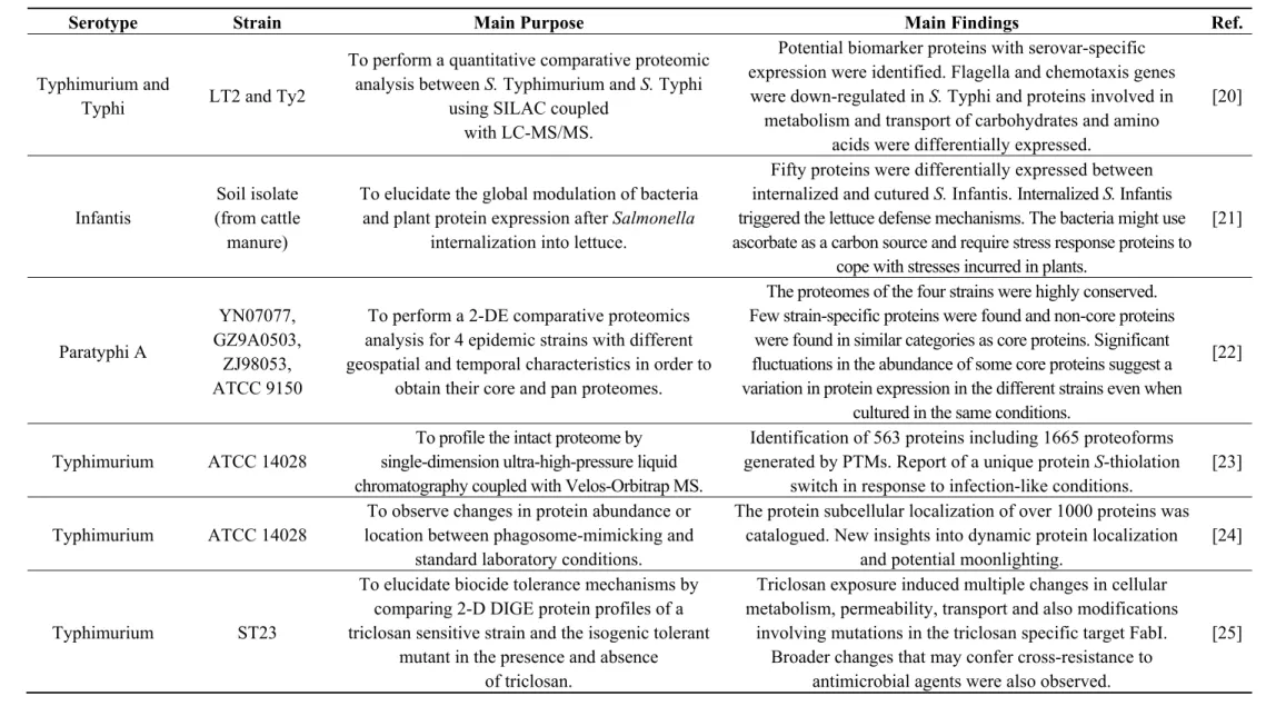
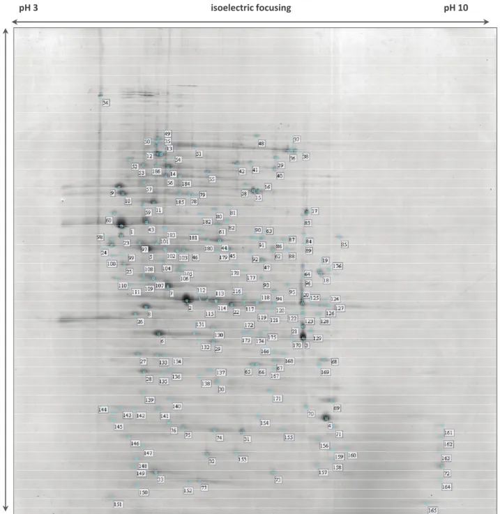
Figure 1. Stained 2-DE (two dimensional gel electrophoresis) gel of total proteins of Salmonella Typhimurium Se20 (phage type DT104B) using IPG (Immobiline™ pH Gradient) strips pH 3–10 NL (non-linear) for the first dimension. Numbered spots were excised for analysis by in-gel digestion and MALDI-TOF MS (matrix-assisted laser desorption/ionization mass spectrometry) identification, described in Table S1.
pH 3 isoelectric focusing pH 10
SDS
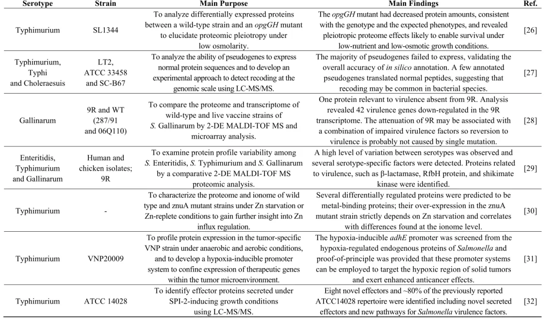
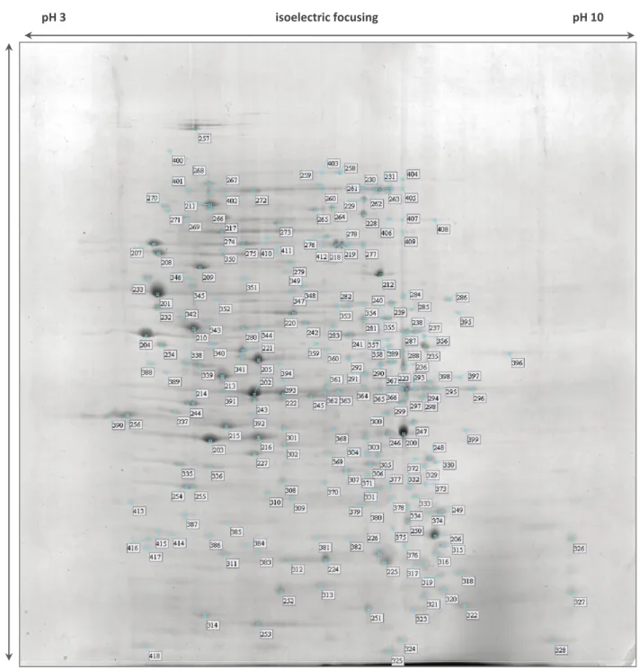
Figure 2. Stained 2-DE gel of total proteins of Salmonella Typhimurium SL1344 using IPG strips pH 3–10 NL for the first dimension. Numbered spots were excised for analysis by in-gel digestion and MALDI-TOF MS identification, described in Table S2.
pH 3 isoelectric focusing pH 10
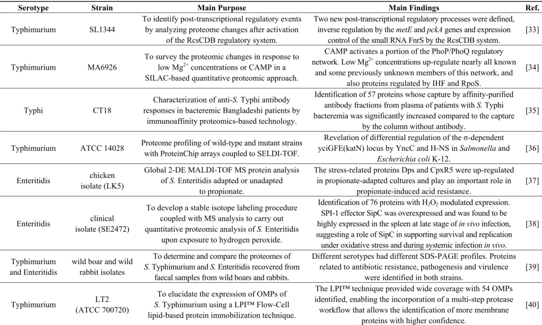
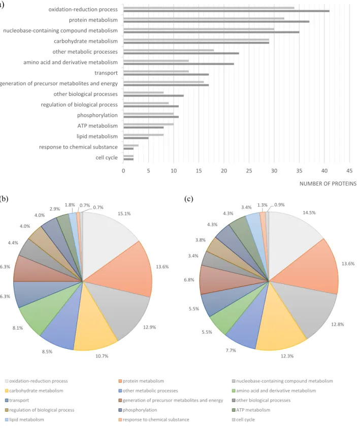
Figure 3. Functional classification of proteins identified in the Se20 and SL1344 strains based on Gene Ontology. (a) Number of proteins in each category for Se20 (light gray) and SL1344 (dark gray); Relative percentages of protein functions in (b) Se20 and (c) SL1344. As this classification reflects the fact that single proteins can be involved in more than one process, the sum of proteins in all categories is higher than the total number of unique proteins identified.
0 5 10 15 20 25 30 35 40 45
cell cycle response to chemical substance lipid metabolism ATP metabolism phosphorylation regulation of biological process other biological processes generation of precursor metabolites and energy transport amino acid and derivative metabolism other metabolic processes carbohydrate metabolism nucleobase-containing compound metabolism protein metabolism oxidation-reduction process
NUMBER OF PROTEINS
(a)
15.1%
13.6%
12.9%
10.7% 8.5%
8.1% 6.3% 6.3%
4.4% 4.0%
4.0%
2.9% 1.8% 0.7% 0.7%
oxidation-reduction process protein metabolism nucleobase-containing compound metabolism carbohydrate metabolism other metabolic processes amino acid and derivative metabolism transport generation of precursor metabolites and energy other biological processes regulation of biological process phosphorylation ATP metabolism lipid metabolism response to chemical substance cell cycle
14.5%
13.6%
12.8%
12.3% 7.7%
5.5% 5.5% 6.8%
3.4% 3.8%
4.3% 4.3%
3.4% 1.3% 0.9%
Table 2. List of some relevant proteins exclusively identified either in Salmonella Typhimurium strain Se20 or in Salmonella Typhimurium strain SL1344.
Strain Spots Protein Gene Biological Process
Se20
24/98 Flagellin fljB ciliary or bacterial-type
flagellar motility
75 aminoglycoside 6'-N-acetyltransferase
type Ib-cr, AAC(6')-Ib-cr4 aac(6')-Ib-cr4 metabolic process
99 ethanolamine ammonia-lyase heavy subunit eutB cellular amino acid metabolic process
106 ATP-dependent protease hslU
ATP catabolic process, proteolysis, response to stress, protein unfolding
134 universal stress protein E uspE response to stress
142/143 aminoglycoside resistance protein A strA response to antibiotic
148 Chain E, Alkyl Hydroperoxide Reductase C
(Substrate-Ready Conformation) ahpC
response to oxidative stress, oxidation-reduction process
182 5'-nucleotidase ushA dephosphorylation,
nucleotide catabolic process
SL1344
205/341 arginine deiminase arcA protein citrullination
215 ornithine carbamoyltransferase arcB ornithine metabolic process
225 fumarate reductase iron-sulfur subunit frdB tricarboxylic acid cycle
227 carbamate kinase arcC arginine metabolic process
237/238/287 glycerol-3-phosphate dehydrogenase glpD glycerol-3-phosphate metabolic process
240 inosine 5'-monophosphate dehydrogenase guaB purine nucleotide
biosynthetic process
259 NADH dehydrogenase subunit G nuoG ATP synthesis coupled
electron transport
296 molecular chaperone DnaJ dnaJ response to stress
332 Hydrogenase -
-344 Phosphoglucomutase pgm carbohydrate metabolic
process
346 oligopeptidase A prlC proteolysis
378 exonuclease III xth DNA catabolic process,
exonucleolytic
396 serine endoprotease htrA proteolysis
406 cell invasion protein SipA sipA pathogenesis
The 225 amino acid protein here detected was predicted by ORF (open reading frame) Finder analysis to have a longer N-terminal length when comparing to other previously described functional aac(6')-Ib variants [66]. The position of spot 75 matches the theoretical molecular weight (MW) of 25031 Da and isoelectric point (pI) value of 5.2 estimated for AAC(6')-Ib-cr4. The AAC(6')-Ib-cr protein is a variant of the widespread aminoglycoside acetyltransferase AAC(6')-Ib that is usually responsible for resistance to amikacin, kanamycin and tobramycin. The AAC(6')-Ib-cr variant also acetylates ciprofloxacin and norfloxacin, but less efficiently than aminoglycoside substrates [70]. Acetylation occurs at the amino nitrogen on the piperazinyl substituent, so only fluoroquinolones with an unsubstituted piperazinyl group are substrates of AAC(6')-Ib-cr. Even though the presence of the aac(6')-Ib-cr gene confers only low-level resistance to substrate fluoroquinolones, it may facilitate the survival of target-site mutants with a 10-fold increase in their mutant prevention concentration [71].
The aminoglycoside resistance protein A, coded by the previously detected strA gene, is an aminoglycoside 3'-phosphotransferase that catalyzes the transfer of the gamma-phosphoryl group from ATP to aminoglycoside antimicrobials, inactivating them [72]. Theoretically this protein has a MW of
30,474 Da and a pI value of 4.7. In the 2-DE gel, the two corresponding spots have a MW similar to
the theoretical value, however spot 142 is slightly more basic than spot 143 (Figure 1). Single proteins separated by 2-DE frequently exhibit multiple spots in the first dimension. These so-called “charge trains” can be caused by isoform differences and post-translational modifications (PTMs). Some PTMs, such as phosphorylation, deamidation, desulfuration or acylation, can lead to electrical charge heterogeneity with minor modifications in molecular weight. Cysteine oxidation has also been reported to be responsible for pI basic shifts [15]. Nontheless, “charge trains” can also be considered artifacts due to the sample treatment and analytical procedures employed, such as carbamylation in the presence of urea or acrylamide adduct formation during electrophoresis [73].
The two strains analysed in this study present phenotypic resistance to sulphonamides. The target of sulfonamide antimicrobials and the basis for their selective effect on bacteria is dihydropteroate synthase (DHPS) in the folic acid pathway [74]. DHPS is a functional homodimer that, in prokaryotes, catalyzes the condensation of p-aminobenzoic acid (PABA) in the de novo biosynthesis of folate, an essential cofactor in protein and nucleic acid biosynthesis [72]. Higher eukaryotes are able to utilize dietary folate, so they do not have DHPS enzymes. Sulfonamides act either by competitively inhibiting DHPS by structural similarity with the PABA substrate or by functioning as alternative substrates for DHPS, forming pterin adducts that are unable to participate in folate biosynthesis [74]. DHPS was identified in both Se20 (spot 154, Figure 1, Table S1) and SL1344 (spot 382, Figure 2, Table S2) strains. In enteric Gram-negative bacteria, sulfonamide clinical resistance is plasmid-mediated by genes such as sul1 and sul2, which encode alternative drug-resistance variants of DHPS that show high insensitivity to sulfonamide drugs but bind normally to the PABA substrate [74]. The DHPS identified in this study (AC: S5HED7) is plasmid-encoded and shows a 100% sequence identity with the S. Typhimurium SL1344 DHPS type-2 (AC: H8WV44), which is present on the pRSF1010SL1344 plasmid. The sul2 gene, which encodes the type-2 DHPS, was previously reported in the Se20 strain [18] and also in the SL1344 strain [75].
cell invasion, bacteriophage binding, and conjugation [76]. OmpA was detected in two different gel locations in both strains. The more abundant spots, 6 for Se20 (Figure 1) and 203 for SL1344 (Figure 2), were found where expected for proteins with theoretical MW of 37,606 Da and pI of 5.5. The less
abundant spots, 141 for Se20 (Figure 1) and 386 for SL1344 (Figure 2), had the same pI but a lower MW of approximately 30 kDa. However, these results are not unexpected as OmpA is known to run
anomalously in SDS-PAGE [76]. β-barrel structures of bacterial outer membrane proteins are usually very stable and survive the SDS denaturing treatment at room temperature. As a result, native and denatured forms of many OMPs migrate at two different apparent molecular weights in SDS-PAGE. The OmpA protein was previously reported to migrate at 30 kDa in its native compacted form [77].
The porin outer membrane protein C (OmpC) was also identified. Antimicrobials such as ciprofloxacin, norfloxacin, cefepime and ceftriaxone strongly interact with OmpC, and so their translocation through this channel is facilitated [78]. The ion channel protein Tsx, which is also likely to play a role in antimicrobial resistance [59], was also identified. TolB, a periplasmic protein associated with the outer-membrane protein Pal, was detected. TolB belongs to the Tol-Pal system that is well conserved among Gram-negative bacteria and plays several roles, including lipopolysaccharide O-antigen surface expression, outer membrane stability, uptake of filamentous phage DNA, resistance to detergents and virulence [79].
The majority of the proteins identified in this study are involved in oxidation-reduction processes (Figure 3). One of the proteins identified in this class was the alkyl hydroperoxide reductase subunit C (AhpC), also named alkyl hydroperoxide reductase protein C22 (spot 148). In bacteria, this enzyme is responsible for hydrogen peroxide removal, a response to oxidative stress. The peroxide-reducing activities of AhpC help to protect pathogenic bacteria from the host immune response [80]; therefore the identification of this protein in the Se20 strain is in accordance with its host-adapted phenotype. The AhpC protein has recently been considered as a possible target for the development of new antimicrobial agents [80]. Other stress response proteins were also identified, namely the heat shock chaperone proteins DnaK, DnaJ, HtpG, HslU, HtrA, GroL, the protein disaggregation chaperone and the universal stress protein E (UspE). Another heat shock protein identified was the Lon protease (HAMAP-Rule MF_01973), which is required for cellular homeostasis and for survival from DNA damage and developmental changes induced by stress.
Bactericidal antimicrobials can induce cell death by stimulating the production of reactive oxygen species, principally O2−, which induces oxidative damage [81]. Superoxide dismutases are responsible
for the destruction of these superoxide anion radicals. In addition to their detoxifying function, bacterial superoxide dismutases have also been shown to be important virulence factors [82]. In S. Typhimurium, SodA and SodB are cytoplasmic superoxide dismutases that require manganese and iron respectively as cofactors [83]. Some studies show that the expression of superoxide dismutase enzymes increases in response to antimicrobial stress [81]. Here, both cytoplasmic superoxide dismutases, SodA (AC: P43019) and SodB (AC: P0A2F5), were identified.
for in vivo multiplication, confer an advantage in the early stage of infection allowing rapid invasion of host cells, and also activate the host immune system while inactivating epithelial cell apoptosis [84]. Individual Salmonella serotypes usually alternate between the production of two antigenic forms of flagella, phase I and phase II, each specified by separate structural genes, fliC and fljB. Our results show that although the phase II flagellin seems to be higly expressed in the Se20 clinical strain and absent from SL1344, the phase I flagellin middle domain variant C12 was identified in both strains (spot 100 and spot 204), and shows a considerable higer expression in the SL1344 reference strain.
An additional protein identified in the clinical strain that may contribute to the pathogenic phenotype of DT104 is the ethanolamine ammonia-lyase (spot 99). Ethanolamine can be readily derived from cell membranes and therefore is available in the large intestine due to enterocyte turnover. Some bacteria, including Salmonella, are able to use ethanolamine as a source of carbon and/or nitrogen in a process that involves the conversion of ethanolamine into acetaldehyde and ammonia by an ethanolamine ammonia lyase [85]. Evidence was provided that in the inflamed intestine, S. Typhimurium has a growth advantage due to its ability to respire ethanolamine that is not utilizable by competing bacteria, showing a direct link between ethanolamine utilization and bacterial pathogenesis [86].
Further, concerning the Se20 clinical strain, the 5'-nucleotidase UDP (uridine diphosphate)-sugar hydrolase (UshA), was identified in spot 182, and appears to be absent in the reference strain. In Escherichia coli, UshA has an important function in nucleotide salvage. However, UshA can also function as a phosphate starvation-induced 5'-nucleotidase, being required for growth when nucleotides are provided as the only source of phosphate [87]. This condition is likely to be significant for bacterial growth in the wild [87], which may play a role in the worldwide dissemination on this strain.
The majority of proteins identified exclusively in SL1344 also reflect the virulence characteristics of this strain. The proteins arginine deiminase (ADI), ornithine carbamoyltransferase and carbamate kinase, constitute the ADI system that, besides its metabolic functions, has also been associated with virulence in some pathogens. These three proteins were identified in the high intensity spots 205, 215 and 227, respectively. It was previously established that the ADI pathway contributes to Salmonella pathogenesis and that arginine deiminase activity has an active role in the successful infection of mammalian hosts by S. Typhimurium [88].
Another high intensity spot was identified as the fumarate reductase iron-sulfur subunit (spot 225). A recent study provided evidence that fumarate reductase is associated with the bacterial flagellar switch complex, which determines the direction of flagellar rotation and is essential for chemotaxis. Fumarate influences the interaction of fumarate reductase with the FliG switch thus affecting flagellar assembly and rotation [89].
The study of specific proteins participating in de novo purine synthesis have shown that the absence of key enzymes in the pathway, namely the inosine 5'-monophosphate dehydrogenase GuaB, can severely attenuate growth rates and directly affect virulence in S. Typhimurium [90]. GuaB was only identified in spot 240 of SL1344.
The NADH dehydrogenase I coded by the nuoG gene (spot 259) is induced under microaerophilic and stationary-phase growth conditions. Mutations in nuo genes affect several mechanisms of microbial physiology and biochemistry which have direct consequences in Salmonella virulence [92].
Hydrogenase, identified in spot 332, has also been described to be essential to virulence in S. Typhimurium. The usage of respiratory hydrogen as a critical growth substrate for energy production allows colonization of the animal host and subsequent virulence during infection. Therefore, hydrogenases can represent potential therapeutic targets to combat Salmonella infections [93].
The phosphoglucomutase enzyme (spot 344) is important in the virulence of numerous pathogens and was recently reported to be required by S. Typhimurium for O-antigen production, resistance to antimicrobial peptides and in vivo fitness [94]. Oligopeptidase A, which is involved in degradation of signal peptides after they are released from precursor forms of secreted proteins, is also a virulence factor and heat shock protein that was identified only in the reference strain (spot 346) [95].
The effector SipA protein identified in SL1344 spot 406 is secreted by the centisome 63 type III secretion system encoded by Salmonella pathogenicity island 1 and is known to be a key factor in the invasion of epithelial cells by S. Typhimurium [96].
Finally, the glycerol-3-phosphate dehydrogenase GlpD was identified in spots 237, 238 and 287 of the SL1344 strain. In a recent study in E. coli, GlpD overexpression resulted in high persisters, i.e., in a bacterial subpopulation capable of surviving antimicrobial exposure or other lethal treatments [97].
Effective therapies to treat resistant bacteria are urgently needed. We must understand the mechanisms underlying antimicrobial drug resistance in more detail, as no single bacterial strain can truly represent its species [7]. In this proteomic analysis we provide a physiological map and an overview of global protein expression of Salmonella Typhimurium Se20 (phage type DT104B) and SL1344 strains under normal growth conditions [14].
3. Experimental Section
3.1. Bacterial Strains and Growth Conditions
Two strains of S. Typhimurium, Se20 [18] and SL1344 [19], were included in this study. Se20 (phage type DT104B) is a previously characterized strain that was recovered from a faecal sample of an elderly patient who was admitted to a Spanish hospital with acute gastroenteritis. The patient was treated for 7 days with ciprofloxacin, and in vivo selection of quinolone resistance was observed post-treatment [18]. Frozen cell stocks of S. Typhimurium Se20 and SL1344 were streaked onto LB (Luria-Bertani) agar (Miller, Scharlau Chemie, S.A. Barcelona, Spain) plates and grown overnight at 37 °C. Pre-cultures were prepared by inoculation of 10 mL of LB broth (Miller, Scharlau Chemie, S.A.) with single colonies of each strain with further overnight incubation at 37 °C. Pre-cultures were diluted to an optical density at 600 nm (OD600) of 0.02 in a final volume of 10 mL of LB broth, and
incubated at 37 °C for 5 h. 3.2. Protein Extraction
Cultures were harvested in the late exponential phase (OD600 of 0.5) by centrifugation at 10,000× g
Bacterial cell pellets were suspended in 0.2 mL of solubilization buffer (10% (w/v) SDS and 12% (w/v) Tris) and lysed by sonication (4 × 10 s, 20 kHz, 100 W) at 4 °C. Cell debris were removed by centrifugation at 14,000× g for 30 min at 4 °C and proteins were further precipitated with cold trichloroacetic acid (TCA) at a final concentration of 20%. The proteins were recovered by centrifugation at 15,000× g for 25 min at 4 °C and washed twice by centrifugation in 0.3 mL of cold acetone for 10 min. Protein pellets were left to air-dry at room temperature. Proteins were extracted from three independent cultures andquantified by the Bradford method [98].
3.3. Two-Dimensional Gel Electrophoresis
Two-dimensional gel electrophoresis (2-DE) was performed according to the principles of O’Farrell [99] but with Immobiline™ pH Gradient (IPG) technology [100]. For isoelectric focusing, precast 13-cm IPG strips with a non-linear gradient from pH 3 to pH 10 (pH 3–10 NL, Amersham Biosciences, GE Healthcare, Uppsala, Sweden) were passively rehydrated overnight (16 h) at room temperature in a reswelling tray with 250 μL of rehydration buffer (8 M urea, 1% CHAPS (3-[(3-cholamidopropyl)-dimethylammonio]-propane-sulfonate), 0.4% DTT (dithiothreitol), 0.5% carrier ampholyte IPG buffer pH 3–10), covered with Dry-Strip Cover Fluid (Plus One, Amersham Biosciences, GE Healthcare). The protein samples (100 μg) were cup-loaded onto the rehydrated IPG strips [101] and focused at 500 V for 1 h, 1000 V for 8 h, 8000 V for 3 h and finally 8000 V incremented to 21,881 Vh in an Ettan™ IPGPhor II™ apparatus (Amersham Biosciences, GE Healthcare). Before the second dimension of electrophoresis, the focused IPG strips were equilibrated twice, each time for 15 min as follows. For the first equilibration, 1% DTT was added to equilibration stock buffer (6 M urea, 30% (w/v) glycerol, 2% (w/v) SDS in 0.05 M Tris–HCl buffer pH 8.8) and in the second equilibration, 4% iodoacetamide was added to equilibration stock buffer. Bromophenol blue was also added to both solutions. The equilibrated IPG strips were then gently rinsed with SDS electrophoresis buffer, blotted to remove excessive buffer, and then applied to 12.52% polyacrylamide gels in a Hoefer™ SE 600 Ruby® (Amersham Biosciences, GE Healthcare) unit. The Laemmli SDS-PAGE technique was used with some modifications [102]. After the second dimension of separation, the 2-DE gels were fixed in a 40% methanol/10% acetic acid solution for 1 h with agitation, then stained overnight in Coomassie Brilliant Blue G-250 with agitation [103]. Gels were rinsed twice with 40% methanol for 45 min to remove excess staining and scanned on a flatbed scanner (Umax PowerLook 1100, Fremont, CA, USA). At least three 2-DE gels were run per protein sample. Images were analyzed using Image Master 5.0 software (Amersham Biosciences, GE Healthcare).
3.4. Tryptic Digestion of In-Gel Proteins
was stopped with 25 μL of 5% formic acid solution and the liquid mixture was collected. Finally, 25 μL of 50% (v/v) ACN/0.1% (v/v) TFA (trifluoroacetic acid) solution was added to the remaining gel pieces to increase the recovery of peptides. The extracted fractions were combined and dried in a Speed-Vac.
3.5. Peptide Mass Fingerprinting
Prior to protein digest analysis, each tryptic peptide mixture was ressuspended in 10 μL of 0.3% formic acid. Then, 1 μL of the resuspension was hand-spotted onto a MALDI target plate (384-spot ground steel plate), overlaid with 1 μL of α-cyano-4-hydroxycinnamic acid matrix solution (7 mg/mL in 0.1% (v/v) TFA/50% (v/v) ACN/8 mM ammonium phosphate) and dried under ambient conditions. All mass spectra were generated on a MALDI-TOF/TOF mass spectrometer Ultraflex (Bruker Daltonics, Bremen, Germany), operating in positive ion reflectron-mode. Spectra were acquired in the m/z range of 600–3500. A total of 500 spectra were acquired for each sample at a laser frequency of 50 Hz. External calibration was performed with the [M + H]+ monoisotopic peaks of bradykinin 1–7 (m/z 757.3992), angiotensin II (m/z 1046.5418), angiotensin I (m/z 1296.6848), substance P (m/z 1758.9326), ACTH clip 1–17 (m/z 2093.0862), ACTH18–39 (m/z 2465.1983) and somatostatin 28 (m/z 3147.4710). The MASCOT search engine was used to match the determined peptide masses to two customized databases: Salmonella Typhimurium from NCBI RefSeq (National Center for Biotechnology Information, U.S. National Library of Medicine, Bethesda, MD, USA), comprising 231,752 entries (Release 62); and Salmonella spp. from Swiss-Prot (Swiss Institute of Bioinformatics, Geneva, Switzerland; The EMBL Outstation—The European Bioinformatics Institute, Cambridge, UK), comprising 12,772 entries (Release 2013_11). The Max Planck Institute of Biochemistry, Martinsried, common contaminants collection (MPI) was included in both databases in order to avoid misleading matches in the presence of contaminant proteins. The search criteria adopted were: (i) proteolytic enzyme, trypsin/P; (ii) one missed cleavage allowed; (iii) fixed modifications, carbamidomethylation; (iv) variable modifications, methionine oxidation; and (v) a peptide tolerance error window up to 50 ppm. A match was considered significant when the probability of it being a random event was below the default significance threshold used (p < 0.05), i.e., with a frequency less than 5%.
4. Conclusions
Acknowledgments
We would like to thank Derek Pickard (Welcome Trust Sanger Institute, Cambridge, UK) for generously providing reference strain SL1344. Susana Correia (SFRH/BD/75160/2010), Júlio D. Nunes-Miranda (SFRH/BD/80496/2011) and Luís Pinto (SFRH/BD/81307/2011) are supported by PhD fellowships granted by FCT (Fundacão para a Ciência e a Tecnologia) and POPH/FSE (Programa Operacional Potencial Humano/Fundo Social Europeu).
Author Contributions
This study was conducted in partial fulfillment of Susana Correia (S.C.) PhD thesis. S.C. performed most of the laboratory work (sample preparation, SDS-PAGE, 2-DE and MS analysis) and manuscript preparation. S.C., J.D.N.-M., H.M.S. and J.L.C. carried MS/MS and bioinformatics analysis. L.P. helped in SDS-PAGE and 2-DE. S.C., M.T., Y.S., C.T. and P.P. were responsible for the microbiology task, and contributed substantially to help the research. G.I., responsible for the Functional Genomics and Proteomics Laboratory, conceived, designed, implemented and coordinated the study. All authors read and also reviewed the article and approved the final version.
Conflicts of Interest
The authors declare no conflict of interest. References
1. Pacheco, R.; Correia, S.; Poeta, P.; Pinto, L.; Igrejas, G. The role of proteomics in elucidating multiple antibiotic resistance in Salmonella and in novel antibacterial discovery. In Salmonella—Distribution, Adaptation, Control Measures and Molecular Technologies; Annous, B.A., Gurtler, J.B., Eds.; InTech: Rijeka, Croatia, 2012; pp. 187–220.
2. Su, L.H.; Wu, T.L.; Chiu, C.H. Development of carbapenem resistance during therapy for non-typhoid Salmonella infection. Clin. Microbiol. Infect. 2012, 18, E91–E94.
3. Mather, A.E.; Reid, S.W.; Maskell, D.J.; Parkhill, J.; Fookes, M.C.; Harris, S.R.; Brown, D.J.; Coia, J.E.; Mulvey, M.R.; Gilmour, M.W.; et al. Distinguishable epidemics of multidrug-resistant Salmonella Typhimurium DT104 in different hosts. Science 2013, 341, 1514–1517.
4. Marathe, S.A.; Kumar, R.; Ajitkumar, P.; Nagaraja, V.; Chakravortty, D. Curcumin reduces the antimicrobial activity of ciprofloxacin against Salmonella Typhimurium and Salmonella Typhi. J. Antimicrob. Chemother. 2013, 68, 139–152.
5. Bumann, D. Pathogen proteomes during infection: A basis for infection research and novel control strategies. J. Proteomics 2010, 73, 2267–2276.
6. Humphrey, S.; Clark, L.F.; Humphrey, T.J.; Jepson, M.A. Enhanced recovery of Salmonella Typhimurium DT104 from exposure to stress at low temperature. Microbiology 2011, 157, 1103–1114.
8. Threlfall, E.J. Epidemic Salmonella Typhimurium DT 104—A truly international multiresistant clone. J. Antimicrob. Chemother. 2000, 46, 7–10.
9. Mendoza Mdel, C.; Herrero, A.; Rodicio, M.R. Evolutionary engineering in Salmonella: Emergence of hybrid virulence-resistance plasmids in non-typhoid serotypes. Enferm. Infect. Microbiol. Clin. 2009, 27, 37–43.
10. Giraud, E.; Baucheron, S.; Virlogeux-Payant, I.; Nishino, K.; Cloeckaert, A. Effects of natural mutations in the ramRA locus on invasiveness of epidemic fluoroquinolone-resistant Salmonella enterica serovar Typhimurium isolates. J. Infect. Dis. 2013, 207, 794–802.
11. Research Topic in Antimicrobials, Resistance and Chemotherapy. Available online: http://www.frontiersin.org/antimicrobials,_resistance_and_chemotherapy/researchtopics/proteom ics_of_antimicrobial_re/1620 (accessed on 28 March 2014).
12. Fernandez-Reyes, M.; Rodriguez-Falcon, M.; Chiva, C.; Pachon, J.; Andreu, D.; Rivas, L. The cost of resistance to colistin in Acinetobacter baumannii: A proteomic perspective. Proteomics 2009, 9, 1632–1645.
13. Becker, D.; Selbach, M.; Rollenhagen, C.; Ballmaier, M.; Meyer, T.F.; Mann, M.; Bumann, D. Robust Salmonella metabolism limits possibilities for new antimicrobials. Nature 2006, 440, 303–307.
14. Lima, T.B.; Pinto, M.F.; Ribeiro, S.M.; de Lima, L.A.; Viana, J.C.; Gomes Junior, N.; Candido Ede, S.; Dias, S.C.; Franco, O.L. Bacterial resistance mechanism: What proteomics can elucidate. FASEB J. 2013, 27, 1291–1303.
15. Kleinert, P.; Kuster, T.; Arnold, D.; Jaeken, J.; Heizmann, C.W.; Troxler, H. Effect of glycosylation on the protein pattern in 2-D-gel electrophoresis. Proteomics 2007, 7, 15–22.
16. Lee, A.Y.; Park, S.G.; Jang, M.; Cho, S.; Myung, P.K.; Kim, Y.R.; Rhee, J.H.; Lee, D.H.; Park, B.C. Proteomic analysis of pathogenic bacterium Vibrio vulnificus. Proteomics 2006, 6, 1283–1289.
17. Vranakis, I.; Goniotakis, I.; Psaroulaki, A.; Sandalakis, V.; Tselentis, Y.; Gevaert, K.; Tsiotis, G. Proteome studies of bacterial antibiotic resistance mechanisms. J. Proteomics 2014, 97, 88–99. 18. De Toro, M.; Rojo-Bezares, B.; Vinue, L.; Undabeitia, E.; Torres, C.; Saenz, Y. In vivo
selection of aac(6')-Ib-cr and mutations in the gyrA gene in a clinical qnrS1-positive Salmonella enterica serovar Typhimurium DT104B strain recovered after fluoroquinolone treatment. J. Antimicrob. Chemother. 2010, 65, 1945–1949.
19. Hoiseth, S.K.; Stocker, B.A. Aromatic-dependent Salmonella Typhimurium are non-virulent and effective as live vaccines. Nature 1981, 291, 238–239.
20. Wang, Y.; Huang, K.Y.; Huo, Y. Proteomic comparison between Salmonella Typhimurium and Salmonella Typhi. J. Microbiol. 2014, 52, 71–76.
21. Zhang, Y.; Nandakumar, R.; Bartelt-Hunt, S.L.; Snow, D.D.; Hodges, L.; Li, X. Quantitative proteomic analysis of the Salmonella-lettuce interaction. Microb. Biotechnol. 2014, doi:10.1111/1751-7915.12114.
23. Ansong, C.; Wu, S.; Meng, D.; Liu, X.; Brewer, H.M.; Deatherage Kaiser, B.L.; Nakayasu, E.S.; Cort, J.R.; Pevzner, P.; Smith, R.D.; et al. Top-down proteomics reveals a unique protein S-thiolation switch in Salmonella Typhimurium in response to infection-like conditions. Proc. Natl. Acad. Sci. USA 2013, 110, 10153–10158.
24. Brown, R.N.; Sanford, J.A.; Park, J.H.; Deatherage, B.L.; Champion, B.L.; Smith, R.D.; Heffron, F.; Adkins, J.N. A Comprehensive Subcellular Proteomic Survey of Salmonella Grown under Phagosome-Mimicking versus Standard Laboratory Conditions. Int. J. Proteomics 2012, 2012, 123076.
25. Condell, O.; Sheridan, A.; Power, K.A.; Bonilla-Santiago, R.; Sergeant, K.; Renaut, J.; Burgess, C.; Fanning, S.; Nally, J.E. Comparative proteomic analysis of Salmonella tolerance to the biocide active agent triclosan. J. Proteomics 2012, 75, 4505–4519.
26. Cooper, B.; Chen, R.; Garrett, W.M.; Murphy, C.; Chang, C.; Tucker, M.L.; Bhagwat, A.A. Proteomic pleiotropy of OpgGH, an operon necessary for efficient growth of Salmonella enterica serovar typhimurium under low-osmotic conditions. J. Proteome Res. 2012, 11, 1720–1727. 27. Feng, Y.; Chien, K.Y.; Chen, H.L.; Chiu, C.H. Pseudogene recoding revealed from proteomic
analysis of Salmonella serovars. J. Proteome Res. 2012, 11, 1715–1719.
28. Kang, M.S.; Kwon, Y.K.; Kim, H.R.; Oh, J.Y.; Kim, M.J.; An, B.K.; Shin, E.G.; Kwon, J.H.; Park, C.K. Comparative proteome and transcriptome analyses of wild-type and live vaccine strains of Salmonella enterica serovar Gallinarum. Vaccine 2012, 30, 6368–6375.
29. Sun, J.S.; Hahn, T.W. Comparative proteomic analysis of Salmonella enterica serovars Enteritidis, Typhimurium and Gallinarum. J. Vet. Med. Sci./Jpn. Soc. Vet. Sci. 2012, 74, 285–291.
30. Ciavardelli, D.; Ammendola, S.; Ronci, M.; Consalvo, A.; Marzano, V.; Lipoma, M.; Sacchetta, P.; Federici, G.; di Ilio, C.; Battistoni, A.; et al. Phenotypic profile linked to inhibition of the major Zn influx system in Salmonella enterica: Proteomics and ionomics investigations. Mol. Biosyst. 2011, 7, 608–619.
31. Chen, J.; Wei, D.; Zhuang, H.; Qiao, Y.; Tang, B.; Zhang, X.; Wei, J.; Fang, S.; Chen, G.; Du, P.; et al. Proteomic screening of anaerobically regulated promoters from Salmonella and its antitumor applications. Mol. Cell. Proteomics: MCP 2011, 10, M111 009399.
32. Niemann, G.S.; Brown, R.N.; Gustin, J.K.; Stufkens, A.; Shaikh-Kidwai, A.S.; Li, J.; McDermott, J.E.; Brewer, H.M.; Schepmoes, A.; Smith, R.D.; et al. Discovery of novel secreted virulence factors from Salmonella enterica serovar Typhimurium by proteomic analysis of culture supernatants. Infect. Immun. 2011, 79, 33–43.
33. Paradela, A.; Mariscotti, J.F.; Navajas, R.; Ramos-Fernandez, A.; Albar, J.P.; Garcia-del Portillo, F. Inverse regulation in the metabolic genes pckA and metE revealed by proteomic analysis of the Salmonella RcsCDB regulon. J. Proteome Res. 2011, 10, 3386–3398.
34. Yu, J.L.; Guo, L. Quantitative proteomic analysis of Salmonella enterica serovar Typhimurium under PhoP/PhoQ activation conditions. J. Proteome Res. 2011, 10, 2992–3002.
36. Beraud, M.; Kolb, A.; Monteil, V.; D’Alayer, J.; Norel, F. A proteomic analysis reveals differential regulation of the σS-dependent yciGFE(katN) locus by YncC and H-NS in Salmonella and Escherichia coli K-12. Mol. Cell. Proteomics: MCP 2010, 9, 2601–2616.
37. Calhoun, L.N.; Liyanage, R.; Lay, J.O., Jr.; Kwon, Y.M. Proteomic analysis of Salmonella enterica serovar Enteritidis following propionate adaptation. BMC Microbiol. 2010, 10, 249. 38. Kim, K.; Yang, E.; Vu, G.P.; Gong, H.; Su, J.; Liu, F.; Lu, S. Mass spectrometry-based
quantitative proteomic analysis of Salmonella enterica serovar Enteritidis protein expression upon exposure to hydrogen peroxide. BMC Microbiol. 2010, 10, 166.
39. Pinto, L.; Poeta, P.; Vieira, S.; Caleja, C.; Radhouani, H.; Carvalho, C.; Vieira-Pinto, M.; Themudo, P.; Torres, C.; Vitorino, R.; et al. Genomic and proteomic evaluation of antibiotic resistance in Salmonella strains. J. Proteomics 2010, 73, 1535–1541.
40. Chooneea, D.; Karlsson, R.; Encheva, V.; Arnold, C.; Appleton, H.; Shah, H. Elucidation of the outer membrane proteome of Salmonella enterica serovar Typhimurium utilising a lipid-based protein immobilization technique. BMC Microbiol. 2010, 10, 44.
41. Di Pasqua, R.; Mamone, G.; Ferranti, P.; Ercolini, D.; Mauriello, G. Changes in the proteome of Salmonella enterica serovar Thompson as stress adaptation to sublethal concentrations of thymol. Proteomics 2010, 10, 1040–1049.
42. Charles, R.C.; Harris, J.B.; Chase, M.R.; Lebrun, L.M.; Sheikh, A.; LaRocque, R.C.; Logvinenko, T.; Rollins, S.M.; Tarique, A.; Hohmann, E.L.; et al. Comparative proteomic analysis of the PhoP regulon in Salmonella enterica serovar Typhi versus Typhimurium. PLoS One 2009, 4, e6994.
43. Encheva, V.; Shah, H.N.; Gharbia, S.E. Proteomic analysis of the adaptive response of Salmonella enterica serovar Typhimurium to growth under anaerobic conditions. Microbiology 2009, 155, 2429–2441.
44. Kim, S.R.; Kim, H.T.; Park, H.J.; Kim, S.; Choi, H.J.; Hwang, G.S.; Yi, J.H.; Ryu do, H.; Kim, K.H. Fatty acid profiling and proteomic analysis of Salmonella enterica serotype Typhimurium inactivated with supercritical carbon dioxide. Int. J. Food Microbiol. 2009, 134, 190–195.
45. Osman, K.M.; Ali, M.M.; Radwan, M.I.; Kim, H.K.; Han, J. Comparative proteomic analysis on Salmonella Gallinarum and Salmonella Enteritidis exploring proteins that may incorporate host adaptation in poultry. J. Proteomics 2009, 72, 815–821.
46. Shi, L.; Chowdhury, S.M.; Smallwood, H.S.; Yoon, H.; Mottaz-Brewer, H.M.; Norbeck, A.D.; McDermott, J.E.; Clauss, T.R.; Heffron, F.; Smith, R.D.; et al. Proteomic investigation of the time course responses of RAW 264.7 macrophages to infection with Salmonella enterica. Infect. Immun. 2009, 77, 3227–3333.
47. Shi, L.; Ansong, C.; Smallwood, H.; Rommereim, L.; McDermott, J.E.; Brewer, H.M.; Norbeck, A.D.; Taylor, R.C.; Gustin, J.K.; Heffron, F.; et al. Proteome of Salmonella enterica serotype Typhimurium grown in a Low Mg/pH medium. J. Proteomics Bioinform. 2009, 2, 388–397.
49. Sonck, K.A.; Kint, G.; Schoofs, G.; Vander Wauven, C.; Vanderleyden, J.; de Keersmaecker, S.C. The proteome of Salmonella Typhimurium grown under in vivo-mimicking conditions. Proteomics 2009, 9, 565–579.
50. Webber, M.A.; Coldham, N.G.; Woodward, M.J.; Piddock, L.J. Proteomic analysis of triclosan resistance in Salmonella enterica serovar Typhimurium. J. Antimicrob. Chemother. 2008, 62, 92–97.
51. Singh, R.; Shasany, A.K.; Aggarwal, A.; Sinha, S.; Sisodia, B.S.; Khanuja, S.P.; Misra, R. Low molecular weight proteins of outer membrane of Salmonella Typhimurium are immunogenic in Salmonella induced reactive arthritis revealed by proteomics. Clin. Exp. Immunol. 2007, 148, 486–493.
52. Hu, W.S.; Lin, Y.H.; Shih, C.C. A proteomic approach to study Salmonella enterica serovar Typhimurium putative transporter YjeH associated with ceftriaxone resistance. Biochem. Biophys. Res. Commun. 2007, 361, 694–699.
53. Nunn, B.L.; Shaffer, S.A.; Scherl, A.; Gallis, B.; Wu, M.; Miller, S.I.; Goodlett, D.R. Comparison of a Salmonella Typhimurium proteome defined by shotgun proteomics directly on an LTQ-FT and by proteome pre-fractionation on an LCQ-DUO. Brief. Funct. Genomics Proteomics 2006, 5, 154–168.
54. Shi, L.; Adkins, J.N.; Coleman, J.R.; Schepmoes, A.A.; Dohnkova, A.; Mottaz, H.M.; Norbeck, A.D.; Purvine, S.O.; Manes, N.P.; Smallwood, H.S.; et al. Proteomic analysis of Salmonella enterica serovar Typhimurium isolated from RAW 264.7 macrophages: Identification of a novel protein that contributes to the replication of serovar Typhimurium inside macrophages. J. Biol. Chem. 2006, 281, 29131–29140.
55. Coldham, N.G.; Randall, L.P.; Piddock, L.J.; Woodward, M.J. Effect of fluoroquinolone exposure on the proteome of Salmonella enterica serovar Typhimurium. J. Antimicrob. Chemother. 2006, 58, 1145–1153.
56. Adkins, J.N.; Mottaz, H.M.; Norbeck, A.D.; Gustin, J.K.; Rue, J.; Clauss, T.R.; Purvine, S.O.; Rodland, K.D.; Heffron, F.; Smith, R.D. Analysis of the Salmonella Typhimurium proteome through environmental response toward infectious conditions. Mol. Cell. Proteomics: MCP 2006, 5, 1450–1461.
57. Encheva, V.; Wait, R.; Gharbia, S.E.; Begum, S.; Shah, H.N. Proteome analysis of serovars Typhimurium and Pullorum of Salmonella enterica subspecies I. BMC Microbiol. 2005, 5, 42. 58. Agudo, D.; Mendoza, M.T.; Castanares, C.; Nombela, C.; Rotger, R. A proteomic approach to
study Salmonella typhi periplasmic proteins altered by a lack of the DsbA thiol: Disulfide isomerase. Proteomics 2004, 4, 355–363.
59. Coldham, N.G.; Woodward, M.J. Characterization of the Salmonella Typhimurium proteome by semi-automated two dimensional HPLC-mass spectrometry: Detection of proteins implicated in multiple antibiotic resistance. J. Proteome Res. 2004, 3, 595–603.
60. Yoon, H.; Lim, S.; Heu, S.; Choi, S.; Ryu, S. Proteome analysis of Salmonella enterica serovar Typhimurium fis mutant. FEMS Microbiol. Lett. 2003, 226, 391–396.
62. Adams, P.; Fowler, R.; Kinsella, N.; Howell, G.; Farris, M.; Coote, P.; O’Connor, C.D. Proteomic detection of PhoPQ- and acid-mediated repression of Salmonella motility. Proteomics 2001, 1, 597–607.
63. O’Connor, C.D.; Farris, M.; Fowler, R.; Qi, S.Y. The proteome of Salmonella enterica serovar Typhimurium: Current progress on its determination and some applications. Electrophoresis 1997, 18, 1483–1490.
64. Qi, S.Y.; Moir, A.; O’Connor, C.D. Proteome of Salmonella Typhimurium SL1344: Identification of novel abundant cell envelope proteins and assignment to a two-dimensional reference map. J. Bacteriol. 1996, 178, 5032–5038.
65. Hogg, G.; Dimovski, K.; Hiley, L.; Holt, K.E. Draft Genome Sequences for Ten Salmonella enterica Serovar Typhimurium Phage Type 135 Variants. Genome Announc. 2013, 1, doi:10.1128/genomeA.00293-13.
66. De Toro, M.; Rodriguez, I.; Rojo-Bezares, B.; Helmuth, R.; Torres, C.; Guerra, B.; Saenz, Y. pMdT1, a small ColE1-like plasmid mobilizing a new variant of the aac(6')-Ib-cr gene in Salmonella enterica serovar Typhimurium. J. Antimicrob. Chemother. 2013, 68, 1277–1280. 67. Rankin, J.D.; Taylor, R.J. The estimation of doses of Salmonella Typhimurium suitable for the
experimental production of disease in calves. Vet. Rec. 1966, 78, 706–777.
68. Stecher, B.; Robbiani, R.; Walker, A.W.; Westendorf, A.M.; Barthel, M.; Kremer, M.; Chaffron, S.; Macpherson, A.J.; Buer, J.; Parkhill, J.; et al.Salmonella enterica serovar Typhimurium exploits inflammation to compete with the intestinal microbiota. PLoS Biol. 2007, 5, 2177–2189.
69. Supek, F.; Bosnjak, M.; Skunca, N.; Smuc, T. REVIGO summarizes and visualizes long lists of gene ontology terms. PLoS One 2011, 6, e21800.
70. Poirel, L.; Cattoir, V.; Nordmann, P. Plasmid-Mediated Quinolone Resistance; Interactions between Human, Animal, and Environmental Ecologies. Front. Microbiol. 2012, 3, 24.
71. Cattoir, V.; Nordmann, P. Plasmid-mediated quinolone resistance in gram-negative bacterial species: An update. Curr. Med. Chem. 2009, 16, 1028–1046.
72. Marchler-Bauer, A.; Zheng, C.; Chitsaz, F.; Derbyshire, M.K.; Geer, L.Y.; Geer, R.C.; Gonzales, N.R.; Gwadz, M.; Hurwitz, D.I.; Lanczycki, C.J.; et al. CDD: Conserved domains and protein three-dimensional structure. Nucleic Acids Res. 2013, 41, D348–D352.
73. Righetti, P.G. Real and imaginary artefacts in proteome analysis via two-dimensional maps. J. Chromatogr. B, Anal. Technol. Biomed. Life Sci. 2006, 841, 14–22.
74. Skold, O. Sulfonamide resistance: Mechanisms and trends. Drug Resist. Updates 2000, 3, 155–160.
75. Kroger, C.; Dillon, S.C.; Cameron, A.D.; Papenfort, K.; Sivasankaran, S.K.; Hokamp, K.; Chao, Y.; Sittka, A.; Hebrard, M.; Handler, K.; et al. The transcriptional landscape and small RNAs of Salmonella enterica serovar Typhimurium. Proc. Natl. Acad. Sci. USA 2012, 109, E1277–E1286. 76. Lal, A.; Vela, J.; Yee, J.; Yuan, E. Rescuing the ompA Deletion Mutant Escherichia coli
JW0940 by Reintroducing ompA in the TOPO Cloning Vector pBAD. J. Exp. Microbiol. Immunol. 2012, 16, 108–112.