Catarina Luísa Cortes Pereira
Licenciada em Arte - Conservação e Restauro especialização em Pintura pela Universidade Católica do Porto – Escola das Artes
Application of ionic liquids and enzymes for the
removal of proteinaceous layers from
polychrome of works of art and evaluation of
the cleaning effectiveness
Dissertação Apresentada na Faculdade de Ciências e Tecnologia da Universidade Nova de Lisboa para obtenção do grau de Mestre
em Ciências da Conservação, especialização em Pintura.
Orientador: Doutora Irina Sandu, Investigadora da FCT-UNL
Co-orientador: Doutor Luís C. Branco, Investigador FCT-UNL
Co-orientador: Doutor Tito Busani, Investigador do CENIMAT
da FCT-UNL
Júri:
Presidente: Doutor Fernando Jorge Pina, Professor Catedrático da FCT-UNL Arguente: Doutor António Jorge Parola, Professor Associado da FCT-UNL
Application of ionic liquids and enzymes for the removal of proteinaceous layers from polychrome
of works of art and evaluation of the cleaning effectiveness
Catarina Luísa Cortes Pereira©
FCT/UNL©
UNL©
A Faculdade de Ciências e Tecnologia e a Universidade Nova de Lisboa têm o direito, perpétuo e
sem limites geográficos, de arquivar e publicar esta dissertação através de exemplares impressos
reproduzidos em papel ou de forma digital, ou por qualquer outro meio conhecido ou que venha a
ser inventado, e de a divulgar através de repositórios científicos e de admitir a sua cópia e
distribuição com objectivos educacionais ou de investigação, não comerciais, desde que seja dado
Acknowledgments
This Project could not be possible without the precious contribution and help from this big list of
persons from the Paintings laboratory; Metals laboratory, Electronic and Optoelectronic Materials &
Devices laboratory; Nanofabrication laboratory & Photochemistry laboratory at FCT-UNL;
Bromatology laboratory at FFUP; Biochemistry & Microbiology laboratory at VŠCHT:
Alexandra Costa; Ana Serafim; Ana Teresa Lopes; Anabela Costa; Andreia Forte; Armindo Melo;
Artur Moro; Catarina Petisca; Elsa Vieira; Isabel Ferreira; Juliana Teixeira; Leslie Carlyle; Luís
Branco; Michaela Crhová; Olga Viegas; Petra Junková; Peter Konik; Raquel Gavara; Sandra Dias;
Sara Fragoso; Shari Almeida; Štěpánka Kučková; Susana Sá and Tito Busani.
To all of them my sincere thanks.
To some thank you for the opportunity you gave me to participate in this project;
To others thank you for letting me work a little with you;
To others thank you for sharing your space with me;
To others thank you for sharing with me your knowledge;
To others thank you for helping me with equipment, reagents and other tips;
To others thank you for the patience;
To others thank you for your minds that help me figure out solutions;
To others thank you for all of the above.
Very little in the field of conservation generates as much controversy as the cleaning of surfaces
Resumo:
Este trabalho apresenta a utilização inovadora de líquidos iónicos como solventes alternativos
para enzimas em tratamentos de limpeza e remoção de materiais proteicos presentes em
superfícies policromadas ou douradas. Os líquidos iónicos são solventes potencialmente verdes, e
apresentados como alternativos para tratamentos de restauro. São também chamados “designer
solvents”, pois as suas características podem ser ajustadas através da seleção de diferentes
combinações entre catião e anião.
Dois líquidos iónicos foram selecionados: IL1) Tetrafluoroborato de 1-butil-3-metilimidazólio
([BMIM][BF4]) e IL2) etil sulfato de 1-etil-3-metilimidazólio ([EMIM][EtSO4]). As formulações foram
preparadas com estes líquidos iónicos e duas proteases diferentes: uma ácida (pepsina) e uma
alcalina (de Aspergillus sojae), adicionalmente formulações aquosas em gel foram preparadas
com estas enzimas para fins de referência. Uma terceira enzima fornecida pelo Departamento
Bromotolagia da Faculdade de Farmácia da Universidade do Porto foi testada apenas na
formulação em gel, a fim de avaliar a sua potencial utilização em tratamentos de limpeza.
Para compreender a atividade enzimática destas formulações e prever a sua capacidade como
agentes de limpeza foram realizadas análises por espetroscopia no ultravioleta/visível (UV-Vis), e
cromatografia líquida de alta eficiência (HPLC). Isto numa fase preliminar e após a limpeza
realizaram-se ainda testes com espetrometria de massas por dessorção/ionização a laser
assistida por matriz com analisador de tempo-de-voo (MALDI-TOF-MS).
Estas formulações foram testadas em reconstruções de superfícies, de pintura a têmpera e óleo e
de douramento, preparadas de acordo com fontes históricas das técnicas artísticas.
Foi também desenvolvido, um protocolo de análise de superfície não invasivo, não destrutivo e a
multi-escala para avaliação da eficácia da limpeza e caracterização de superfície antes e após o
tratamento. As diferentes técnicas complementares de análise adotadas foram: a
estereomicroscopia, microscopia ótica (OM) com a luz visível e de fluorescência, microscopia de
força atómica (AFM), microscopia eletrónica de varrimento (SEM) e colorimetria (CIE L*a*b*).
Este protocolo de análise de superfície mostrou-se adequado, não apenas, para monitorizar o
processo de limpeza, mas também para a caracterização completa da superfície, antes e após o
tratamento, incluindo informação sobre a presença de resíduos e possível deterioração da
superfície.
Também se comprovou que as formulações de líquidos iónicos podem ser usadas com sucesso
para a remoção de material proteico, como alternativa às formulações em gel.
Estudos futuros deverão ser realizados no sentido de determinar qual o líquido iónico ou grupo de
líquidos iónicos mais adequado, as principais preocupações deverão focar-se em melhorar
aspetos como a compatibilidade com os outros materiais presentes na superfície, e possíveis
efeitos a longo prazo de resíduos após limpeza.
Abstract:
A novel use of ionic liquids as alternative solvents for enzymes in cleaning treatments for the
removal of proteinaceous materials from painted or gilded surfaces is presented. The ionic liquids
are potentially green solvents to be applied in restoration treatments being also called designer
solvents, because of their peculiar properties which can be adjusted by selecting different
cation-anion combinations.
Two ionic liquids were selected: IL1)1-butyl-3-methylimidazolium tetrafluoroborate ([BMIM][BF4])
and IL2) 1-ethyl-3-methylimidazolium ethylsulfate ([EMIM][EtSO4]). Formulations were prepared
with these ionic liquids and two different proteases: one acid (pepsin) and one alkaline (from
Aspergillus sojae). Additionally aqueous gel formulations were prepared with these enzymes for
reference purpose. A third enzyme provided by the Bromatology Department at the Faculty of
Pharmacy from the Porto University was tested only in gel formulation in order to assess its
potential use in cleaning treatments.
To understand the enzyme activity of these formulations and predict their ability as cleaning
agents, analyses were performed with ultraviolet–visible (UV-Vis) spectroscopy and
high-performance liquid chromatography (HPLC) prior cleaning; and with matrix-assisted laser
desorption/ionization time of flight mass spectrometry (MALDI-TOF-MS) after cleaning.
These formulations were tested on mock-up samples prepared in accordance with documented
and historical sources of artistic techniques of egg tempera and oil painting, and gilding.
A non-invasive non-destructive multi-scale analytical protocol was carried out for cleaning
effectiveness evaluation and surface characterization before and after treatment. Different surface
analytical techniques were adopted to this purpose: stereomicroscopy (SM), optical microscopy
(OM) with visible and fluorescence light, atomic force microscopy (AFM), scanning electron
microscopy (SEM) and electron dispersive spectroscopy (EDS) and colorimetry (CIE L*a*b*
system).
The surface analytical protocol proved to be adequate, not only, for monitoring the cleaning
process but also for complete characterization of the surface, before and after treatment, including
information on the presence of residues and possible surface deterioration.
It was also proved that the formulations of enzymes combined with ILs can be used successfully for
the removal of proteinaceous material as alternatives to gel formulations.
More studies should be conducted to determine the most suitable IL or group of ILs, the main
concern should focus on improving aspects such as compatibility with other surface materials, and
possible long-term effects of residues after cleaning.
Index:
Acknowledgments ... iii
Resumo: ...v
Abstract: ... vii
1. Introduction: ... 1
1.1. Concepts review: ... 2
1.2. Surface characterization and treatment monitoring ... 6
2. Materials and methods ... 9
2.1. Materials ... 9
2.2. Mock up samples from documented reconstructions ... 9
2.3. Cleaning formulations ... 10
2.4. Cleaning protocol ... 11
3. Analytical protocol and Instruments: ... 13
3.1. Enzymatic activity measurements ... 13
3.2. High-performance liquid chromatography (HPLC) ... 13
3.3. Cross-sections and staining test ... 13
3.4. Surface characterization, assessment and control of the cleaning process ... 14
3.5. Other techniques - MALDI-TOF Mass Spectrometry ... 16
4. Results ... 17
5. Discussion ... 23
5.1. Enzyme activity ... 23
5.2. Other considerations on the new formulations ... 24
5.3. Surface monitoring protocol ... 26
5.4. Peptide analysis - MALDI-TOF-MS ... 35
6. Conclusion ... 39
7. Future trends ... 41
8. Bibliography ... 43
Figures Index:
Figure 1.1: Examples of documented reconstructions as it would be observed in a cross-section, with the identification of the different types of layers; a) painting on canvas support, b) gilding on wood support. ... 2
Figure 1.2: a) General structure of an amino acid; b) Peptide bond between amino acids. ... 3
Figure 1.3 – Diagram of the “lock and key" hypothesis of enzyme action. ... 5
Figure 2.1 – Overall view of the initial condition of the samples used in this project, Grouped vertically: a) Tempera painting with egg white and fish glue varnishes; b) Oil painting with egg white and fish glue varnishes; c) Oil painting with casein varnish; d) Gilding with animal glue varnish; a & b prepared on canvas mounted on glass slides and c & d prepared on wood. ... 9
Figure 2.2: methylimidazolium cation [R-MIM]+ and some examples of anions in ionic liquids: ... 11
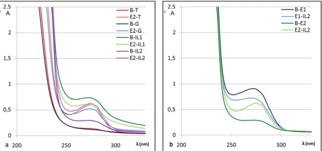
Figure 4.1 - UV spectrometry compared results with reference blank solutions (B) for the alkaline enzyme (E2) with every formulation buffer (T), gel (G) and ionic liquids (IL1 & IL2). ... 18
Figure 4.2 – UV spectrometry compared results: a) for the alkaline enzyme (E2) with every formulation buffer (T), gel (G) and ionic liquids (IL1 & IL2); b) tests for IL2 with enzymes E1 & E2.18
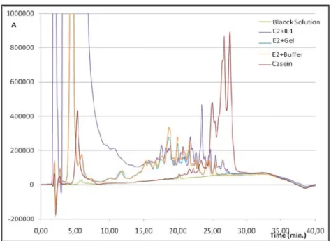
Figure 4.3 – HPLC chromatograms, for casein catalysis with E2 in different solutions: Tris HCL 8.4pH buffer (T), 8.4pH gel (G) and ionic liquid IL1. ... 20
Figure 5.1 – Cross-section images of the sample GG-CA with OM at 100x with visible light... 26
Figure 5.2 – Cross-section images of the sample OG-Ca with OM at 100x with visible light ... 26
Figure 5.3 - 100x OM image of the sample EG-S-EW with visible light, showing craquelure pattern. ... 27
Figure 5.4 - 100x OM images with visible light, the effect of varnish: a) OG-S, unvarnished surface; b) OG-S-Ca, sample with casein varnish. ... 28
Figure 5.5 - 100x OM UV fluorescence images of the different materials used in the samples: a) EG-YO-EW Egg white varnish over a egg tempera paint sample; b) EG-YO unvarnished egg tempera paint sample; ... 28
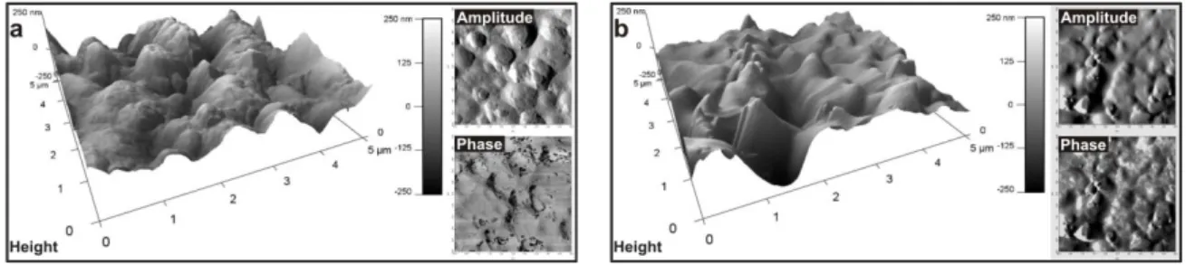
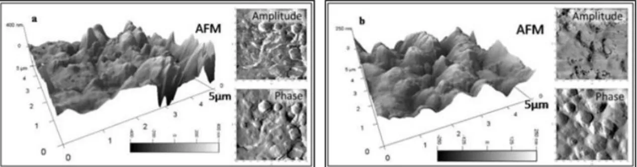
Figure 5.6 - 2D and 3D rendering of the AFM image set of height/topography, amplitude and phase (a) Unvarnished reference sample of tempera painting; (b) Unvarnished reference sample of oil painting. ... 29
Figure 5.7 - 3D and 2D AFM height images, surface features measured on the obtained section and: a) tempera painting without varnish, pigment grain size measurements of. b) tempera painting e with egg white varnish, measurement of the height of the varnish layer in a gap area. ... 29
Figure 5.8 2D and 3D rendering of the AFM image set of topography, amplitude and phase (a) Tempera painting sample after cleaning; (b) Unvarnished reference sample of tempera painting. 31
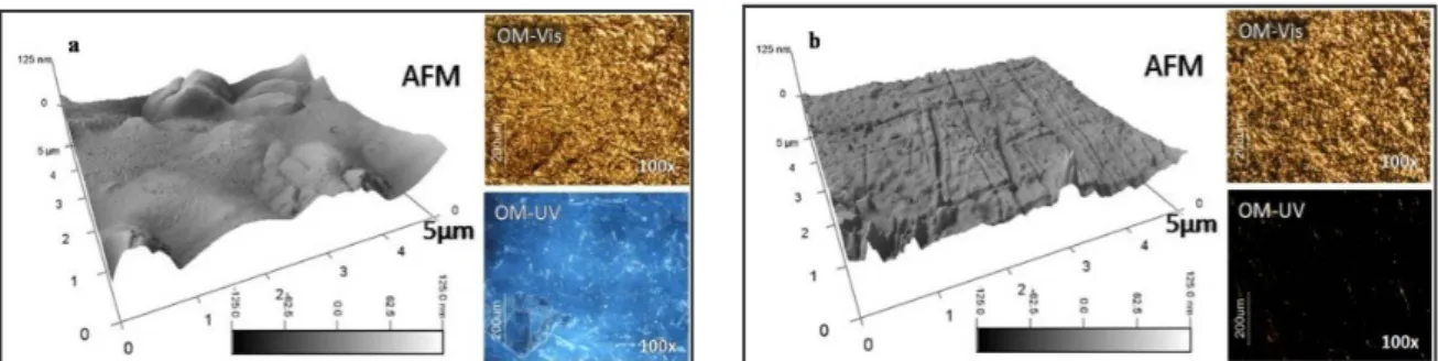
Figure 5.9 - 3D rendering of the AFM topographic image and OM with visible light and UV light, images set of animal glue coating from a gilded surface sample (a) Before cleaning ... 32
Figure 5.11 – OG-S-CP sample: OM images with visible light and UV light after removal with IL1+E1, and 3D rendering of the AFM image and, after removal of isinglass varnish. ... 33
Figure 5.12 – EG-S-EW. Tempera painting sample after removal of egg white varnish with cleaning formulation: IL1+E1 (a) Elemental mapping with SEM/EDX, (b) 2D and 3D rendering of the AFM image set on the same area. ... 33
Figure 5.14 – AFM images for topography, amplitude and phase of a reference tempera painting surface with fish glue varnish sample cleaned with IL2 [EMIM][EtSO4]. ... 34
Figure 5.13 – Cotton swab collection after cleaning of two samples. ... 34
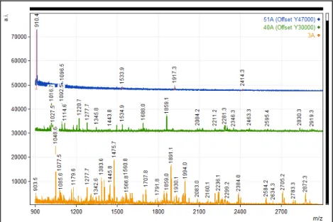
Figure 5.15 – MALDI-TOF-MS analysis of the material retrieved from the cotton swabs used on samples of egg white varnish on oil painting (OG-EW) samples, with E1 formulations (3A-Gel, 40A-IL1 and 51A-IL2). ... 35
Figure 5.16 – MALDI-TOF-MS analysis of the material retrieved from the cotton swabs used on samples of egg white varnish on oil painting (OG-EW) samples, with Gel formulations (4A-E1, 17A-E2 and 28A-E3). ... 36
Tables Index:
Table 2.1 – Final formulations composition summary. ... 11
Table 4.1 – Results of the removal tests obtained for different periods of time for the four proteinaceous varnishes with the three enzyme+gel formulations. ... 17
Table 4.2 – UV Spectrometry absorbance for enzyme activity: ... 17
Abbreviations list:
IL – Ionic liquids
IL1 – [BMIM][BF4] 1-butyl-3-methylimidazolium tetrafluoroborate
IL2 – [EMIM][EtSO4] 1-ethyl-3-methylimidazolium ethylsulfate
RTIL – Room temperature ionic liquids
E.C. – Enzyme classification
E1 – Acidic protease
E2 – Basic protease
E3 – Yeast extract
EG – Egg tempera
OG – Oil paint
GG – Gold leafing - gilding
S – Sinopia
YO – Yellow ochre
EW – Egg white varnish
CP – Fish glue varnish
Ca – Casein varnish
CA – Animal glue varnish
G – Aqueous cellulose based buffered gel
UV-Vis – Ultraviolet–visible spectroscopy
HPLC – High-performance liquid chromatography
MALDI-TOF-MS – Matrix-assisted laser desorption/ionization time of flight mass spectrometry
SM – Stereomicroscopy
OM – Optical microscopy
AFM – Atomic force microscopy
SEM – Scanning electron microscopy
EDS – Electron dispersive spectroscopy
1. Introduction:
Although cleaning is a frequent treatment in the restoration field, and it is mostly agreed on why
should be performed, it is never fully accepted without controversy on how, as it goes against the
major ethical principles of conservation: it is completely irreversible and causes the biggest visible
alteration to the object. A way to justify the need of such treatment is accepting its benefits and
choosing the material and techniques that are most compatible, with the lowest risk for the object
and no long term consequences.
There is always a search for new methods as none of the known ones are suitable for every
situation. The use of enzymes was a breakthrough in the cleaning process as they are highly
selective to the material to be removed not affecting surrounding materials of different nature [1, 2].
Usually they are used with an aqueous system but [1], this cannot be applied for every situation. An
innovative solution is related with the possible use of ionic liquids (ILs) as alternative solvent
systems.
Similar problems and considerations that are usually addressed in any cleaning treatment as
summarized by Khandekar [2], on the subject of aqueous gel cleaning on painted and varnished
surfaces, should be considered also for the use of ionic liquids. Firstly the efficiency of the
treatment: what is being cleaned and the active components, the clearance process, residues (of
the varnish and cleaning solution) associated problems of compatibility and long term effects.
Secondly, as stated by Khandekar, there is a need to develop ‘an adequate experimental
methodology’.
The experimental project developed here has the main objective of giving a scientific groundwork
for the novel use of combined ionic liquids and enzyme formulations for the cleaning and removal
of proteinaceous material (varnishes) in conservation and restoration surface treatments.
Another objective is to establish valid treatment monitoring and effectiveness assessment protocol,
using non-invasive surface analytical techniques. The experimental protocol should be also
suitable for other surface treatments and surface characterization on cultural objects.
The experimental work was mainly divided in four steps:
a) Formulation selection and establishment of the cleaning protocol. Two proteases
(appendixes I and II), commercially available for restoration treatments, were acquired; and a
third one was provided by the Bromatolagy Department of the Faculty of Pharmacy from the
Porto University. Several ionic liquids, selected from those available and recommended by
relevant bibliography, were tested in preliminary trials for evaluation of enzymatic activity by
ultraviolet–visible (UV-Vis) spectroscopy and high-performance liquid chromatography
(HPLC); from these two ionic liquids were selected. Finally seven enzyme formulations were
prepared with the two ionic liquids or gel and the three enzymes. The preliminary tests also
b) Complete surface characterization Mock-up samples were prepared as documented
reconstructions of oil and tempera painting and gilding, with four proteinaceous varnishes
(egg white, isinglass, animal glue and casein). The second step was a complete surface
characterization of these samples by different analytical microscopy techniques, namely
Stereomicroscopy (SM), Optical Microscopy (OM), Scanning Electron Microscopy (SEM) and
Atomic Force Microscopy (AFM); and also Colorimetry (CIE L*a*b*), in order to describe the
surface and conservation state before the treatment.
c) Experimental cleaning tests on the mock up samples with the enzyme formulations
prepared with both ionic liquids and, for reference, with gel.
d) Evaluation of the effectiveness assessment, by surface characterization analysis with the
techniques mentioned in step b); in addition, residue identification measurements were done
with Matrix-Assisted Laser Desorption/Ionization Time of Flight Mass Spectrometry
(MALDI-TOF-MS).
1.1. Concepts review:
The documented reconstructions (mock-up samples) are prepared duplicating the stratigraphy (structure and materials) of artistic techniques as described in relevant literature, such as historical
art treatises or manuals and other sources1. For this project oil (OG) and egg tempera (EG)
painting and gilding (GG) were prepared following the structure represented in Figure 1.1.
Figure 1.1: Examples of documented reconstructions as it would be observed in a cross-section, with the identification of the different types of layers; a) painting on canvas support, b) gilding on wood support.
The varnish, usually the top layer of such composites, provides protection of the surface against foreign degradation elements, and also enhances optical properties of the polychrome surface
such as glow and color saturation. So it is expected of the material to be the more transparent,
elastic and unaltered as possible with time2 [1]. Different materials have been used through time for
the preparations of varnishes: natural resins, proteins, even oil and more recently synthetic
materials.
Painting materials and particularly varnishes suffer deterioration due to ageing (natural material
degradation) enhanced by external factors (e.g.: light, humidity, microbial degradation, deposition
of pollutants and others). When the deterioration reaches the point where the varnish no longer
fulfills its use i.e., does not protect the surface and/or alters its perception, needs to be removed
1
There are several ancient treatises and more recent studies and compilations on the artistic materials and techniques, here more than a specific replica the intended was a generic painting structure.
2
and replaced. Here it will be addressed the problem of the removal of four protein varnishes as
they become highly insoluble with time and can hardly be removed. The varnishes studied in this
project are: egg white (EW), animal glue (CA), isinglass (fish glue, CP) and casein (Ca).
The preparation of these proteins as filmogenic materials to be applied as varnish is described in
many ancient recipes and art treatises [3, 4].
Cennino Cennini3 advises the use of animal glue as a pre-varnish for the pigment terre-verte; and
egg white4 as “a sort of varnish” to make the work seem “stronger”5; or for wood and stone
sculptures to be applied over the skin areas of hands and face [3]. Other authors advise the use of
egg white as a temporary protection varnish, Palomino6 in the eighteen century already focusing on
the conservation of paintings, points out the advantage of this varnish as a sacrificial layer because
it can easily be removed with sponge and water when its dirty and again replaced with new one [4].
Other protein varnishes are found in painting surfaces as original varnish or in later additions such
as in conservation treatments. For instance on tempera paintings, sometimes done without varnish
layer, fish glue varnish was used as a “reviving” technique popularized by De Mayerne7 in the
seventeen century; according to him the tempera paintings would, “look as if they were painted in
oil” [1].
Proteins are bio-functional8 polymeric chains composed of amino acids, amphoteric compounds9, containing both functional carboxylic acid group (-COOH) and basic amine (-NH2) and a
side chain (-R) that is specific to each amino acid (Figure 1.2a) [5].
Figure 1.2: a) General structure of an amino acid; b) Peptide bond between amino acids.
There are twenty naturally occurring amino acids and a protein can have chains in the order of the
thousands amino acids linked together by –NHCO– the peptide bond (Figure 1.2b). The number of
amino acids and the order in which they are aligned in the chain identifies and gives specific
structure and function to the proteins. Amino acids aligned themselves in chains, the primary
3
Cennino d'Andrea Cennini (c. 1370 – c. 1440), Italian painter and author of the “Il libro dell'arte”.
4
Also historically referred as glair.
5
These were pre-varnishes as the final varnish should be applied only after one year of the painting execution [1].
6
Acislo Antonio Palomino de Castro y Velasco (1653 – 1726), Spanish painter and author of the “El Museo Pictórico y Escala Óptica”.
7
Théodore Turquet de Mayerne (1573 – c.1655) was a Swiss physician and author.
8
Compounds that participate in reactions inside living cell, or have structural and mechanical function in the living tissues.
9
Capable of reacting chemically either as an acid or as a base substance. C
R
N+H3 COO
-H
α carbon
Side chain carboxyl terminal group
Amine terminal group
a C OH O R H
H2N
R ' H
H2N C
C NH H R ' COOH O H2N
R H
H2O O
OH
structure of the protein. A spatial arrangement of the chain as helixes, sheets and turns allows for
interactions between non sequenced amino acids stabilized by hydrogen bonds (secondary
structure). The third order of structure for the proteins is the functional shape of the secondary
structure, polypeptides within the protein folds in a unique three-dimensional way [6]. With time
denaturation occurs, i.e. unfolding of the natural three-dimensional configuration of the protein
chain, this is what makes the surface of the protein varnish to become more opaque, hardened and
brittle and the protein less soluble. Considerable more stable to oxidation processes than other
materials of the painted surface, proteins are however very susceptible to humidity, being this the
main factor for their deterioration [7]. The other important factor is related with their biological
degradation as proteins are a food source to microorganisms.
Common proteins used as artistic and restoration materials, and considered in this study, are:
Egg white, contains about 87% water, 2% of other materials and 12% of protein mixture, of the
latter about 65% is albumin, a class of proteins that are present both in egg white and egg yolk and
other materials. The alteration in the egg white varnish properties was already recognized in the
nineteen century, and its use inadvisable in conservation treatments since no satisfying
effectiveness of the removal of these varnishes was achieved by conventional methods [8].
Gelatins are the group of proteins found in biological tissues and main constituent of animal and
fish glues, the purity of the glue is related with the quality and amount of other materials. The fish
and animal glue are differentiated by the specific amino acid proportions; in general the fish glues
are also more pure which results in a clearer and stable glue.
Caseins are proteins obtained from precipitation of milk and may contain different impurities such
us lactose, acids and butter. The casein is differentiated from the other proteins because it belongs
to the phosphoproteins group, which are proteins that are physically bond to substances containing
phosphoric acid [6, 9]. For this they are insoluble in water and more susceptible to deterioration by
oxidation processes. The conservation practice reported some difficulty in removing the
proteinaceous materials [7, 8,] and therefore, required the use of more aggressive treatments,
usually toxic for the conservator, that endangered the other layers. The ideal was to find a specific
treatment to proteins that did not affect the rest of the materials. This would be only achieved with
the introduction of enzymes in the cleaning process [1, 2] a major breakthrough as enzymes are
catalysts very specific to substrates and can be used in moderate conditions. Early works of the
mid-twentieth century already reports the use of enzymes [10] albeit with doubtful approaches, and
in the late 1960s Banks [11] refers the use of collagenase10, to remove glues, with some success
and conscious of the needed cares and possibilities
Enzymes are themselves proteins with specific amino acids sequences and unique three-dimensional configuration that allows the interactions with very specific materials in certain active
areas, that have been described many times as a lock and key system [6, 12] Figure 1.3.
10
Figure 1.3 – Diagram of the “lock and key" hypothesis of enzyme action.
Enzymes of interest to conservators are those that facilitate degradation processes acting as
catalyst11 in hydrolytic reactions12, in this case the hydrolysis of proteinaceous material specifically
known as proteases or peptidases. In the international enzyme classifications it is the group
E.C.3.4.13, where E.C. stands for enzyme classification; 3. for hydrolysis and 4. for the affected
substrate: peptides or proteins.
As early as 1977 Hatton [13] proposes the use of the enzymes as cleaning agent in cellulose
based gel for the first time. More recently Wolbers [14] popularized the use of aqueous cleaning
systems for conservation purpose, as a viable, less toxic alternative to common free organic
solvents, including the preparation of enzyme in gel mixtures. Another author that disseminated the
use of enzymes in conservation treatments was Cremonesi [15].
But there are situations where these aqueous systems do not give the best results. The works
mentioned [14, 15] show recipes of complex mixtures with over 4 components, this high number
makes difficult to fully predict the interaction of the mixture, its interaction with the treated surface
and possible long term deterioration problems.
The use of organic solvents that are compatible with the enzymes is not advised as they are
usually highly toxic and strong solvents in themselves establishing interactions with the other
materials of the painted surface. A possible alternative could be related with the use of “green
solvents”, mainly based on ionic liquids.
Ionic liquids (ILs) can be defined as organic salts with a melting point below 100ºC, or more interesting as liquids at room temperature (room temperature ionic liquids; RTIL). In the last 20
years the interest from both academia and industry has gained significant impact mainly due to
their environmental friendly characteristics as green14 alternative for traditional volatile organic
solvents (VOCs) and because of their wide range of physicochemical properties. There are a large
number of organic cations and anions that can be combined to form different ionic liquids, and this
is one of the most important features of these compounds.
11
A catalyst is a substance that without being consumed, or change the reaction equilibrium, accelerates its process and/or allows it to occur in more desirable conditions, such as lower temperature.
12
Includes lipase, amylase and peptidase enzymes that catalyses the hydrolytic breakdown of lipids, sugars and proteins respectively.
13
Enzyme classification as recommended by the International Union of Biochemistry and Molecular Biology and available at http://www.chem.qmul.ac.uk/iubmb/.
14
ILs have been also described with some peculiar properties such as their high conductivity,
variable range of density and viscosity values, tunable polarity and solubility as well as their high
thermal and chemical stability. Overall, the number of possible ionic liquids is estimated around the
ten to the eighteenth (1018), whereas the number of traditional solvents widely used in industry
accounts for a few hundred.
Ionic liquids are also called “designer solvents” because, by changing cation-anion combinations
[16], they can be adjusted to specific situations or requirements such as stability, solubility,
viscosity, improvement of enzyme cleaning effectiveness, surface compatibility, and lower toxicity
behavior [20]. The previous work of Pacheco [21] reports on the use of IL as a potential new
material for heritage conservation treatments; it is focused on the use of the IL as alternative
solvents and cleaning agents for resinous material, natural and synthetic, with some encouraging
results.
The use of enzymes in IL media it is not an old science, and is still an unstudied area in
conservation science with a large number of possibilities. The first report of the use of IL for
biocatalysis with enzymes has just a little more than 10 years [22] but, rapidly started to be a major
interest area of research. Kragl & coworkers [23] and Roosen & coworkers [24] show the
production of articles registered on the web of science on the subject of biocatalysis using ILs in
the past years. Fred van Rantwijk & coworkers [25] and Moniruzzaman & coworkers [26] reported
reviews about biocatalysis using ionic liquids.
In the present work, the ILs will be addressed from the point of view of compatibility in restoration
treatments when combined with enzymes, as an alternative to traditional gel formulation.
1.2. Surface characterization and treatment monitoring
The characterization of the materials present in a cultural heritage object is the first step before any
conservation or restoration procedure [1, 27]. In a painted surface besides the rheological and
other physical-chemical properties of paint materials, the applications method, number of paint and
varnish layers and their thickness are important issues to consider.
To fully understand the object’s surface characteristics and its state of conservation analytical tools
are needed and in some cases sampling of the objects is required. The sampling is always a
problem to consider as it means the loss of material, even if the information retrieved is considered
most valuable and the sample is minute in size. But in the case of surface treatments such as the
removal of varnish layers this problem can be overcome by applying noninvasive and
nondestructive surface techniques.
Surface properties of cultural heritage artifacts have been approached in recent years by
conservation and material scientists and physicists who applied non-invasive analytical tools to
describe the behavior of the polychrome and gilding surfaces, according to the optical, mechanical,
physical and chemical characteristics [28-31]. The analytical data obtained from a multiscale and
multiple technique approach is useful for the practitioner (conservator, curator or art historian) to
to predict future changes of the surface with regard to the choice of best preservation conditions or
of the right restoration treatment (for example: cleaning, consolidation or varnishing) [5, 29, 32].
The techniques chosen herein have been previously reported for their usefulness:
stereomicroscopy (SM), optical microscopy (OM), scanning electron microscopy (SEM-EDX),
atomic force microscopy (AFM), colorimetry (CIE L*a*b*) [28, 29, 31, 33-36]. AFM was recently
reported in researches relating to the study of the surfaces of gilding layers and the nano-scale
effectiveness assessment of cleaning treatments [31, 37-39]. This technique has a great potential
for monitoring cleaning interventions on paintings or polychromed surfaces of works of art, and
could bring an important contribution in assessing the presence of residues of the cleaning
formulation at a nano-scale.
It will be demonstrated that the use and combination of different surface analytical techniques
returns an extensive and complete set of information that fully characterize the surface at different
scales (from macro- through micro- to nano-scale) in order to assist the proposed treatment.
The evaluation of the formulations behavior, specifically the activity of the enzymes, was monitored
before and after its use in the cleaning process. The analytical techniques used are those
commonly reported in proteomic science: UV-Vis spectroscopy [40], HPLC [41] and
2. Materials and methods
2.1. Materials
Most of the reagents used in this work were provided by Sigma-Aldrich. Acetonitrile and
trifluoroacetic acid were supplied by Merck. Antares supplied two enzymes as commercial ready to
use gel formulations (G+E1, pepsin; and G+E2, from Aspergillus sojae), also Tylose powder, White
Spirit, Shelsol, cotton and proteinaceous glues. The sequencing grade modified trypsin, was
provided by Promega; The ZipTips, pipette tips for sample preparation were supplied by Millipore.
Ionic liquids were provided by Solchemar; embedding polyester resin for cross-sections is
distributed by MR Dinis dos Santos.
2.2. Mock up samples from documented reconstructions
To better understand the possible use and effectiveness of the formulations suggested here, tests
were done on documented reconstructions of polychromed surfaces prepared during 2009 and
March of 201115.
Mock-up samples of tempera (EG) and oil (OG) painting on canvas and wood supports and also
traditional water gilding (GG) over bole ground were replicated in accordance with historical and
artistic sources [3, 44, 45] following the general scheme from figure 1.
Figure 2.1 – Overall view of the initial condition of the samples used in this project, Grouped vertically: a) Tempera painting with egg white and fish glue varnishes; b) Oil painting with egg white and fish glue varnishes; c) Oil painting with casein varnish; d) Gilding with animal glue varnish; a & b prepared on canvas
mounted on glass slides and c & d prepared on wood.
15
Except for the gilded samples all of the others were previously prepared. The choice of using these samples was their availability and a way to have a less recent support as it would not be done any sort of artificial ageing at this point.
d
c b
The Figure 2.1 shows the initial aspect of the prepared mock up samples. For each technique a
type of protein vrnish – egg white (EW), fish glue (CP), animal glue (CA) and casein (Ca) – was
selected and applied at the end of 2010 and in April 201116. Except for the gilded surfaces all
samples were duplicated in two different colors of iron oxide based pigments – yellow ochre and
sinopia – to control possible darkening or blanching17 of the surface after treatment. A small area of
the surface in each artistic technique was preserved in order to be tested with every formula (44
areas for testing the four new IL+Enzyme formulations and other 33 for reference gel formulations);
also other areas were left without varnish to serve as reference surfaces.
Although mock-up samples are not a real situation of an ancient paint or polychrome, their use
allows the test of the cleaning formulations in a setting where multiple factors influence the removal
process but, the precise composition is known. As pointed out by Phenix & Sutherland [46] this can
be accepted as a stepping stone and a good compromise when developing new methods.
Preliminary cleaning tests were developed to establish the cleaning protocol and IL selection for
this, thin layers of the same four varnishes were prepared over microscope glass slides.
2.3. Cleaning formulations
Two commercially available enzymes, used in conservation for the removal of proteinaceous
materials, were acquired: an acid enzyme (E1), pepsin (appendix I) a protease of the pig’s stomach
mucosa and an alkaline enzyme (E2) from Aspergillus sojae (appendix II). A third enzyme (E3) is an extract from yeast [40] provided by the Bromatology department of the Faculty of Pharmacy
from the University of Porto, to be tested as a new alternative enzymatic agent, active at pH 7, and
potentially less expensive. Because E3 has not only one active agent but, is an extract with three
different enzymes [40] it is expected a potentially more effective performance with a higher range of
catalytic interactions. However, because is not in a purified form at this moment; the enzyme
activity may vary from batch to batch.
For E1 and E2 the supplier provides also ready-to-use cellulose based buffered gel to the specific
conditions of each enzyme. For the third enzyme however a gel was prepared following Cremonesi
[15] instructions.18 The enzymes were chosen according to optimal performance in the different pH
ranges. Depending on the desired work conditions in a restoration treatment a basic or an alkaline
or a neutral enzyme can thus be selected.
The imidazolium based ILs (figure) are by far the most common ILs used in enzymatic reactions [20, 24, 47-49]. But the ILs general properties, although follow some pattern, varies substantially
with specific anion- cation- combinations [17, 49, 50]. Park [20] also makes notice that although
polar solvents inactivate the enzymes, ionic liquids does not.
16
We would like to acknowledge at this point the collaboration of Michaela Crhová who helped with the preparation of the samples.
17
Blanching – A local, opaque, whitish discoloration on the surface of a painting. Blanching occurs because of a loss of or an alteration in the binding medium in an oil film, or because of the partial dissolution of a varnish. In accordance with the definition at the Canadian Conservation Institute - CCI Notes 10/11 [http://www.cci-icc.gc.ca/publications/notes/10-11-eng.aspx].
18
Figure 2.2: methylimidazolium cation [R-MIM]+ and some examples of anions in ionic liquids: Trifluoroacetate [TFA]-, Trifluoromethanesulfonate [TFO]-, Tetrafluoroborate [BF4]
-, bis(trifluoromethylsulfonyl)imide [NTF2]-, Ethylsulfate [EtSO4]-, Hexafluorophosphate [PF6]-.
In order to select among the numerous IL possibilities preliminary tests for enzyme activity and
cleaning capability were done with ten ionic liquids, Figure 2.1 shows common possible anions.
The different ILs were chosen in accordance with literature review for bio-compatibility, availability
at the lab and predicted compatibility with the painted surfaces [21]. From the ten ionic liquids two
with the best results in the preliminary tests were chosen and labeled as IL1 and IL2 (appendix III):
1-butyl-3-methylimidazolium tetrafluoroborate (IL1 – [BMIM][BF4]) and 1-ethyl-3-methylimidazolium
ethylsulfate (IL2 – [EMIM][EtSO4]).
Seven formulations were prepared: four innovative formulations using two commercial proteases
(E1, E2) and two ionic liquids (IL1, IL2) and three reference formulations with the three enzymes
(E1, E2 and E3) in the traditional cellulose gel mixture (table 2.1 ).
Table 2.1 – Final formulations composition summary.
Formulation Enzyme Solvent pH
E1+G
Enzyme + solvent (≅ 1g / 100ml)
Pepsin Commercial cellulose based gel 5
E2+G from Aspergillus sojae Commercial cellulose based gel 8.4
E3+G Yeast extract phosphate buffer3% Tylose / 7pH Prepared cellulose based gel 7
E1+IL1 Pepsin
20% water v/v
[BMIM] [BF4] -
E2+IL1 from Aspergillus sojae [BMIM] [BF4] -
E1+IL2 Pepsin [EMIM] [EtSO4] -
E2+IL2 from Aspergillus sojae [EMIM] [EtSO4] -
E1 = acidic protease; E2 = basic protease; E3 = yeast protease (Porto); G =gel; IL1 = [BMIM] [BF4]; IL2 = [EMIM] [EtSO4].
2.4. Cleaning protocol
For the new formulations an optimized cleaning protocol needed to be developed. The cleaning
process is in itself a complex issue. There are a great number of variables to be considered in
particular: type of substrate that is to be removed, its nature and state of conservation. The
materials present in the formulation, including their proportions and possible interactions; the
needed conditions to activate the formulation, mixing, temperature, pH, to name a few; and the
cleaning, namely the manner and time of application, as well as the removal and clearance steps. N
N
R
CH3
[R MIM]
+N S S O O O O F F
F F F
F NTF2
-S
O
O O O
EtSO4
-P F F F F F F
PF6
-B
F
F
F
F
BF
4-S
O O O F F FTFO
-F FF
TFA
-O
For an innovative method such as the one proposed here it was necessary to simplify whenever
possible. Basically the protocol would follow the indications of use for the enzymes with gel
formulations as described by Wolbers [14], Cremonesi [15] and in the suppliers’ material data sheet
(appendix I and II). The enzyme is added to the solvent in accordance with the supplier indications
in a proportion of approximately 1g of the enzyme to 100ml of the proper gel (buffered at the ideal
pH for each enzyme) and the same proportions were used for the ionic liquids. The mixture, after
stirring, is left to stand for 20 minutes in a double-boiler set at 37oC, this step allows for some of the
air introduced during the mixing to be released and to activate the enzyme. The mixture is then
applied with a soft brush and let it be for different amounts of time. As this step depends upon the
type of material present, the preliminary tests helped to establish the contact time for each
formulation and varnish. Following, the cleaning material is removed with dry cotton swabs and in
sequence, allow drying in between, two damp cotton swabs and one with white spirit19 was used.
This is the clearance phase, the first swabs are to remove all residual products and cleaning
material and the last one is to inactivate possible enzyme residues on the surface, as
recommended by the supplier instructions (appendix I and II) and bibliography [14, 15].
Following this cleaning protocol, different times of contact with the varnish surface (from 30
seconds to 35 minutes) were considered and tested for each enzyme + gel formulation and each
protein (egg white, isinglass, animal glue and casein) on the prepared glass slides. When
necessary, in order to avoid drying of the gel on the surface, the area tested was covered with a
transparent polyester film.
With the application times established another round of test were done, then using different ionic
liquids to choose the ones that presented, at least, similar behavior as the gel formulation, i e, that
needed the same amount of time to obtain observed similar cleaning efficiency.
19
3. Analytical protocol and Instruments:
3.1. Enzymatic activity measurements
For the measurements of enzymatic activity the Kunitz method was followed for liquid solutions as
described by Whitaker [51] and also adapted for gel solutions. Initially E1 and E2 were tested, as
recommended, in buffered solutions. Stock solutions of acetate buffer (pH=5) and Tris HCL buffer
(pH=8.4) were prepared as described by Gomori [52]. The enzymatic activity is obtained by
quantifying the products, specifically the aromatic amino acids, in solution after a certain amount of
time with UV absorbance spectroscopy and comparing these values to reference blank solutions.
It was verified that the enzyme activity protocol can be used in a similar approach with the gel
formulations for the three enzymes; the same was done for the ILs with E1 and E2. This test
excluded all the ILs that showed enzyme inactivation, i.e., not compatible for these enzymes.
Two UV-Vis spectrometers (Shimadzu UV-1800 UV-VIS Spectrophotometer and Varian Cary 100
Bio UV-Visible Spectrophotometer) were used for the absorption studies.
As the specific enzyme activity was not the purpose of these tests no comparison was done
between the obtained values and known standards20.
3.2. High-performance liquid chromatography (HPLC)
The HPLC at the Bromatology Department of the Faculty of Pharmacy from the Porto University
was used to study some of the formulations for enzymatic activity and formulations shelf-life; these
tests were done following the protocol as described by Mota & coworkers [41]. The enzymatic
activity was followed by HPLC spectra with the indication of peptide separation different from the
blank samples and by comparison of the peak areas. For measuring shelf-life the test was done at
time intervals of the same preparation and peaks area was compared for interpretation of the
possible loss of enzyme activity. Due to work constrictions HPLC was not used extensively, on all
samples.
3.3. Cross-sections and staining test
Although in the present work the varnishes in the samples are well known it is suggested the
preparation of cross-sections for the identification of the painting structures and generic qualitative
protein identification/mapping by means of staining tests21. Cross-sections were prepared from
minute fragments from a set of representative samples with and without varnish and before and
after treatment. These fragments are imbedded in a polyester resin, followed by polishing with
successive finer grain of micro-mesh paper. Images were obtained by optical microscopy (OM)
technique with visible and fluorescent-light sources. For the identification of the protein material
20
The specific enzyme activity for E1 and E2 is included in the product data sheet in appendixes I and II. For E3 further studies are to be carried out at University of Porto (Faculty of Pharmacy), but reference values are described by Teixeira [40].
21
and its location in the surface structure a spot test was done using Sypro® Ruby Protein Stain dye,
this identifies and maps the distribution of the proteinaceous material by an orange color observed
under green emission of fluorescence22.
3.4. Surface characterization, assessment and control of the cleaning process
To monitor and assess effectiveness of the treatment an analytical protocol was established and
used before and after the removal of the protein-based varnish. This protocol is based on the
complementary use of different microscopy techniques, at a multi-scale, non-invasive and with little
or no sample preparation: the stereomicroscope (SM) at 10x magnifications, then the optical
microscope (OM) 50x to 200x magnifications, and scanning electron microscope (SEM) on
uncoated samples allowing good resolution imaging up to 3000x magnification. The atomic force
microscopy (AFM) was introduced at an image size of 5 micrometers.
Also measurements of colorimetric values (CIE L*a*b*) were performed. The comparison of the
images and values obtained before and after treatment of the same surface allowed the surface
characterization, but also, to gather information about cleaning effectiveness, possible residues,
and other features in useful time.
3.4.1. Stereomicroscopy and Optical Microscopy
An Olympus stereomicroscope system SZX12, mounted on an extendable arm SZ-STU2 with a
digital camera DP-12 and an independent light source HighLight 3100 was used to record the
sample areas at 10x, 32x and 90x magnifications. Then Axioplan Zeiss 2 imaging binocular
microscope (at 50x, 100x and 200x), with both visible (dark field) and fluorescent radiation, coupled
to a Nikon DXM1200F digital camera was used to image the surfaces, before and after the removal
of the varnish layers. The blocks of filters used for observing the fluorescence were: BP 300-400,
FT 395 and LP 420 (filter 8); BP 450-490, FT 510 and LP 515 (filter 6).
3.4.2. Scanning Electron Microscopy
SEM images were acquired from backscattered electrons and low vacuum secondary electrons
with an Auriga CrossBeam Workstation (SEM-FIB) operating at 1-5 kV, with a typical working
distance (WD) 4.5-9.6 mm, aperture size 30µm, magnifications: from 200x up to 3000x. Due to
work constrictions SEM was not used extensively, on all samples.
With similar operating conditions Electron Scanning Microscopy with Energy Dispersive X-ray
Spectroscopy (SEM/EDX), was also conducted to selected samples for the purpose image
processing and elemental mapping of the surfaces after treatment.
22
3.4.3. Atomic Force Microscopy
MFP-3D Asylum instrument was used in non contact imaging mode (AC mode) using Si probes
Tap300Al from Budget Sensors (cantilevers with 300 KHz resonance frequency and 0.2 N/m spring
constant). The scan size was 5 μm2 with a resolution of 512 by 512 lines. Before AFM
measurements, the samples were cleaned with a soft N2 flow to remove free dust contaminations.
The images record the height mapping, this results in 2D images, that throughout this work are,
shown in a gray scale, where brighter areas represent higher areas, from these 3D images are
easily obtained using Igor Pro 6.2 Software. Images were recorded at 3 different locations, for each
investigated area, in order to verify the repeatability and statistical significance of the observed
features. Measurements of surface roughness, the average value of the three scanned areas, were
obtained with the software. Other images were also recorded: amplitude images, which registered
the tip amplitude changes at the given frequency, and phase images, which measure the
dissipative forces between the tip and the samples.
In a non contact mode, the amplitude is set at constant oscillation amplitude maintained by
feedback loop. When there is a height variation, in order to maintain the average tip-to-sample
distance the oscillation amplitude is adjusted. The amplitude images are the result of the amplitude
oscillation adjustments of the cantilever.
The phase is a qualitative measurement of the interaction forces between the tip and the sample.
Even if, no amplitude changes are monitored, the phase can show regions where the sample tends
to interact with the tip in a non-conservative regime: when it shows high contrast between the white
areas (positive) due to attractive interactions and dark areas (negative) due to repulsive
interactions. This interaction with the tip can be particularly relevant indicating, for example, the
presence of different materials or just the same material with different molecular arrangement
(amorphous/crystalline), for example. Notice that these are false colors and can be adjusted to
different hue/tones with different software settings, in this project it was set to a gray scale.
3.4.4. Colorimetry
For the colorimetric measurements a Datacolor International Microflash colorimeter, with a Xe lamp
was used. The measuring area has a diameter of 18mm (22mm illuminated area), the geometry
was 0°/0° and the light source D65.
The colorimetric characterization for each investigated area was performed through the
measurement of L*a*b* parameters (using CIE L*a*b* 1976 system) [34]. Calibration was carried
out before each set of measurements using a pure white standard (calibration values: L= 96.43,
a=-0.33, b= 1.27). Understanding possible variations of each component is important: L* value
measures “lightness” from 0 to 100 corresponding to a variation from black to white. If variation of
L* (∆L*) is positive this means that the sample is lighter after the treatment than before, and when
the value is negative indicates an increase in darkness. a* values ranging from positive to negative
represent color changes from magenta to green, while b* variation indicates changes from yellow
and the average values were calculated together with the standard deviation from the mean. Delta
E was finally calculated from the L*, a*, b* values before and after the removal of the coating layer
and compared with reference samples.
3.5. Other techniques - MALDI-TOF Mass Spectrometry
Following the cleaning procedures each third cotton swab was analyzed for the presence of
peptides, by MALDI-TOF mass spectrometry in accordance with the protocol described by Kučková
[42]. This allowed for the identification of peptides, demonstrating and comparing the activity of the
enzymes in the different formulations, and some interpretation could be made with respect to
4. Results
Table 4.1 shows the results summary of the preliminary tests done for the two commercial
enzymes (E1 and E2) and E3 in the gel formulation to establish contact time.
From the available ILs, ten structures based on imidazolium cation family were selected and tested in the same way. Appendix III summarizes the main characteristics of the ILs and preliminary tests
observations.
Table 4.1 – Results of the removal tests obtained for different periods of time for the four proteinaceous varnishes with the three enzyme+gel formulations.
Cleaning formulation
Effectiveness/time of application
EW CP CA Ca E1+gel
(pH = 5)
++/3 min ++/3-9 min ++/9 min +/35 min
+/30 seconds +/30 seconds -/30 seconds - -/30 seconds
E2+gel (pH = 8.4)
++/30 seconds ++/30 seconds ++/3-9 min ++/27 min
- - +/30 seconds - -/30 seconds
E3+gel (pH = 7)
++/3 min ++/3 min ++/9 min +/35 min
+/30 seconds +/30 seconds -/30 seconds - -/30 seconds
Legend:
++ Easy removal (the varnish layer is easily removed with the removal of excess formula); + Removal (after gel application the varnish is swollen and this allow a mechanical removal);
- Difficult removal (after gel application the varnish is swollen and this allows a mechanical removal, leaving residues);
- - No removal (the varnish could not be visibly removed).
Simultaneously, enzyme activity tests with the UV spectrometer technique23 were done for the
formulations, once more using enzymes E1 and E2 as reference, first just in buffered aqueous
solution24 (table 4.2), then in gel and finally with the most promising ILs after preliminary cleaning.
Table 4.2 – UV Spectrometry absorbance for enzyme activity: Measurements for casein hydrolysis in buffered aqueous solutions.
Test ID Protein pH Enzyme at 280 nm Abs.
B5E1
Casein solution 50mg/ml
pH5
Acetate Buffer E1 0.598
B8E2 pH8,4
Tris HCL Buffer E2 1.33
B7E3 pH7
Phosphate Buffer E3 0.530
Procedure notes: Adaptation of Whitaker’s procedures [51], according with the work done by Teixeira [41]
Absorbance at 280nm represents one of the standard measurements in order to indicate enzyme
activity, the results were obtained for the buffer aqueous solutions relates with concentration of
aromatic amino acids present in solution which can be converted to enzyme activity units by
23
Procedure notes: 3 tubes, one blank and 2 for testing enzymatic activity; the catalysis occurred during 30min in a double-boiler at 37ºC; after that it was added 1ml of triichloroacetic acid (TCA) to stop the enzymatic activity (to the blank tube, 1ml TCA was added before the addition of the enzyme solution); waited 30min; after centrifuge at 10000xg for 10min the supernatant was measure for absorption considering the absorption of the blank as zero [51].
24
comparison with standard solutions with known concentrations25, this however was not the
objective at this point. Firstly because E1 and E2 have known activity values from the supplier26,
and for E3, as it is an extract that varies from batch to batch27; and secondly because when
changing the conditions, changing the solvent, the results are no longer comparable. However this
is important in order to understand if the enzyme is active in the solution (when the conditions are
similar the results can be compared).
Figure 4.1 - UV spectrometry compared results with reference blank solutions (B) for the alkaline enzyme (E2) with every formulation buffer (T), gel (G) and ionic liquids (IL1 & IL2).
Figure 4.2 – UV spectrometry compared results: a) for the alkaline enzyme (E2) with every formulation buffer (T), gel (G) and ionic liquids (IL1 & IL2); b) tests for IL2 with enzymes E1 & E2.
25
“One unit of is defined as the amount of enzyme which yields absorbance of 1mmole of tyrosine per minute under the conditions used” [51]
26
See appendix 1 and 2 for material characteristics data sheets.
27
Complete study of its characteristics is being done at Bromatology Department of the Porto University (Faculty of Pharmacy), and results cannot be presented here.
A
Enzymes need a minimum amount of available water to be active [20, 53-55]. Additional UV
absorbance tests were performed at different water/IL (v/v) proportions in order to identify the
minimum amount of water required, that was 20%, this high amount could be related to the water
affinity of the IL itself, leaving less available water for the enzymes [53, 54].
Figure 4.1 shows the complete set of results for E2; it was detected that in the IL tests the blank
solution (B), measurements before enzyme catalysis show some absorbance. This observation
could be due to the fact that ILs have higher viscosity and for its characteristics able to retain some
amount of protein after centrifugation.
Figure 4.2 shows results for IL2 with the enzymes E1 and E2. No control of pH was done because
during the preliminary cleaning tests both enzymes were active with both selected ILs. However,
for the enzyme activity tests an associated problem was identified related with the preparation of
protein solution with casein in alkaline medium. For the IL and E1 with affinity to acid medium, the
absorption curve shows anomalies, as the blank solution (B) presented higher absorbance (Figure
4.2).
From these tests, the precise compositions of the formulations to be tested were set and also
established the application times for each protein: approximately 30seconds to 1minute for fish
glue and egg white varnish; 3minutes for animal glue varnish and 30minutes for casein varnish. In
total seven formulations were prepared to be experimented at 1g/100ml (enzyme/solution), and for
E3 1ml/100ml (extract/solution); for the IL, the formulation has 20% v/v of water content: IL1E1,
IL1E2, IL2E1, IL2E2, GE1, GE2 and GE328 (table 2.1).
For the HPLC the same catalytic protocol29 was followed as it was done for the UV spectrometer
technique and as described by Mota [41]. Due to time restrictions, a complete study was done only
for formulation IL1+E2. Figure 4.2 shows that the enzyme is fully active in the IL formulation,
although the peptide distribution is different. The elution of peptides is done by their polarity and
this is related with size of molecular chain; first to be eluted are the most polar components (the IL
is the first component to be separated), and last the proteins30, as polarity decreases with
increasing size and secondary interaction of non-sequenced amino acids by hydrogen bonds [12].
The peak area relates with the quantity of peptides, therefore adding all peak areas gives us the
amount of eluted peptides.
When comparing the three groups of peptides, for the different formulations (figure 4.3) it is
observed that the cleavage in the IL, for the same amount of time results in predominantly bigger
sized peptides.
28
E1 = acidic protease; E2 = basic protease; E3 = neutral, yeast protease (Porto Faculty of Pharmacy); G =gel; IL1 and 2 = the selected ionic liquids.
29
See footnote 23.
30
Figure 4.3 – HPLC chromatograms, for casein catalysis with E2 in different solutions: Tris HCL 8.4pH buffer (T), 8.4pH gel (G) and ionic liquid IL1.
For the same formulation a shelf-life test was done, i e, determining how long the formulation would
be active after mixing all the ingredients31. For gel formulations it is common practice to use the
same formula for just one working week, this is because the cellulose based gels are susceptible to
contamination by bacterial growth and inactivation of the enzyme with time. This is less likely with
IL because of toxicity levels32, but inactivation can occur naturally, most commonly due to self
cleavage processes and possible external contaminations. Tests show (table 5.1) that after one
week there was still 85% activity measured in amount of peptides. The anomaly observed, for the
second day measurements, is because it was not observed the waiting time33 required for enzyme
activation before adding the protein.
Enzymes need a medium that allows them to have the three-dimensional configuration required to
establish an interaction with the substrate. This occurs in the presence of water; as the water
molecules interacts with the enzyme maintaining the tertiary structure The protein is breakdown in
peptides through the rupture of the peptide bond consuming water, which means that a small
amount of water should always be added to the solution. Preliminary cleaning tests and enzyme
activity tests showed that at least 20% v/v (water/IL) was needed. Such high amount can be
explained because the ILs selected are hydrophilic and can strip the enzymes of the water needed
for the reaction to occur and for the maintenance of the tertiary structure [20, 53].
A set of images was acquired for each sample area with all the surface analytical techniques34,
these studies gave us a surface characterization for each artistic technique and materials;
appendix IV show representative examples of all the samples, varnished and unvarnished.
31
Formulation kept in a refrigerated environment, at 4oC.
32
The toxicity in the imidazolium ionic liquids family is related to the ring in the structure and tends to increase with the increase in the alkyl chain length [17].
33
After refrigeration it is recommended by the supplier to wait at room temperature at least 15min.
34

![Figure 2.2: methylimidazolium cation [R-MIM] + and some examples of anions in ionic liquids:](https://thumb-eu.123doks.com/thumbv2/123dok_br/16626520.740475/31.892.116.787.106.320/figure-methylimidazolium-cation-mim-examples-anions-ionic-liquids.webp)