João Miguel Rocha Salazar
2019
Optimization of the production of fucoxanthin-rich fractions
from the microalga Isochrysis galbana
Optimization of the production of fucoxanthin-rich fractions from
the microalga Isochrysis galbana
Master Degree in Marine Biology
Thesis
2017/2018
Author: João Miguel Rocha Salazar
Student nº 56417
Supervisors
Prof.a Dra. Luísa Paula Viola Afonso Barreira
Optimization of the production of fucoxanthin-rich fractions from
the microalga Isochrysis galbana
Declaração de autoria de trabalho
Declaro ser o autor deste trabalho, que é original e inédito. Autores e trabalhos consultados estão devidamente citados no texto e constam da listagem de referências incluída.
Copyright
A Universidade do Algarve reserva para si o direito, em conformidade com o disposto no Código do Direito de Autor e dos Direitos Conexos, de arquivar, reproduzir e publicar a obra, independentemente do meio utilizado, bem como de a divulgar através de repositórios científicos e de admitir a sua cópia e distribuição para fins meramente educacionais ou de investigação e não comerciais, conquanto seja dado o devido crédito ao autor e editor respetivos.
i Agradecimentos
Um especial obrigado à professora Luísa Barreira e ao professor João Varela pelo voto de confiança. Pelas oportunidades que me deram ao longo deste ano, pela disponibilidade e pela transmissão constante de conhecimento, levo isso comigo para sempre. Não tenho palavras para expressar a gratidão que sinto pelos vossos conselhos cirúrgicos e pela vossa amizade, sinto-me um verdadeiro privilegiado.
Um especial obrigado ao João Navalho e a toda a equipa da Necton S.A. por me terem recebido de braços abertos. Sinto que estou em constante aprendizagem nesta casa e que fortaleço laços de amizade diariamente. Um abraço especial ao Pedro Mendonça pela disponibilidade em explicar todo o processo industrial com um fairplay tremendo e à Inês Povoa por me dar as primeiras diretrizes de gestão industrial com uma boa disposição constante.
Um especial abraço e obrigado à Tamára Santos por ser uma pessoa fantástica dum altruísmo singular. Devo-lhe imenso pela disponibilidade total, pelo conhecimento de laboratório que me transmitiu e pela amizade. Esta experiência não seria possível sem o auxílio diário da melhor coordenadora de laboratório que alguém podia ter.
Um especial abraço e obrigado ao Hugo Pereira acima de tudo pela amizade e pela disponibilidade total na coordenação deste projecto. Aprendi imenso e sinto-me um privilegiado por poder continuar a construir uma relação profissional e simultaneamente de amizade contigo.
Um grande abraço ao meu amigo Katkam pelas horas que passamos juntos no laboratório. Sinto-me um sortudo pelo voto de confiança que me deu e pela boa disposição diária. Um especial obrigado à Inês Maia e a toda a equipa do Marbiotech por serem os melhores partners de laboratório que alguém podia ter.
Um grande obrigado ao Stefan, à Ana e à Raquel por serem os melhores parceiros nesta aventura. Que seja apenas o começo de uma longa carreira profissional para todos é tudo o que vos desejo, mesmo. Vocês são os maiores!
Um grande abraço aos meus Rapazes por estarem comigo sempre e pelo apoio fundamental que me deram numa das piores fases da minha vida. Seria impossível concluir este projecto a tempo e horas sem o apoio de todos eles. À nossa!
À minha família em especial à minha mãe e irmãos pelo apoio constante, pela palavra na hora certa e por serem o pilar mais importante da minha vida. Esta é para ti Pai!
Sem vocês, nada disto seria possível. Obrigado!
ii Resumo
As microalgas são organismos microscópicos uni- ou multicelulares com a habilidade de converterem energia solar em energia química num processo conhecido por fotossíntese. Estes seres vivos autotróficos sintetizam ácidos gordos polinsaturados (PUFA) como os acidos eicosapentoenoico (EPA) ou docosahexaenoico (DHA) conhecidos pelas suas aplicações terapêuticas em ensaios clínicos como agentes antioxidantes, anticancerígenos ou no combate à obesidade. Apesar dos benefícios conhecidos do EPA e DHA os seres humanos e outros vertebrados como os peixes não têm capacidade para os sintetizarem e por isso existe uma grande necessidade de encontrar novas fontes sustentáveis destes compostos de maneira a aliviar a pressão sobre os stocks de pesca continuando a dar resposta a um mercado em crescimento. As microalgas utilizam a sua pigmentação não só como forma de capturarem a luz solar, mas também, como uma ferramenta no combate ao stress oxidativo. O pigmento em destaque nesta tese é a fucoxantina, uma xantofila com capacidade para desempenhar as duas funções acima descritas dependendo da espécie de microalga. As bioactividades associadas à fucoxantina fazem com que seja alvo de inúmeros ensaios clínicos demonstrando ser não só um poderoso agente antioxidante bem como um promissor agente no combate a inflamações, diabetes e obesidade, tendo por isso tem um valor de mercado acrescido uma vez que pode ser utilizado em diversas áreas com implicações muito revelantes na saúde humana. A variabilidade da biomassa de microalgas atraiu nos últimos anos inúmeras indústrias que vêm nestes seres vivos a solução para diversos problemas como: a redução de CO2
atmosférico; uma fonte sustentável de biodiesel devido ao alto nível lipídico de algumas espécies; uma nova fonte promissora de proteína ou o potencial para isolar novas moléculas com relevância nas áreas das ciências biomédicas, nas indústrias cosméticas ou farmacêuticas. Na última década a União Europeia tem reunido esforços e financiado projectos nas mais diversas áreas de caracterização de microalgas sendo promotora da aprovação das espécies Chlorella e Tetraselmis para consumo humano. O desafio prende-se agora com o estabelecimento de protocolos universais que permitam aos diversos países da UE comparar resultados e criar novos consensos levando à regulamentação de cada sector e de um mercado global em crescimento. Assim sendo, a microalga haptofita Isochrysis galbana foi escolhida com base no seu perfil bioquímico como uma fonte promissora para fucoxantina, um carotenoide com elevada actividade antioxidante e DHA. Nas instalações da Necton, os ALGEM® environmental labscale photobioreactors foram usados para averiguar quais as melhores condições de crescimento celular da
iii
cultura bem como de acumulação de fucoxantina e DHA. Estes reactores permitem submeter as culturas às condições abióticas desejáveis tendo a capacidade de simular: fotoperíodo, intensidade luminosa (LED), temperatura e agitação. Os ensaios nos reactores ALGEM® revelaram que a estação do ano com maior acumulação de fucoxantina foi o Outono (6.4 ± 0.1 mg/g peso seco) e que o pH mais propicio para a bioacumulação deste carotenoide é pH 7.5 (7.88 ± 0.05 mg/g peso seco). O perfil de ácidos gordos realizado no GC-MS identificou a estação do ano Outono e o pH 8.5 como aqueles que produziram maiores níveis de DHA com 11.9 % e 16.7 % do total de ácidos gordos, respectivamente. A biomassa produzida nos reactores tubulares da Necton continha 17.69 ± 1.06 mg/g peso seco de fucoxantina e foi usada para identificar o melhor método de extracção testando as condições: rácio de biomassa/solvente e tempo de extracção. Os resultados identificaram a proporção 1/2 (10g biomassa humida/ 20 mL etanol) com 1 hora de agitação como a melhor para extrair fucoxantina. Com base neste resultado, vários extractos foram preparados para testar a extracção líquido-líquido (LTPS) na recolha de fucoxantina e DHA. Esta nova técnica permite o fracionamento de um extracto de microalgas separando compostos de elevado valor como o DHA, a fucoxantina e outros ácidos gordos, por isso a eficácia da técnica foi averiguada testando diferentes proporções de hexano e água (H/W: 1/2, 1/1, 2/1) e quantificando a quantidade de fucoxantina e DHA em cada sistema. O LTPS demonstrou ser uma técnica apropriada para a extração destes compostos, com o ratio 2/1 (160/80, H/W) a extrair mais fucoxantina, enquanto a maior quantidade de DHA foi recolhida no sistema 1/1 (120/120, H/W). Por estas razões, conclui-se que o sistema de solventes mais promissor para o LTPS foi o ratio 1/1 (H/W) assumindo a combinação de critérios: quantidade de fucoxantina; quantidade de DHA; peso da fração coloidal. Idealmente, o melhor sistema de solventes será aquele que conseguirá extrair a maior quantidade de fucoxantina sem comprometer significativamente a quantidade de DHA e de fase coloidal. Novos ensaios serão preparados para testar esta e outras condições capazes de influenciar a dinâmica das emulsões.
iv Summary
Microalgae are microscopic unicellular and multicellular organisms capable of harvesting sunlight and convert it to chemical energy in a process known as photosynthesis. These photoautotrophs synthesize PUFA, including eicosapentaenoic (EPA) and docosahexaenoic (DHA) acids known for their therapeutic applications such as antioxidant, anti-cancer or anti-obesity effects. Despite the benefits of these molecules, humans and other vertebrates like fish lack the metabolic pathways to be able to biosynthesize them, therefore, there is an urgent need to find new sustainable sources for antioxidant agents, DHA and other essential PUFAs. The haptophyte Isochrysis galbana was chosen based on its biochemical profile, as a promising new feedstock for fucoxanthin and DHA. In Necton facilities, the ALGEM® environmental labscale photobioreactors were used to assess the ideal abiotic conditions for the cultivation of I.galbana and its intracellular accumulation of fuoxanthin and DHA. The ALGEM® annual variation trial revealed a significant higher bioaccumulation of fucoxanthin during Autumn season while the pH trial identified the highest value of fucoxanthin at pH 7.5 (7.88 mg/g DW). The fatty acid profile analysis of each Algem® trial identified Autumn season and pH 8.5 as the ones accumulating more DHA, 11.9 % TFA and 16.7 % TFA, respectively. The biomass from the tubular photobioreactors contained 17.69 ± 1.06 mg/g DW of fucoxanthin and it was used to identify the best extraction conditions focusing on: biomass/solvent ratio and time of extraction. The results identified the proportion 1/2 (10g WB/ 20 mL ethanol) at 1 hour mixing as the best for the extraction of fucoxanthin. Several extracts were prepared in this way to test the efficiency of the LTPS in extracting fucoxanthin as well as DHA. This novel liquid-liquid extraction system allows to fractionate a microalgae extract effectively separating high-value compounds such as fucoxanthin, DHA and other PUFA, therefore, the efficiency of LTPS was assessed by testing different solvent systems using hexane and water (H/W: 1/2, 1/1, 2/1) and quantifying the amount of DHA and fucoxanthin in each fraction of all solvent systems. The LTPS revealed to be a suitable technique with the solvent system 2/1 (160,80 H/W) being the one that extracted more fucoxanthin, while the majority of DHA was collected in the ratio 1/1 (120/120, H/W). For these reasons, the best LTPS solvent system was selected based on a combination of the following criteria: quantity of fucoxanthin; amount of DHA and yield of colloidal fraction. The most promising solvent system was considered to be the ratio 1/1 (H/W). Despite that, new trials are being study aiming to
v
determine a solvent proportion that extracts the highest possible amount of fucoxanthin without significantly compromise the DHA content and the amount of colloidal fraction. Although more trials should be performed, this work revealed to be an important step towards the confirmation of the LTPS technique as a promising protocol for a biorefinery concept.
vi Theme Justification
Optimizing an extraction protocol of fucoxanthin and associated high-value molecules such as fatty acids from Isochrysis galbana empowers the endorsement of this haptophyte in industrial scale production with a multipurpose application of its biomass. The microalgal carotenoid market is on the rise revealing to be a multi-million dollars industry, which raises the need to understand the biochemistry of these compounds from their production by microalgae to their industrial extraction/purification. Currently, microalgae biomass is applied in food industries mainly for aquaculture purposes but the bioactivity of some of their molecules scratches the surface of more challenging applications such as anti-cancer, anti-obesity or anti-oxidant therapies. The challenge starts by identifying, which abiotic factors provide more intra-cellular accumulation of carotenoids, mainly fucoxanthin, and ω-3 PUFA followed by the need to develop a feasible extraction protocol that can be applied at industrial scale. The application of such method would provide to the applicant the tools to pursue a leading market position by supplying an extremely desirable compound enriched with high-value low molecular weight molecules. The haptophyte Isochrysis galbana was chosen based on its biochemical richness with regards to fucoxanthin and DHA contents.
vii Glossary
ABS – Absorbance
ATP – Adenosine Triphosphate BHT - Butylated Hydroxytoluene DHA – Docosahexaenoic acid DW – Dry Weight
EPA – Ecosapentaenoic acid FA - Fatty Acid
FAME – Fatty Acid Methyl Ester
GC-MS – Gas Chromatography – Mass Spectrometer HPLC – High Performance Liquid Chromatography H/W – Hexane/Water
LTPS - Liquid Three Phase System MUFA – Mono Unsaturated Fatty Acid PBR - Photobioreactor
PUFA - Polyunsaturated Fatty Acid R2 – Coefficient of Correlation SFA – Saturated Fatty Acid TAG – Triacylglycerol TFA – Total Fatty Acid WB – Wet Biomass
1 Index
Agradecimentos ... i
Resumo ... ii
Summary ...iv
Theme Justification ...vi
Glossary ... vii
1. Introduction: ... 1
Microalgae ... 1
1.2 Biochemistry of Microalgae ... 2
1.2.1 Lipids ... 2
1.2.2 Amino acids and Vitamins ... 3
1.2.3 Carbohydrates ... 4
1.2.4 Pigment composition ... 4
1.2.4.1 Fucoxanthin ... 5
1.3 Microalgae Fields of Application ... 7
1.3.1 Market Value and Industrial Approaches ... 8
1.3.2 Emulsions: Liquid Three Phase System (LTPS) ... 10
1.4 Isochrysis galbana ... 13
1.4.1 Biochemical Profile ... 14
1.4.2 Pigment profile ... 17
2. Objectives ... 19
3. Methodology ... 20
3.1 ALGEM®: Environment Labscale Photobioreactors ... 20
3.1.1 Pigment Extraction and Analyzes ... 24
3.1.2 Determination of fucoxanthin by HPLC ... 24
3.1.4. Gas Chromatography – Mass Spectrometer ... 26
3.2 Biomass production: Necton’s photobioreactors (PBR) ... 28
3.2.1 Extraction Optimization: Effect of Biomass/Solvent Ratio and Time ... 30
3.2.2. Liquid Three Phase System (LTPS) ... 31
4. Results and Discussion ... 35
5. Conclusions ... 48
1 1. Introduction
Microalgae
The term “microalgae” refers to all microscopic unicellular and multicellular organisms capable of harvesting sunlight and convert it to chemical energy in a process known as photosynthesis (Hemaiswarya et al., 2013; Metting, 1996). Consensus still needs to be achieved regarding the inclusion or exclusion of blue-green algae (Cyanophyta) within this term, mainly because they belong to the prokaryotic bacterial domain (Singh et al., 2005; Williams et al., 2010). Even so, it is unanimous that they played a crucial role in the evolutionary path of eukaryotic microalgae (Keeling, 2004). Photoautotrophic microorganisms are the oceans primary producers; they are present throughout the water column and are provided with exceptionally cellular diversity which allows them to survive in both marine and freshwater environments living either isolated or in colonies ( Ahlgren et al., 1992; Kirst, 1990; Renaud et al., 1999; Takabayashi et al., 2006). Different cellular shapes and sizes provide dissimilar advantages or disadvantages mostly regarding nutrient uptake, predator avoidance, light harvesting and position in the water column (Takabayashi et al., 2006). Such differences in cellular size and shape of diatoms, coccolithophores and dinoflagellates translates in different sinking rates in open ocean with implications on the organic matter flow between pelagic and benthic layers (Smayda, 1971).
The cellular variability of these phototrophs is the key to their taxonomy classification, often based on pigment composition (Hemaiswarya et al., 2013), but which can also be assembled according to fatty acid content (Mourente et al., 1990) or thylakoid’s membrane organization (Gualtieri, 2001; Metting, 1996). According to evolutionary criteria, microalgae can be divided into cyanobacteria, a prokaryotic phylum of photosynthetic phycobilin-containing bacteria (Lewin, 2002; Newcomb et al., 1975), and several eukaryotic lineages, the most important being Glaucophyta (phycobilin-containing freshwater microalgae), Rhodophyta (red algae), Cryptophyta (cryptophytes or cryptomonads), Chlorophyta (green algae), Euglenophyta (euglenoids), Dinoflagellata (dinoflagelates), Chrysophyceae (golden-brown algae), Bacillariophyta (e.g. diatoms) and Haptophyta (algae with haptonema) (Gualtieri, 2001; Hemaiswarya et al., 2013; South et al., 1987). Several of the above-mentioned microalgae have received widespread attention over the last decades due to the identification of novel molecules and their applications in numerous fields, which will be further explained below.
2 1.2 Biochemistry of Microalgae
Autotrophic microalgae require inorganic C, N, P, and S sources combined with sunlight to photosynthesize organic molecules and generate ATP that will provide energy to the chemical reactions within the metabolic pathways of the growing process increasing their biomass while sustainably generating lipids, proteins, amino acids and carbohydrates (Pignolet et al., 2013).
1.2.1 Lipids
Microalgal lipids are versatile in their biochemical structure and composition, existing as neutral lipids (e.g. triacylglycerol, TAG) and polar lipids (e.g. phospho- and glycolipids), forming fatty acid chains ranging from medium (C10-C14) to long (C16-18) or very-long (>C20) and occurring either in unsaturated or saturated forms (Hu et al., 2008; Li et al., 2014). Neutral lipids such as mono-, di- or triacylglicerides are the cells energy sources while polar lipids such as phospholipids are the building blocks of cellular membranes. Thus, it is not surprising that these lipid classes have been long studied due to their chemical composition, molecular dynamics and metabolic role (Heinzelmann et al., 2014).
Chemically, phospholipids are assembled by attaching a polar phosphate head-group to a non-polar hydrocarbon chain, and the classification of these molecules depends mainly on the functional groups linked to the phosphate head. The five major classes are: phosphatidylcholine (PC), phosphatidylethanolamine (PE), phosphatidylserine (PS), phosphatidylinositol (PI), and phosphatidylglycerol (PG), each one containing a group of species that differ on the fatty acyl group (Pichot et al., 2013; Yan et al., 2010). The molecular behavior of phospholipids is intrinsically related to their physical properties, mostly known for their cellular role in biological bi-layer membranes as a result of their amphiphilic nature (Bloom et al., 1991).
Metabolically, the synthesis of polar lipids and TAGs can take place either at the endoplasmatic reticulum (ER) membrane or at the chloroplast envelope (Schüler et al., 2017). Microalgae ω-3 PUFA, assembled in the ER membrane, include eicosapentaenoic (EPA) and docosahexaenoic (DHA) acids known for their therapeutically applications in the treatment of inflammatory, cardiovascular and autoimmune diseases, as well as, their essential role in the nervous system in cellular membranes of the brain, fetal development and healthy aging (Dunstan et al., 2007; Schüler et al., 2017; Singh et al., 2005; Swanson et al., 2012). Despite the benefits of these molecules, humans and other vertebrates like
3
fish lack the metabolic pathways to be able to biosynthesize them and meet the nutritional requirements of these organisms for these biomolecules (Vanthoor-Koopmans et al., 2013). Therefore, there is an urgent need to find sustainable sources for EPA, DHA and other essential PUFA. Structurally, microalgae lipids are present not only as an essential part of the cell and organelle (e.g. chloroplast, thylakoid, mitochondria) membranes in the form of phospholipids, glycerides and glycolipids, but also as an energy and carbon reservoir in the form of TAG, stored in lipid droplets, created as a product of the fatty acid synthesis (Guckert et al., 1990; Radakovits et al., 2010). Microalgae are known to shift from starch to lipid biosynthesis depending on environmental stresses (e.g. nutrient limitation, high light intensity, nitrogen depletion), increasing the concentration of TAG when conditions are unfavorable (Guckert et al., 1990). They do so, at the expense of energy used for growth translating into a biomass productivity decline (Wijffels et al., 2010).
1.2.2 Amino acids and Vitamins
Lipid biosynthesis is not the only metabolic pathway undergoing readjustments as a consequence of environmental conditions. In fact, amino acid, protein and vitamin contents are also known to vary between species and strains, depending on environmental parameters, such as pH, temperature, nutrient composition, harvesting stage and photoperiod (Brown, 1991; Juneja et al., 2013).
The amino acid and protein composition of microalgal biomass is extremely rich and versatile and for that reason its potential is not yet fully explored. Cyanobacteria produce a powerful anti-HIV protein (Cyanovirin-N) which acts also as multi-drug resistance reverser, as well as lipopeptides with cytotoxic, antitumor and cholesterol regulation properties, all of them with great impact on human health (Burja et al., 2001; Singh et al., 2005). Moreover, microalgal biomass is also abundant in essential and non-essential amino acids: aspartic acid, serine, glycine, leucine, tyrosine, histidine, arginine and alanine, in different concentrations, depending on the culture medium, abiotic factors and genetics (Ahlgren et al., 1992; Enright et al., 1986). Biochemical composition of these microorganisms also includes vitamins (e.g. biotin, niacin, A, C, E, B2, B6 and B12)
and minerals, which can represent as much as 30% of the microalgae dry weight composed mainly of Ca, K, Fe, Mg (Batista et al., 2013; Matos et al., 2017).
4 1.2.3 Carbohydrates
Microalgal carbohydrates are the early product of photosynthesis, having either a structural role in cellular membranes or a metabolic purpose as an energy and carbon source for the biosynthesis of more complex molecules (Hecky et al., 1973; Williams et al., 2010). Carbohydrates can be found as monomers of simple sugars (e.g. glucose), disaccharides (e.g. sucrose) or polysaccharides (e.g. starch, chrysolamin), the last being mostly known for their anticancer, antioxidant and immune stimulatory properties (Chen et al., 2011; Koller et al., 2014; Lordan et al., 2011; Matos et al., 2017). Additionally, micro- and macroalgae polysaccharides can interact with a sulfate anion to form sulfated polysaccharides known for their anti-inflammatory and anti-viral bioactivities (Hasui et al., 1995; Matsui et al., 2003; Wijesekara et al., 2011). Depending on their chemical composition and arrangement, microalgal polysaccharides can either be associated in cell walls to provide structure and participate in extracellular communication or as energy molecules (e.g. starch) to fuel the cellular metabolism (Pignolet et al., 2013; Templeton et al., 2012).
1.2.4 Pigment composition
These micro-photoautotrophs are provided with pigmentation responsible not only for coloration but mainly for light harvesting, CO2 fixation and protection against
excessive illumination by either blocking sunlight or neutralizing reactive oxygen species (ROS) that would produce oxidative damage (Moghadamtousi et al., 2014). There are three major classes of pigments in microalgae: i) Phycobiliproteins, an association of a chromophore called phycobilin that is covalently bound to a protein (Mulders et al., 2014). They are photosynthetic accessory pigments of cyanobacteria and red algae (Rhodophyta), among others, playing a role in the phycobilisomes, water-soluble protein complexes responsible for harvesting visible light and transferring excitation energy to the reaction centers for conversion to chemical energy during photosynthesis (Román et al., 2002); ii) Carotenoids, composed of a single long hydrocarbon lipophilic chain made up with eight isoprene units (Mulders et al., 2014; Varela et al., 2015). This pigment class has two main divisions, the non-polar hydrocarbon carotenes (e.g. β-carotene) and the polar xanthophylls (e.g. fucoxanthin, astaxanthin), which are oxygenated byproducts of carotenes (Crupi et al., 2013; Moghadamtousi et al., 2014). Carotenoids function mainly as secondary pigments - although this classification may differ, depending on the pigment composition of each species - in the photosynthesis light harvesting process, and are also
5
scavengers of free radicals preventing photo-oxidative stress (Gouveia et al., 2010). The antioxidant capacity and the light absorbing properties of these compounds is influenced by the position and number of double blonds within the polyene backbone (Stahl et al., 2003); and finally iii) chlorophylls, often classified as tetrapyrroles due to their molecular arrangement surrounding a magnesium ion with four pyrrole rings forming a large aromatic ring coupled with a hydrocarbon tail (Mulders et al., 2014). They are present in the thylakoids membrane and the differences in their chemical structure are mainly on the aromatic ring, allowing the classification in 3 types: a, b and c (Pignolet et al., 2013). They play a critical role in photosynthesis by selectively absorbing light in the red and blue wavelengths, which gives them the characteristic green color (Hosikian et al., 2010).
All these pigment classes have a high market value due to their multiple applications such as replacing toxic synthetic dyes in food and textile industries (Dufossé et al., 2005); anticancer, antimutagenic and novel drugs with antioxidant properties (Crupi et al., 2013); or as fluorescent dyes bound to antibodies and cellular receptors.
1.2.4.1 Fucoxanthin
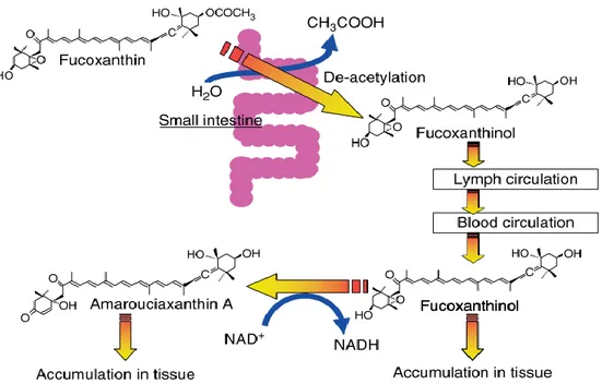
Fucoxanthin is an orange-colored carotenoid that has shown a lot of nutraceutical potential revealing to be a promising molecule with anti-cancer, anti-oxidant, anti-obesity (Peng et al., 2011), anti-inflammatory and anti-diabetic bioactivities, as well as being an active reducer of cardiovascular risk factors such as high blood pressure, high cholesterol and chronic inflammation (D’Orazio et al., 2012). This compound has a carbon backbone with an allenic group bond to a 5,6-monoepoxide and an acetyl group on the terminal ring which confers it a higher antioxidant capacity than their metabolites fucoxanthinol and halocynthiaxanthin (Miyashita et al., 2011; Sachindra et al., 2007). The cellular uptake after ingestion is dependent on the lipophilicity of the carotenoid and its interaction with the phospholipid membrane of the intestinal cells (Sugawara et al., 2001). Hence, the bioactivity of fucoxanthin and its metabolites (Fig. 1) relies on the digestibility of fucoxanthin which starts in the gastrointestinal tract by enzymes such as lipases that hydrolyze it or cholesterol esterase that deacetylate it to fucoxanthinol. Fucoxanthinol then travels to the liver where it is dehydrogenated into amarouciaxanthin A (Asai et al., 2004; Das et al., 2010; Peng et al., 2011). This mammalian metabolic pathway was described in mice after oral ingestion of fucoxanthin and two hours later no presence of it was detected while fucoxanthinol was predominantly present (Asai et al., 2004).
6
The same pathway was likewise proposed by Miyashita et al., (2011) (Fig. 2), whose work has also established the connection between fucoxanthin and its anti-obesity effect by down-regulating the expression of monocyte chemoattractant protein-1 (MCP-1), which promotes accumulation of macrophages in the adipose tissues leading to inflammatory stress.
Figure 1 - Metabolic pathway of fucoxanthin during digestion and intestinal absorption in mice. From Asai et al. (2004).
7
1.3 Microalgae Fields of Application
The variability of microalgal biomass composition has attracted a variety of different industries that now look at it as a reliable and clean multipurpose source for: aquaculture and human nutrition (Hariskos et al., 2014; Muller-Feuga, 2000); sustainable production of biodiesel using lipid-rich microalgae (Chisti, 2008; Wijffels et al., 2010); biological CO2 mitigation from either atmosphere or industrial exhaust gases (Sydney et
al., 2010; Wang et al., 2008); and pharmaceutical and drug development using carotenoids with anticancer, anti-inflammatory or antioxidant effects ( Moghadamtousi et al., 2014; Peng et al., 2011). For aquaculture or mariculture purposes, different microalgae strains are known to be a suitable feedstock to feed larvae of teleosts, crustaceans and mollusks due to their protein, vitamin and PUFA contents, size, lack of toxicity and high digestibility of the cell walls (Brown, 1991; Spolaore et al., 2006). Additionally, some species such as I. galbana contain free fatty acids known for their antibacterial effects against pathogenic microorganisms (e.g. Vibrio strains) decreasing the need for antibiotics. Thebiochemical composition of microalgal biomass is also very desirable for feeding cattle, pigs and poultry (Koller et al., 2014). Although the vast majority of microalgae used as aquaculture and poultry feeds is harmless, toxicity should also play an important role when deciding which species to use, since some strains of
Figure 2 – Metabolic decomposition of fucoxanthin during digestion, intestinal absorption and tissue accumulation in mice. Adapted from Miyashita et al., (2011).
8
diatoms, blue-green algae and dinoflagellates are known to produce secondary metabolites (e.g. domoic acid, saxitoxin, aplysiatoxin) capable of inducing poisoning, paralysis or destruction of important glutamate receptors in the brain of animals and humans (Shimizu, 2003).
Regarding human consumption, microalgae have been used as an alternative food source for hundreds of years mainly to surpass starvation. In the 16th century, before the Spanish conquest of Mexico, populations used to harvest “Spirulina” from the lake Texcoco, dry it in the sun and use it as food paste (Ciferri, 1983). The same habit, dating hundreds to thousands of years, is known to be present in populations of the Republic of Chad and China that live near lakes and ponds where “Spirulina” and Nostoc algae, respectively, were harvested using fine nets with the same purpose of finding another source of protein (Ciferri, 1983; Habib et al., 2008; Spolaore et al., 2006). The struggle to find a food stock that could satisfy the demand felt by these populations made them pioneers in the establishment of a novel food source rich in proteins, lipids and vitamins. Since then, microalgae for human consumption have gained widespread attention, not only because of their higher protein productivities when compared to soybean, rice and/or meat proteins (Kay et al., 2009), but also due to relatively low cost of cultivating them in raceway ponds (Norsker et al., 2011) and their nutrient contents. More recently, the European Union has approved the use of Chlorella sp. and Tetraselmis sp. for human consumption which represents another step towards the valorization of microalgae as a sustainable novel source of protein.
1.3.1 Market Value and Industrial Approaches
Today’s market value of microalgae species varies depending on its chemical and nutrient compositions and production costs, increasing when the concentration of compounds such as carotenoids, PUFAs, amino acids and vitamins is higher. The current market value of carotenoids is projected to reach US$ 1.4 Billion this year (D’Alessandro et al., 2016), which encourages industries to adapt production methods and develop a biorefinery model that allows them to extract high-value products and use the residual biomass to produce clean renewable energy such as biodiesel (Draaisma et al., 2013; Hariskos et al., 2014). The market value for microalgal pigments is on the rise, primarily due to the bioactive properties of compounds such as fucoxanthin or astaxanthin, which are known for their anticancer, antioxidant and skin protection properties, and secondarily, due to the biochemistry developments that highlight the metabolic
9
degradation pathways of these molecules within fish and mammalian organisms, ultimately providing a better understanding of their bioactivities and bioavailability (Asai et al., 2004; Guerin et al., 2003; Miyashita et al., 2011). For these reasons, it is crucial to understand the optimal growth conditions (e.g. light intensity, salinity, nitrogen availability, aeration flow) for the target microalgal strains in order to maximize the intracellular accumulation of these compounds and combine that knowledge with extraction techniques to optimize new industrial procedures.
Micro- and macroalgae have already been compared with results showing that microalgae produce up to fifty times more fucoxanthin (mg/g DW) than macroalgae (Xia et al., 2013), which enhances the need for scalable strategies for the extraction of such compounds. Current methods for carotenoid extraction from freeze-dried biomass include one-phase solvent extractions using ethanol, hexane or acetone with different yields depending on the affinity between fucoxanthin and the solvent chosen (Fig. 3), two-phase liquid systems using polar and non-polar solvents such as water and hexane (Kim et al. 2012), or alcohol-salt aqueous two-phase systems which have shown a 70% recovery of fucoxanthin and over 60% recovery of low molecular weight high-value compounds (Gómez-Loredo et al., 2014, 2015).
In contrast to traditional solvent extractions, techniques such as supercritical CO2
extraction have already proven to be less time-consuming by requiring fewer steps to effectively extract carotenoids from microalgal biomass (Macías-Sánchez et al., 2005). Additionally, the method has already been applied successfully for astaxanthin extraction
Figure 3- Efficiency of solvent extractions of fucoxanthin from freeze-dried biomass of Isochrysis aff.
galbana. Extractions were conducted for 1h at room temperature and analyzed with HPLC. From Kim et al., 2012.
10
from Chlorela vulgaris (Mendes et al., 1995), Chlorophyll a from Synechococcus sp. Sánchez et al., 2007) and carotenoids from Nanochloropsis guaditana (Macías-Sánchez et al., 2005), revealing to be a less toxic approach in comparison to conventional organic solvent extractions. Furthermore, Gilbert-López et al. (2015) successfully demonstrated the efficiency of this technique in extracting fucoxanthin, fucoxanthinol and other fuco-isomers from I. galbana biomass, enhancing the biorefinery strategy of downstream processing of biomass by combining supercritical CO2 extraction followed
by solvent extractions of residual biomass. The disadvantage of this method relies on its high costs of acquisition and operation which ultimately limits its widespread use in comparison with other cheaper and equally effective methods.
1.3.2 Emulsions: Liquid Three Phase System (LTPS)
The Liquid Three Phase System (LTPS) is a liquid-liquid extraction that uses a hexane-water solvent system to create an oil/water emulsion. The stability of the resulting emulsion is greatly enhanced by the presence of amphiphilic compounds such as the phospho- and/or betaine lipids, which are abundant in microalgae. This fact makes the LTPS of great interest in terms of novel techniques for the fractionation of microalgal lipids and the production of extracts rich in high-value compounds such as fucoxanthin and fatty acids.
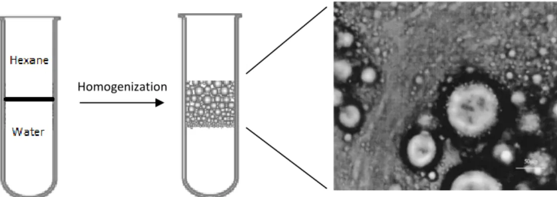
The production of emulsions has long been used to satisfy our daily demands in a variety of fields either in the petroleum industry by manufacturing crude oil or to supply the food markets with elementary products such as butter, mayonnaise, ice-cream, sauces or milk (Gouveia et al., 2010; Tambe et al., 1994). By definition, they are the result of two immiscible liquids combined together, with one liquid being dispersed in colloidal particles named droplets into the other liquid (Mcclements et al., 2007; Novales et al., 2003). The surrounding liquid is often referred as the “continuous phase” while the droplets are often classified as the “dispersed phase”. There are two main types of emulsions: oil droplets dispersed in an aqueous phase, which are classified as oil-in-water emulsions (e.g. milk, ice-cream); and water droplets spread in an oil phase that are classified as water-in-oil emulsions (e.g. butter) (Novales et al., 2003). Therefore, in order to form any type of stable emulsion three conditions must be met: i) the two liquids must be immiscible; ii) agitation must applied to homogenize the mixture; iii) an emulsifier agent of any type must be present (Fig. 4) (Chen et al., 2005).
11
The complexity of the interactions occurring between dispersed and continuous phases is tremendous due to the various physicochemical properties involved (e.g. size of droplets, charge, particle-fluid interface dynamics, droplet density and viscosity of the continuous phase) acting simultaneous and continuously throughout the process ultimately stabilizing or disturbing it (Gao et al., 2014; Walstra, 1993). The size and interaction of the droplets with the continuous phase are key factors for the overall kinetics of any given emulsion, ultimately revealing to be the cause of different types of instability mechanisms, as shown in figure 5 (McClements et al., 2007).
Figure 5- Representation of outcomes for emulsion instability. When colloidal particles have a lower density than the continuous phase they move upwards (creaming) and when they have a higher density, they move downwards (sedimentation). Flocculation is the aggregation of smaller droplets that keep their boundaries intact. By contrast, coalescence is the merging of smaller droplets forming a bigger droplet. The Ostwald Ripening it’s the physical interaction of bigger molecules that gradually increase size at the expense of smaller droplets. Adapted from McClements et al., 2007.
Figure 4- Schematic representation of an oil-water emulsion. The immiscible liquids are homogenized with agitation promoting the formation of colloidal droplets. The emulsifier agent coats the oil droplets enhancing the stabilization of the emulsion. Adapted from McClements et al. 2007 and Huang
et al. 2001.
12
Although emulsions are known to be thermodynamically unstable, since the contact of oil and water molecules is unfavorable, one can promote its stability by either using emulsifiers (e.g. phospholipids, proteins), texture modifiers (e.g. polysaccharides) or manipulating physical properties (e.g. temperature) favoring the emulsification (Chen et al., 2005; Dickinson, 2003; Mcclements et al., 2007; Pichot et al., 2013). Emulsifiers act by adsorbing to the droplet creating an external film layer that minimizes any instability mechanism and facilitates the interaction with the interface layer (Chen et al., 2005; Dickinson, 2003). Texture modifiers such as hydrocolloids formed by polysaccharides (e.g. pectin, fenugreek gum, arabic gum) are often used in food emulsions to increase the viscosity of the continuous phase promoting emulsion stability avoiding flocculation and coalescence, as well as, improving the shelf-life of the final product (Dickinson, 2009; Huang et al., 2001). The manipulation and monitoring of temperature throughout the process can greatly increase the outcome of any given emulsion by shifting the effect of parameters such as: viscosity, cohesive force and surface tension of the continuous phase. In fact, these parameters tend to decrease at temperatures greater than 30 °C, critically affecting emulsification (Chen et al., 2005; Joshi et al., 2012). The biochemical profile of microalgae biomass is known to be rich in phospholipids, proteins and polysaccharides thus making it good candidates as emulsifiers.
Starting with the preparation of an ethanolic microalgal extract, the LTPS technique develops a stable emulsion after the dispersion of the extract, containing mostly lipids, small sugars, peptides and pigments, in between both solvents. Due to the presence and amphiphilic nature of phospho- and/or betaine lipids three phases are formed: hexane, colloidal, water. Non-polar molecules reveal great affinity for the organic solvent phase while polar compounds prefer the aqueous solvent. Here, the suitability of LTPS in extracting the major groups of biomolecules including the carotenoid fucoxanthin and the -3 PUFA from I. galbana biomass is investigated. In addition, a first approach to understand the mechanisms behind the colloidal phase stability are researched by testing different ratios of water and hexane.
Isochrysis galbana was selected due to their nutritional composition with regards to DHA and the carotenoid fucoxanthin (Kim et al., 2012; Goméz-Loredo et al., 2014). Understanding the biochemical dynamics of this species and develop extracts rich in fucoxanthin and other high-value molecules in agreement with that knowledge represents one step further to better appreciate the value of microalgal biomass.
13 1.4 Isochrysis galbana
I. galbana is an unicellular bi-flagellated (Fig. 6) non-coccolith-forming microscopic photosynthetic alga that belongs to the phylum Haptophyta, class Prymnesiophyceae and order Isochrysidales (Kaplan et al., 1986; Parke, 1949; Sánchez et al., 2000; Sorrosa et al., 2005). As a marine microalga, its distribution ranges from European to North American waters crossing the south Iberian coast to the North Atlantic and Irish seas and reaching as far as the Pacific Ocean, from California’s coast up to Australia (Ahmed et al., 2014; Bandarra et al., 2003; Moita et al., 1999; Wang et al., 2014). The cellular architecture of this species is versatile not only in terms of morphological shape, but also on an intracellular level, primarily regarding the position, size and profile of its golden-brown chromatophores (Parke, 1949). Furthermore, as they are photosynthetic, they possess a stigma (or eye-spot apparatus), an organelle that plays an important role in sensing light, which helps the cell guiding the flagella to adjust its position towards or away from it (Parke, 1949). Unique to all haptophytes is the haptonema, a linear or arc-shaped microtubular complex present between the flagella with various roles, depending on the species, providing either sensorial guidance, substrate adherence or an appendix for predation in some mixotrophic strains (Green et al., 1977; Thierstein et al., 2004).
The haptonema of I. galbana is very rudimental and only visible in electron micrographs. Additionally, its multiplication alongside the flagella are the first signs that the cell is initiating mitosis (Fig. 7) that will culminate in two daughter-cells (Andersen, 2004; Hori et al., 1985). Surprisingly, asexual reproduction via cellular maturation of cysts is not the only form of reproduction for Haptophytes (Parke, 1949). In fact, the life cycle of this microflagellates typically includes haploid and diploid phases, each one capable of independent asexual reproduction (Ramawat et al., 2014).
14 1.4.1 Biochemical Profile
I. galbana accumulates lipids (Fig.8), such as TAG, under nitrogen depletion or increased salinities (Breuer et al., 2012; Liu et al., 2001), or synthetize less monounsatured fatty acids (MUFA) under shorter photoperiods or less PUFA under higher temperatures (Bandarra et al., 2003; Zhu et al., 1997). Multi-factor experiments provide an important insight of the fragility and plasticity of these molecular interactions by identifying the biological outcome of any combination of abiotic factors. Biochemically, lipids serve both as structural and fuel molecules and essential, biotechnologically relevant PUFA in this species include mainly EPA and DHA, although other fatty acids are also present in significant amounts, such as myristic, palmitoleic and linoleic acids (Batista et al., 2013).
Figure 6- A- I. galbana under Olympus S20 microscope (100x). Adapted from Gorgônio et al., (2013). B- I.galbana observed with a Nomarski differential interference contrast microscopic. Adapted from Liu et al., 2001. In both images the flagella are visible within the extracellular space surrounding the cells.
Figure 6- A- I. galbana under Olympus S20 microscope (100x). Adapted from Gorgônio et al., (2013). B- I.galbana observed with a Nomarski differential interference contrast microscopic. Adapted from Liu et al., 2001. In both images the flagella are visible within the extracellular space surrounding the cells.
Figure 7- I. galbana cells after completing cytokinesis have their edges covered with scales. The red arrow represents the formation of the first flagellum. n= Nucleus, g= Golgi complex. Adapted from Hori et al., (1985).
15
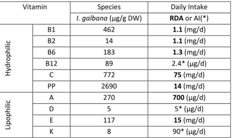
I. galbana has all the essential and non-essential amino-acids with some strains being able to accumulate more histidine, leucine, lysine and methionine. In fact, protein and lipid content of these strains can represent more than 50% of its dry weight (Brown, 1991). Regarding vitamin composition, this species can reveal high values for vitamin PP also known as niacin, vitamin B6 and vitamin C as well as lipophilic vitamins such as A, D, E, and K (Table 1; Roeck-Holtzhauer et al., 1991). Pyridoxine (vitamin B6) serves a very important metabolic pathway in the human organism regarding neurotransmitters and hemoglobin synthesis. Additionally, it is implicated in the prevention of cardiovascular traumas by decreasing homocysteine levels (Thaver et al., 2009). Niacin, is at the genesis of energy for cellular metabolic purposes by being a source for the biosynthesis of NAD+ and NADH which is used in mitochondria for the processes of oxidative phosphorylation and the Krebs cycle, ultimately generating Adenosine Triphosphate (ATP); human deficiencies in this vitamin can culminate in bone marrow leukemia, decreased apoptosis activity and compromised cell cycle regulation (Jacobson et al., 2012).
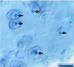
Figure 8 - I. galbana cells stained with Sudan Black B dye showing the lipid droplets accumulated intracellularly (arrows). Scale bar = 5 µm. Adapted from Liu et al. (2001).
16
Vitamin Species Daily Intake
I. galbana (µg/g DW) RDA or AI(*)
H yd ro p h ili c B1 462 1.1 (mg/d) B2 14 1.1 (mg/d) B6 183 1.3 (mg/d) B12 89 2.4* (µg/d) C 772 75 (mg/d) PP 2690 14 (mg/d) Li p o p h ili c A 270 700 (µg/d) D 5 5* (µg/d) E 117 15 (mg/d) K 8 90* (µg/d)
In I. galbana, carbohydrates (Fig. 9) are mainly constituted of monosaccharides such as glucose, mannose and galactose, which interact to form more complex molecules such as chrysolaminarin a polysaccharide formed by glycosidic bonds of glucose (Gorgônio et al., 2013).
Figure 9 – Comparison of the sugar monomers composition between Tetraselmis gracilis (Prasionophyte), Dunaliella tertiolecta (Clorophyta) and Isochrysis galbana (Prymnesiophyte). From Gorgônio et al., 2013.
Table 1- Vitamin composition of I.galbana cultivated in Conway medium at 18 °C, values from
Roeck-Holtzhauer et al., (1991). The human daily intake of vitamins is based either on the Recommended Dietary Allowance (RDA) or on the Adequate Intakes (AI*), the values presented are for the life stage group of Females ranging between 31-50 years old, from Trumbo et al., (2001).
Table 1- Vitamin composition of I.galbana cultivated in Conway medium at 18 °C. Adapted from Roeck-Holtzhauer et al., 1991.
17 1.4.2 Pigment profile
I. galbana is a brown microalga with an intracellular complex pigment composition comprising chlorophyll a and c as well as β-carotene and the polar xanthophylls fucoxanthin, diadinoxanthin and diatoxanthin (Andersen, 2004; Crupi et al., 2013; Takaichi, 2011). Concerning chlorophyll c, I. galbana contains two sub-types: monovinyl chlorophyll c1 and the divinyl chlorophyll c2, both playing a role in light harvesting (Garrido et al., 1997; Zapata et al., 2000). During photosynthesis, chlorophyll a, c and fucoxanthin act as primary pigments transferring light energy to the photosynthetic electron transport chain, while diadinoxanthin, diatoxanthin and β-carotene are secondary pigments acting as quenchers of excessive energy from chlorophyll a, providing photoprotection and preventing the emergence of ROS by releasing the excess energy as heat (Mulders et al., 2014; Takaichi, 2013; Varela et al., 2015; Zapata et al., 2004). Isochrysis galbana survival, as any other species, is dependent of the ongoing relationship between its evolutionary heritage and the environment, resulting in metabolic readjustments as a consequence of an extracellular stimulus (e.g. photoperiod, nutrient depletion/repletion). For that reason, the biosynthesis of pigments and isomers along with carotenoid derivatives (e.g. 13Z-, 13’Z-; all-E – fucoxanthin) is flexible during the life-cycle of this phototrophs (Crupi et al., 2013; Schüler et al., 2017). I. galbana uses fucoxanthin as a primary pigment in the light harvesting complexes of the thylakoid membranes; low light exposure creates a sub-saturating scenario that during acclimation induces carotenoid synthesis to maximize light harvesting, resulting in the intracellular accumulation of fucoxanthin and β-carotene (Gómez-Loredo et al., 2016; Mulders et al., 2014). Additionally, the pigment composition of this species can vary upon strains tested. Ahmed et al., (2014) isolated an Australian strain presenting 3205 µg/g (DW) of a putative violaxanthin and neoxanthin isomer, which is in agreement with the hypothesis developed by Lohr et al. (1999) that emphasizes that every algae species with the diadinoxanthyll cycle also possesses the violaxanthin cycle, the latter possibly acting as the biochemical precursor of the former regulated by the photon flux density (Fig. 10). Although working with diatoms, Lohr et al., (1999) suggested that when the photosynthetic apparatus is exposed to high light, a conversion of diadinoxanthin to diatoxanthin occurs which is later converted back to diadinoxanthin with low light exposure, leading to the synthesis of fucoxanthin. This theory is supported by Obata et al., (2012) which also highlights the complex pigment dynamics of this species, by concluding that the cell is also capable of decreasing its chlorophyll a concentration under
18
photo-acclimation to high light exposure or during the light phase of photosynthesis, and decreasing it again during the dark phase upon cellular multiplication.
Overall, one should keep in mind that the yield of all these biomolecules (lipids, amino-acids, vitamins, pigments and proteins) is entirely dependent on the multifactor interaction between the culture growth phase upon harvesting (e.g. lag, exponential or stationary phases), extraction methods used, the genetic variability, culture conditions, and the biomass drying method (Fabregas et al., 1986; Gouveia et al., 2008; Zhu et al., 1997).
Nowadays, markets have turned to microalgae to provide a sustainable source of fucoxanthin, mainly because the current ones (e.g. brown seaweeds) do not accumulate as much of this pigment and require additional steps in extraction protocols to prevent poisoning by iodine, a byproduct of some macroalgae fucoxanthin extractions (Wu et al., 2016).
Figure 10 - Hypothetical coupling between the xanthophyll- and the diadinoxanthin- cycles leading to synthesis of fucoxanthin in diatoms as a response of photon flux density. HL = High Light LL= Low Light. Adapter from Lohr et al., 1999.
19
2. Objectives
The aim of this master thesis was to develop and optimize an extraction protocol to produce fucoxanthin-rich fractions and associated biomolecules of interest from the microalga Isochrysis galbana. Primarily, the cultures were submitted to a novel technology of environmental modulation, the Algem labscale photobioreactors, to understand the optimal abiotic conditions for maximizing growth rate and infer on the bioaccumulation of these target compounds under different regimes. The carotenoid profile was assessed by diodearray-high performance liquid chromatography (HPLC-DAD) and the ω-3 PUFAs were measured using gas chromatography coupled with mass spectrometry (GC-MS). Additionally, a novel liquid-liquid extraction system (LTPS) was tested using the industrially produced biomass of Necton to infer on its efficiency to extract fucoxanthin and DHA. The goal with the present work was to determine if: i) Isochrysis galbana could be cultivated in Algem labscale photobioreactors? ii) Possible identification of abiotic parameters leading to accumulation of these target compounds? iii) The suitability of the LTPS system in effectively extracting these molecules?
20 3. Methodology
3.1 ALGEM®: Environment Labscale Photobioreactors
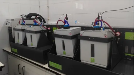
At Necton S.A., a Portuguese microalgae producing company placed in Olhão since 1998, ALGEM® labscale photobioreactors (Fig. 11) were used to test the growth response of I.galbana to different sets of abiotic factors. These systems simulate environmental parameters in an expedite way, allowing an effective control of temperature, photoperiod, light intensity and mixing, while accurately measuring growth rate by spectrophotometry at 740nm. In order to optimize the growth of this strain and to infer on total amount of fucoxanthin, as well as on fatty acid profile, two weekly trials were performed focusing firstly on the annual variations (seasons) followed by testing a pH range of 7 to 8.5. Nutrients (e.g. Nutribloom® solution produced by Necton) were only added at the beginning of the cultivation period. The media concentration of nitrates was 4 mM.
Figure 11- The Algem environment labscale photobioreactors coupled to a dedicated chiller to regulate the temperature inside each chamber.
21
ALGEM® photobioreactors software contains records of environmental conditions for each season worldwide (Fig.12). In the experiments, the GPS coordinates of Necton’s facilities were used, to accurately simulate the seasonal abiotic conditions felt in Belamandil, Olhão. Each chamber was programmed to represent the environmental conditions of each season (Summer, Spring, Autumn, Winter) based on an average of all parameters (e.g. light intensity, temperature, photoperiod) measured in Olhão that are recorded in the database of the ALGEM® software.
The database considered the months from December to February as Winter season; from March to May as Spring season; from June to August as Summer season; and from September to November as Autumn season. During the course of the experiment, the cultures growth was followed spectrophotometrically, on a daily basis. One the second trial,the abiotic conditions used to test the cultures response to the pH range 7-8.5, were the ones that provided a higher growth rate in the first trial; table 2 describes the environmental parameters used in each experiment. In both trials the optical density of the initial inoculum was assessed using a UVmini-1240 spectrophotometer. Afterwards, the inoculum was diluted for a final volume of 2L from which it was equally divided for each chamber, as demonstrated in figure 13. The experiments lasted one week, after which the biomass in each chamber was individually centrifuged and the pellets stored frozen (-20ºC).
22
Figure 12 - ALGEM® photobioreactors software. The equipment has a database of climate records from the last 80 years and the interface allows the user to choose a GPS position. The abiotic parameters are then adjusted according to the chosen location. It also gives the possibility to create an abiotic profile by selecting different types of light (e.g. Red, Blue, White or Total), photoperiod and temperature either constant or in daily cycles. The pH control inside each chamber is individually regulated by CO2 injection. Finally, the interface allows the user to assess the growth rate of the
culture through a graphic representation of the absorbance (at 740 nm) at a desirable interval of time.
Figure 12 - ALGEM® photobioreactors software. The equipment has a database of climate records from the last 80 years and the interface allows the user to choose a GPS position. The abiotic parameters are then adjusted according to the chosen location. It also gives the possibility to create an abiotic profile by selecting different types of light (e.g. Red, Blue, White or Total), photoperiod and temperature either constant or in daily cycles. The pH control inside each chamber is individually
23
Table 2 – The abiotic conditions determined for the trials conducted in ALGEM® labscale photobioreactors. On the first trial (seasons) each chamber comprehended its unique set of environmental conditions. On the second trial (pH) the environmental profile selected was the one that provided the higher growth rate on the first trial.
Trial Seasons pH
Chamber Summer Spring Autumn Winter 7 7,5 8 8,5
Temperature (°C) 20.5 - 28.5 12.1 - 20.2 15.1 - 23.1 7.9 - 13.8 20.5 - 28.5 Light Intensity (µmol m-2 s -1) 0 - 1600 0 - 1200 0 - 980 0 - 650 0 - 1600 Photoperiod (Hours) 15 13 11 9 15 pH 8 Salinity (g/L) 33 33 Mixing (RPM) 60 60
Figure 13 – The different stages of preparation for an Algem incubation. A,B – Dilution of Necton’s culture and inoculation inside a laminar flow chamber. C,D- Setup of Algem lid which is connected to the CO2 injection system, air entrance and exit, as well as a pH probe. From here, the Erlenmeyers were placed inside each chamber to
start the experiment.
24 3.1.1 Pigment Extraction and Analyzes
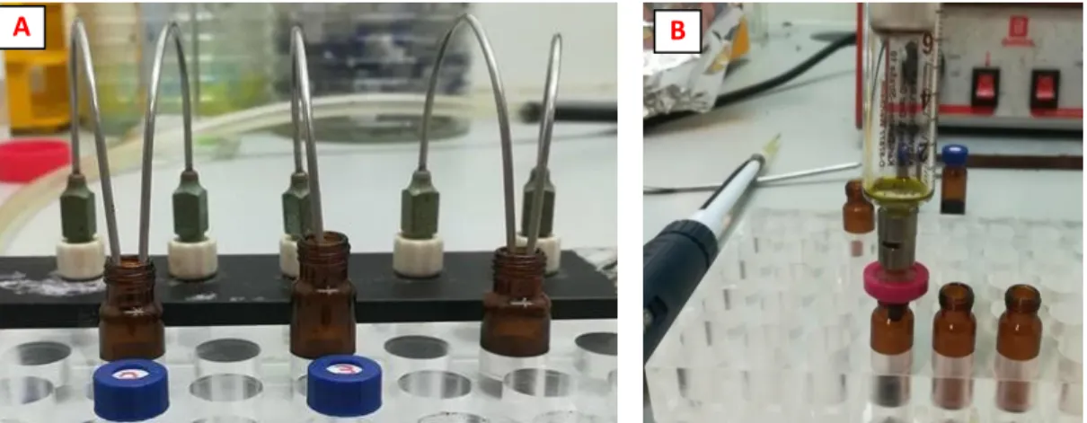
Pigments extraction and analyses was conducted for the biomass resulting from the Algem® trials, as well as, for the biomass produced outdoors in Necton’s photobioreactors (PBR) in order to compare the fucoxanthin content. For this, 20 mL of seawater were added to defrosted biomass and the mixture homogenized for 10 seconds in a vortex homogenizer. From here, a small volume was transferred to new falcon tubes, depending on the overall concentration of each pellet; from the outdoor biomass 0.25 mL were used for the extraction whilst from the Algem® trials 0.5 mL were used. These new falcon tubes were centrifuged at 8000g for 5 minutes at 10ºC and the supernatant -salt water- was discarded. Following that, 3mL of acetone and 0.7g of glass beads (mesh 500-750 µm) were added to all tubes that were subsequently submitted to 2 minutes of vortex in order to promote the breaking of cellular walls by the glass beads while the acetone extracted the pigments. During this process, all tubes were kept on ice to freeze the cell walls and potentially increase the extraction yield. Afterwards, the tubes were centrifuged and the supernatant was collected to different vials. This process was repeated 3 times to ensure full pigment extraction. The vials were later dried in nitrogen gas flow (Fig.14A) and all samples were resuspended in 700 µl of methanol HPLC grade and filtered to new vials using 0.25 µm PTFE filters (Fig. 14B).
3.1.2 Determination of fucoxanthin by HPLC
The vials containing the recently resuspended and filtered samples were analyzed in a Dionex 580 HPLC System (DIONEX Corporation, United States) equipped with a PDA 100 Photodiode-array detector, P680 Pump, ASI 100 Automated Injector and STH 585 column oven, using a LiChroCART® RP-18 (5µm, 250x4 mm, LiChrospher®) column
Figure 14 – A: The process of drying the samples using a gentle flow of Nitrogen. B: The resuspension of the samples using a syringe and a PTFE filter.
Figure 14 – A: The process of drying the samples using a gentle flow of Nitrogen. B: The resuspension of the samples using a syringe and a PTFE filter.
25
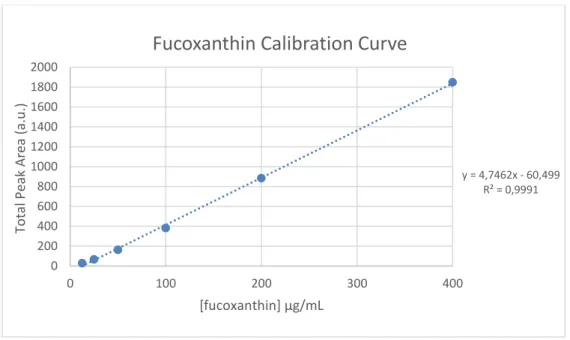
and Chromeleon® software. The mobile phase consisted of 9:1 (v/v) acetonitrile:water as solvent A, and, ethyl acetate as solvent B and the gradient program applied was: 0–16 min, 0–60% B; 16–30 min, 60% B; 30–32 min 100% B and 32-35 min change to 100% A at a flow rate of 1 ml/min. The injection volume was 100 µl and the temperature was maintained at 20°C. Fucoxanthin was detected and quantified at 450 nm using a calibration curve made by a serial dilution (400-12.5 µg/mL) from a fucoxanthin standard (Fig. 15).
To facilitate the evaluation of the extraction efficiency, a spectrophotometric method was developed to estimate the amount of fucoxanthin in the extracts, since spectrophotometry is a cheaper and less time-consuming technique to quantify this pigment. Hence, a calibration between the absorbance of the extracts at 450 nm and the fucoxanthin content of those samples measured by HPLC was performed (Fig. 16). The resulting correlation coefficient was high (R2= 0.96). For this reason, it was decided to quantify the fucoxanthin content of the ethanolic extractions by spectrophotometry.
y = 4,7462x - 60,499 R² = 0,9991 0 200 400 600 800 1000 1200 1400 1600 1800 2000 0 100 200 300 400 To ta l Peak Area (a.u .) [fucoxanthin] µg/mL
Fucoxanthin Calibration Curve
Figure 15 – The calibration curve resulting from the serial dilution of fucoxanthin, measured in HPLC.
26 3.1.4. Gas Chromatography – Mass Spectrometer
Fatty acid methyl esters (FAME) were produced following a modified protocol from Lepage & Roy (1984). The pellets were resuspended in 1.5 mL of methanol with acetyl chloride (20:1, v/v) and 1 mL of hexane, transferred to derivatization vials and homogenized using an Ultra _Turrax in two cycles of 60 and 30 seconds, always on ice. From here the vessels were placed in a water bath at 70°C for 60 minutes (Fig.17).
y = 0,0744x - 0,0019 R² = 0,9603 0 0,5 1 1,5 2 2,5 3 3,5 4 0 10 20 30 40 50 60 Ab s a t 450nm [fucoxanthin] HPLC (µg/mL)
Fucoxanthin Correlation
Figure 17 – A: Derivatization vessels with the samples and the solvents. B: The process of breaking the cells using the Ultra turrax while keeping the samples in ice to prevent excess heat and possible degradation of compounds. C: The hot water bath were the samples were placed after being wrapped in Teflon to minimize losses by evaporation.
Figure 17 – A: Derivatization vials with the samples and the solvents. B: The process of breaking the cells using the Ultra turrax while keeping the samples in ice to prevent excess heat and possible degradation of compounds. C: The hot water bath were the samples were placed after being wrapped in Teflon to minimize losses by evaporation.
A B C
Figure 16 – Correlation between the fucoxanthin present in the samples measured in spectrophotometry at 450 nm and HPLC.
Figure 16 – Correlation between the absorbance of the extracts measured at 450 nm and fucoxanthin content measured by HPLC.
27
Later the samples were allowed to rest in ice and the content transferred to centrifuge tubes. To clean the derivatization vials, 1mL of distilled water and 4 mL of hexane were added and transferred back to the centrifuge tubes. Then, the tubes were centrifuged for 5 minutes at 2000g and the hexane fraction transferred to new glass tubes. This step was performed twice to minimize FAME losses. Following, anhydrous sodium sulfate (Na2SO4) was added to remove water residues and the hexane solution was filtered with
Whatman 0.25 µm PTFE syringe filters (Fig 18A and B). Afterwards the samples were dried under a gentle nitrogen flow and finally resuspended in 500 µl gas chromatography-grade hexane and transferred to vials after filtration.
The FAME profile was assessed using a Bruker GC-MS (Bruker SCION 456/GC, SCION TQ MS, Fig. 19 A,B) equipped with a ZB-5MS capillary column using helium as a carrier gas. The temperature was 60°C (1 min), 30°C min−1 to 120°C, 5°C min−1 to 250°C, and 20°C min−1 to 300°C (2 min). Individual calibration curves were made for each FAME using the standard 37 FAME Mix (Supelco).
A B
Figure 18 – A: The first filtration of the hexane fraction. B: After a gentle flow of nitrogen to dry the samples, they were resuspended and transferred to chromatography vials.
Figure 18 – A: The first filtration of the hexane fraction. B: After a gentle flow of nitrogen to dry the samples, they were resuspended and transferred to chromatography vials.
28 3.2 Biomass production: Necton’s photobioreactors (PBR)
Isochrysis galbana was industrially produced in Necton’s tubular photobioreactors (PBR). Figure 20 summarizes the cultivation and harvesting procedures starting with the inocula which were kept in the laboratory under controlled temperature and supplied with aeration enriched with CO2. From here, it is usually transferred to the
outside green wall systems ranging from 100 to 800 L. Once the desired optical density was achieved for these systems, the cultures were transferred to the 15 m3 tubular PBR. Throughout this scale-up procedure, the cultures were checked daily for contaminations, using a microscope, and for overall healthiness by evaluating cellular structure and motility. Additionally, nutrients such as industrial iron and Nutribloom® were supplied weekly. The harvest procedure was conducted using an industrial centrifuge and the biomass was packed and stored frozen (-20ºC).
Figure 19 – A,B: The gas chromatography equipment with the coupled auto sampler.
Figure 19 – A,B: The gas chromatography equipment with the coupled auto sampler.
A
29
Figure 20– Schematic representation of the industrial process of microalgae production, from the inocula to green wall panels (GWP) and later to tubular photobioreactors (R1-4). The reactors are connected to the harvest tanks (HT1-3), which are in turn connected to the centrifuge. Each reactor and tank has its own pumps and a set of both manual and electronic valves. The aeration system is equipped with a valve and timer that regulates the frequency and duration of CO2 injections. The
ultra-filtration (UF) system purifies the water according to industrial standards. After harvesting, the final product (A: GreenFormula®; B: ICE-Concentrated frozen paste; C: Lyophilized biomass) is packed and stored to be sold.