Technology
Journal
Original Article
ORAL MUCOSAL LESIONS ASSOCIATED WITH THE
USE OF DENTURES: CASE SERIES
LESÕES DA MUCOSA ORAL ASSOCIADAS AO USO DE
PRÓTESES: UMA SÉRIE DE CASOS
Luana Eduarda de OLIVEIRA1, Fabio Augusto ITO2, Ademar TAKAHAMA-JUNIOR3,
Heliton Gustavo de LIMA4
1 Universidade Estadual de Londrina (UEL). Acadêmica do Curso de Odontologia da Universidade Estadual de Londrina. E-mail: [email protected].
2 Universidade Estadual de Londrina (UEL). Possui Graduação em Odontologia pela Universidade Norte do Paraná, Mestrado e Doutorado em Estomatopatologia pela Faculdade de Odontologia de Piracicaba, Universidade Estadual de Campinas (FOP-UNICAMP). Professor Adjunto da área de Estomatologia e Patologia Oral do Curso de Odontologia da
Universidade Estadual de Londrina. E-mail: [email protected].
3 Universidade Estadual de Londrina (UEL). Possui Graduação em Odontologia, Mestrado e Doutorado em Estomatopatologia pela Faculdade de Odontologia de Piracicaba, Universidade Estadual de Campinas FOP-UNICAMP.
Professor Adjunto da área de Estomatologia e Patologia Oral do Curso de Odontologia da Universidade Estadual de Londrina. E-mail: [email protected].
4 Universidade Estadual de Londrina (UEL). Possui Graduação em Odontologia pela Universidade Norte do Paraná, Mestrado e Doutorado em Ciências Odontológicas Aplicadas com área de concentração em Patologia Bucal pela Faculdade de Odontologia de Bauru, Universidade de São Paulo FOB-USP. Professor Adjunto da área de
Estomatologia e Patologia Oral do Curso de Odontologia da Universidade Estadual de Londrina. E-mail: [email protected].
ABSTRACT: The World Health Organization recognizes tooth loss as a social and public
health problem. The edentulism can be defined as total or partial loss of permanent dentition. It occurs as a consequence of several deleterious effects that take place throughout an individual’s lifetime. Thus, the use of denture, for the restoration of masticatory function, is necessary. The poor hygiene and fit of these denture as well as other local and systemic factors may contribute for the appearance of buccal lesions, such as denture stomatitis, traumatic ulcers, angular cheilitis and fibrous hyperplasia. Therefore, this article aims to report a series of three clinical cases of oral mucosa lesions associated with the use of dental prosthesis, in elder ly patients. The reported cases are of female patients aged between 54
and 66 years who were diagnosed and adequately treated for denture stomatitis, angular cheilitis and inflammatory fibrous hyperplasia. Therefore, this study highlights the importance of the dentist in the prevention, diagnosis and treatment of these oral diseases prevalent in dentistry.
Keywords: Geriatric Dentistry. Dental Prosthesis. Aged. Mouth Diseases.
RESUMO: A Organização Mundial da Saúde reconhece a perda dentária como um
problema social e de saúde pública. O edentulismo pode ser definido como a perda total ou parcial da dentição permanente e ocorre como consequência de vários efeitos deletérios que se sucedem durante a vida do indivíduo. Desta forma, a utilização de próteses dentárias, para o restabelecimento da função mastigatória, torna-se necessária. A falta de higienização e adaptação dessas próteses dentárias bem como outros fatores locais e sistêmicos podem contribuir para o aparecimento de lesões bucais como a estomatite protética, úlcera traumática, queilite angular e hiperplasias fibrosas. Portanto, esse artigo tem como objetivo relatar uma série de 3 casos clínicos de lesões bucais associadas ao uso de prótese dentária, em pacientes idosos atendidos no ambulatório de Estomatologia da Clínica Odontológica da UEL. Os relatos apresentados são de pacientes do sexo feminino com a idade entre 54 e 66 anos que receberam diagnósticos e adequados tratamento para estomatite protética, queilite angular e hiperplasia fibrosa inflamatória. Portanto, esse trabalho realça a importância do cirurgião-dentista, na prevenção, diagnóstico e tratamento dessas prevalentes doenças bucais na odontologia.
Descritores: Odontogeriatria. Prótese Dentária. Idoso. Doenças da Boca.
1. INTRODUCTION
The world population has been quickly aging in various regions of the planet, due to the decreased fertility rate and increased life expectancy1. The World Health Organization
(WHO) established that 60-year-old people or over are considered elderly in developing countries2. In the last 200 years, people in all
countries of the world achieved an impressive
health-related progress, which leads to an increased life expectancy. Globally, life expectancy increased to an average of 73 years in 20193.
The elderly are those who most uses dental prostheses, with values of tooth loss that reach 92.4%4. During aging, there is a
decrease in the protective function of the oral mucosa, such as the reduction of oral epithelium proliferation and collagen synthesis
in the connective tissue. Therefore, a decreased tissue regeneration is expected, as well as less resistance to injuries. Alterations in the oral mucosa of these patients can be explained by the interaction of many factors, such as systemic condition, aging, metabolic alterations, nutritional factors, medication use, psychobiological habits, prosthesis use, alcohol and tobacco consumption5-8. The main
mucosal lesions associated to removable prosthesis are denture stomatitis, angular cheilitis, inflammatory fibrous hyperplasia and traumatic ulcers. The presence of candida infection, poor oral hygiene, mechanical trauma, low salivary pH and reduced occlusion vertical dimension (OVD), have been associated with the development of these lesions6,9.
The oral rehabilitation does not eliminate the possibility of new problems arising regarding the biological and prosthetic elements involved10. The maintaining of
proper hygiene of the prostheses has an essential role in preventing lesions related to its use11. Dentists play an important role in
guiding patients on the proper hygiene of their prostheses12.
Constant exposure to various harmful agents predisposes the oral mucosa to various oral diseases13. The use of inadequate
removable prosthesis must be considered an important factor influencing the presence of oral diseases14. Thus, this article reports a
series of three cases of oral mucosa lesions associated with dentures in elderly patients, as well as discusses the etiopathogenesis, prognosis and current treatment for these conditions.
2. CASE REPORT
2.1. Case Report 1
A 54-year-old woman was referred to the Stomatology Service, School of Dentistry, Londrina State University, complaining of “gum pain”. Her medical history included schizophrenia that currently was being managed with chlorpromazine and olanzapine. Extraoral examination revealed a reddish erosion in the labial commissure, bilaterally (Figure 1). On the intraoral examination, a sessile nodule was noted in the right posterior maxillary vestibule, with a smooth surface, with color similar to normal oral mucosa, occasionally symptomatic, measuring about 3.5 cm in diameter (Figure 2). Besides, a diffuse erythematous asymptomatic area in the hard palate and upper alveolar ridge was also observed. Moreover, oral mucosa dryness was verified. According to the clinical features, a diagnosis of angular cheilitis was established for the lesions in the labial commissure; and denture stomatitis for the lesions on the hard palate and upper alveolar ridge. The clinical diagnostic hypothesis for the nodule associated with the border of maxillary denture was inflammatory fibrous hyperplasia (IFH). The proposed treatment to the denture stomatitis consisted in using antifungal for seven days, to which a nystatin oral suspension solution of 100,000 IU/mL was prescribed, recommending the patient to do mouthwash with the medication three times a day, and then swallow it. Regarding the sessile nodule, the proposed treatment was to
discontinue the use of the denture, and a total regression of lesion was noted after 21 days. In addition, the patient received instructions on oral and prosthetic hygiene and she was encouraged to replace her dentures.
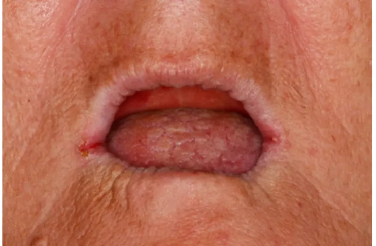
Figure 1 – Clinical aspect of angular
cheilitis characterized by maceration, erythe-ma, and crust formation of corners of mouth.
Figure 2 - Discrete sessile nodule in
the right posterior in alveolar mucosa that had a smooth surface.
2.2. Case Report 2
A 59-year-old woman sought our
ser-vice, complaining that “a fleshy tissue had appeared in mouth”. During anamnesis, she reported to be in treatment for depression with clonazepam, venlafaxine and carbamaz-epine. The patient was a former smoker, hav-ing had stopped 10 years before, and report-ed consumption of alcoholic beverages twice a week. Extraoral examination did not show remarkable features. Upon the intraoral exam-ination, we observed multiple sessile nodules with normochromic colored, irregular surface and shape with distinct borders, located on the anterior maxillary vestibule. The lesion had flaccid consistency and measured about 3 cm (Figure 3). On the basis of these clinical features, the presumptive clinical diagnosis was of IFH, and excisional biopsy under local anesthesia was performed. Histopathological examination revealed a fragment of mucosa lined by a stratified squamous parakeratinized epithelium, showing areas of hyperplasia. In the submucosa, there was a hyperplastic dense fibrous connective tissue with chronic inflammatory infiltrate, confirming the diagno-sis of IFH (Figure 4). One week after the bi-opsy, an optimal tissue repair was observed. Adjustments were performed in the upper denture and instructions concerning the need for replacement by new dentures were given.
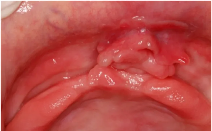
Figure 3 – Multiple erythematous
nod-ules with irregular surface and shape located on the anterior maxillary vestibule.
Figure 4 – Photomicrograph of
biop-sy specimen showing a fragment of mucosa lined by a stratified squamous parakeratinized epithelium, with areas of hyperplasia. Submu-cosally, hyperplastic dense fibrous connective tissue showed discreet chronic inflammatory infiltrate. (HE, 100x).
2.3. Case Report 3
A 60-year-old woman sought our ser-vice, complaining of “bleeding gum”. In the an-amnesis, she reported having stopped using
the prosthesis due to the discomfort and pain that caused it. Her medical history included hypertension and hypothyroidism that current-ly was being managed with spironolactone, levothyroxine and omeprazole. Extraoral ex-amination found no abnormalities. Intraoral inspection, revealed areas with diffuse ery-thematous spots, smooth-surfaced and as-ymptomatic in the hard palate and upper al-veolar ridge (Figure 5). Poor oral hygiene as also observed with presence of coated tongue and intense halitosis. Based on the clinical as-pects observed, the clinical diagnosis was of denture stomatitis. Thus, the patient received instructions on oral and prosthetic hygiene and to remove the dentures, especially during sleep. The therapy established was of nystatin oral suspension of 100,000 IU/mL, with similar recommendations to case 1. After one week, there were regression of lesions in hard palate and oral and prosthetic hygiene significant-ly improved. The patient was advised of the need to replace her prostheses.
Figure 5 – Areas with diffuse
erythem-atous spots, smooth-surfaced in the hard pal-ate and upper alveolar ridge.
3. DISCUSSION
The poor hygiene of the dentures as-sociated with trauma act as predisposing factor to the appearance of oral mucosal le-sions, its development depending on the pa-tient’s health conditions15. In this case series,
all patients presenting oral mucosa lesions associated with the use of prosthesis and were female with aged from 54 to 60 years. It is known that women seek dental treatment more often than men, allowing lesions to be detected, besides postmenopausal hormonal changes that make the mucosa more suscep-tible to hyperplastic reactions16.
Denture stomatitis (DS) is a widely prev-alent inflammatory disease among prosthesis wearers. It affects mostly the palatal mucosa and is strongly associated with poor denture hygiene17. Multiple etiological factors
contrib-ute to denture stomatitis, such as microor-ganisms Candida sp. and Gram-negative an-aerobes bacteria; impaired salivary flow and salivary gland function; trauma from ill-fitting dentures; poor denture and oral hygiene; and impaired immune response secondary to sys-temic conditions. Certain strains of Candida, specifically hyphal-forming Candida albicans, are more commonly found in candidal infec-tions in DS patients. These virulent strains are capable of epithelial binding, disruption of epithelial integrity, and invasion18-19. The
pro-gression of DS without an adequate treatment can lead to a systemic infection, especially in immunocompromised patients20. Most red oral
lesions are of inflammatory nature, but some are potentially malignant, especially oral
eryth-roplakia21. This lesion has a high probability of
showing signs of dysplasia or malignancy at the time of diagnosis. Thus, the biopsy is man-datory in cases of doubt. Other rare lesions, that must be excluded during the diagnosis are: Kaposi´s sarcoma, erosive lichen planus, and discoid and systemic lupus erythemato-sus22-23.
Studies have demonstrated that many denture wearers attempt to maintain denture hygiene only by brushing, as one would brush natural dentition; however, this is inadequate for maintaining proper denture hygiene, and other methods, such as use of commercial dis-infectant solutions, or immersion of dentures in dilute sodium hypochlorite, are required as part of daily and routine denture maintenance, as well as removing it during the night24-25 in
order to give rest to the supporting tissues26.
Sodium hypochlorite 1%, has been accept-ed by the American Dental Association as a prosthesis cleaning and disinfection agent; its advantages include a wide antimicrobial spectrum27. The oil from the seeds of Ricinus
communis (castor bean) has detergent
prop-erties and is able to damage the fungal cell wall, leading to cytoplasm extravasation and consequent cell death28. Mechanical brushing
with mild soap associated with the disinfection with sodium hypochlorite is still one of the most efficient ways to proceed with the hygiene, due to its simplicity, low cost and efficiency in removing the biofilm. However, in older adults who depend on others, it is advised that such cleaning procedure should be done by a fami-ly member or caregiver29. With the purpose of
improving hygiene through adequate cleaning of the prosthesis, some recommendations are
included, as the regular and frequent use of soft brushes, antifungal therapy, mouthwash with anticandidal activity, including triclosan, chlorhexidine and gluconate, as well as the hygiene of the soft tissues that are in contact with the prosthesis30.
In the cases here presented, all the den-tures were in unfavorable hygiene conditions. Thus appropriated instructions about oral and denture hygiene were given by the dentists. In addition, it was prescribed antifungal for sev-en days (nystatin oral suspsev-ension solution of 100,000 IU/mL). After one week, it was pos-sible to observe regressions in lesions on the hard palate and a significant improvement in oral and denture hygiene. Patients were also advised to remove their dentures during sleep and to replace them with new dentures. Brantes et al. demonstrated that the habit of night use of the denture is considered an inde-pendent risk factor for the development of oral lesions. Furthermore, they also found that the longest period of use of the same denture and biofilm had statistically significant relationship to oral lesions9.
Another disease reported in our cases was angular cheilitis. Clinically, it is a frequent condition characterized by erythema, crack-ing, fissurcrack-ing, and maceration of the lip corners and the adjacent skin, either in one commissure or both.31. The angular cheilitis
is usually related to one or more factors, in-cluding: infectious agents (Staphylococcus
aureus, Streptococci and Candida),
dermato-logical diseases, nutritional deficiency, immu-nodeficiency, hypersalivation and mechanical factors, leading to the loss of the OVD32. Some
systemic disorders as the Down syndrome,
in-flammatory bowel diseases (such as Crohn’s disease or ulcerative colitis) can be associat-ed with angular cheilitis33-35.
In our case, the presence of angular cheilitis was related to loss of OVD caused by edentulism and wearing the same denture for a long time. Occasionally, this type of cheilitis can be miss diagnosed for other less frequent lesions, which can have a similar clinical ap-pearance, such as herpes simplex, impetigo, and pemphigus vulgaris. The treatment often requires a multidisciplinary approach. The main factor for a successful treatment is iden-tifying each case’s correct etiological factors. The most common treatment is based on an-tifungals. However, local antibiotics and corti-coids, vitamin complexes, hygiene of the pros-theses, and allergen prevention has also been used36-37. In our case, due to the large time
using the same denture, the treatment estab-lished was the replacement of the prosthesis to reestablish the OVD and reduce the forma-tion of prominent folds at the corners of the mouth, which provides accumulation of saliva and the skin may become cracked and infect-ed secondarily. Moreover, it was prescribinfect-ed topical antifungals and oriented about the cor-rect oral and prosthetic hygiene.
Acute and chronic irritation from defec-tive or ill-fitting dentures may injure the oral mucosa, resulting in the formation of traumatic ulcers or hyperplastic tissues folds. IFH is the most frequent lesion among inflammatory/re-active lesions, and its development correlates directly to denture use38. The prevalence rate
of IFH ranges from 5% to 20% of all the oral biopsies and is observed in 65% of the lesions in denture users39. The IFH is a
non-neoplas-tic proliferative lesion resulting from a hyper-plastic reaction of the fibrous connective tis-sue, and it develops in association with the borders of poorly fixed total or partial remov-able prosthesis40. In our cases of IFH, the
pa-tients were wearing poorly adapted dentures for more than 19 years. In addition, dentures also showed deterioration, irregular edges, rough and cracked surfaces. Clinically, IFH is asymptomatic with multiple folds that can be detected on the alveolar mucosa. Ulcerations are occasionally observed in the bottom of fis-sures of the lesion. This lesion usually occurs in middle-aged and older adults who wear re-movable dentures for a long time, with a high-er prevalence rate in women41. Histologically,
it is a dense fibrous connective tissue with variable chronic inflammatory infiltrate42.
This lesion presents excessive colla-gen deposition, being responsible for the in-creased volume43.The treatment consists in
surgical removal of the hyperplastic tissue, and adjustment or replacement of the prosthe-sis to prevent recurrences. The red and small lesions, which are mostly inflamed lesions, completely disappear when the denture is re-moved or its flanges are shortened. In one of our cases the proposed treatment was to dis-continue the use of the denture for a certain period of time and in the other case excisio-nal biopsy. It is now known that other lesions
of appearance nodule may mimic the IFH, such as benign mesenchymal neoplasms, as well as non-neoplasic proliferative processes, being of is important the correlation of clinical and microscopic aspects.
The prevalence of lesions in oral mu-cosa related to the use of dentures can be reduced through adequately instructing the patients; the preservation of oral hygiene; and cares by dentist in the various stages of mak-ing the prostheses, resultmak-ing in stable occlu-sion, good fixation of its basis to the mucosa, and adequate peripheral sealing, within the limits of the basal area44. In addition, the
pa-tient must also understand the maintenance requirements, and need to alter personal be-haviours which compromise oral health45.
4. CONCLUSIONS
Based on this serie of cases, the oral lesions found were associated to poor oral and denture hygiene, in addition to several years of using the same prosthesis and their deficiency of adaptation. Considering the sys-temic and oral consequences of these factors and the great number of patients wearing den-tures, a special attention should be given by clinicians and public health officials to these denture related problems.
5. REFERENCES
1. Kowal P, Goodking D, HE W. An Aging World: 2015. Washington, DC, 2016.
2. WHO. The uses of Epide-miology in the Study of the Elderly. Tech-nical Report Series 706. Geneva: WHO; 1984.
3. Roser M, Ortiz-Ospina E, Hannah Ritchie. Life Expectancy. J Our World in data 2019.
4. Ministry of health. SB zil 2010- National oral health policy. Bra-sília, 2012.
5. Mello dos Santos C, Bal-binot J, Pereira D, Neves F. Denture stomatitis and its risk indicators in south Brazilian older adults. Gerodontology. 2010; 27: 134– 140.
6. Jainkittivong A, Aneksuk V, Langlais R. Oral Mucosal lesions in denture wearers. Gerodontology.2010; 27: 26–32.
7. Leal SC, Bittar J, Portugal A, Falcão DP, Faber J, Zanotta P. Med-ication in elderly people: its influence on salivary pattern signs and symptoms of dry mouth. Gerodontology. 2010; 27: 129–133.
8. Bof F, de Franca A,
Ma-kumbundu P. Relationship between oral health, nutrient intake and nutritional sta-tus in a simple of Brazilian elderly peo-ple. Gerodontology. 2009; 2: 40–45.
9. Brantes MF, Azevedo RS, Rozza-de-Menezes RE, Póvoa HC, Tucci R, Gouvêa AF, et al. Analysis of risk factors for maxillary denture-related oral mucosal lesions: A cross-sectional study. Med Oral Patol Oral Cir Bucal. 2019; 1 (24): 3.
10. Gray JC, Navarro-Coy N, Pavitt SH, Hulme C, Godfrey M, Crad-dock HL, et al. Improvdent: Improving dentures for patient benefit. A crossover randomissed clinical trial comparing impression materials for complete den-tures. BMC Oral Health. 2012; 12: 37. doi: 10.1186/1472-6831-12-37.
11. Collis JJ, Stafford Tafford GD. A survey of denture hygiene in pa-tients attending Cardiff dental hospital. Eur J of Prosthodont Restor Dent. 1994; 3 (2): 67-71.
12. Kulak-Ozkan Y, Kazazoglu E, Arikan A. Oral hygiene habits, den-ture cleanliness, presence of yeasts and stomatitis in elderly people. J Oral Reha-bil. 2002; 29: 300-304.
13. Shulman JD, Beach MM, Rivera-Hidalgo F. The prevalence of oral mucosal lesions in U.S. adults: data from the Third National Health and Nutrition
Examination Survey. J Am Dent Assoc. 2004; 135: 1279-1286.
14. Espinoza I, Rojas R, Aran-da W, Gamonal J. Prevalence of oral mucosal lesions in elderly people in Santiago. J Oral Pathol Med. 2003; 32: 571-575.
15. Mandali G, Sener ID, Turk-er SB, Ulgen H. Factors affecting the distribution and prevalence of oral mu-cosal lesions in complete denture wear-ers. Gerodontology. 2011; 28: 97-103.
16. Coelho CMP, Sousa TCS, Dare AMZ. Denture-related oral muco-sal lesions in a Brazilian school of den-tistry. J Oral Rehabil. 2004; 31: 135-139. 17. Gendreau L, Loewy ZG. Epidemiology and etiology of denture stomatitis. J Prosthodont. 2011; 20: 251-260.
18. Salerno C, Pascale M, Contaldo M. Candida-associated den-ture stomatitis. Med Oral Patol Oral Cir Bucal. 2011;16: 139-143.
19. Altarawneh S, Bencharit S, Mendoza L, Curran A, Barrow D, Barros S,et al. Clinical and Histological Findings of Denture Stomatitis as Related to In-traoral Colonization Patterns of Candida albicans, Salivary Flow, and Dry Mouth. J Prosthodont. 2013; 22: 13-22.
20. Hadjieva H, Dimova M, To-dorov S. Stomatitis Prosthetica-A polye-tiologic. J of IMAB. 2006 12.
21. McNamara KK, Kalmar JR. Erythematous and Vascular Oral Mucosal Lesions: A Clinicopatholog-ic Review of Red Entities. Head Neck Pathol. 2019; 13 (1): 4-15. doi: 10.1007/ s12105-019-01002-8.
22. Scully C, Porter S. Swell-ings and red, white, and pigmented lesions. BMG. 2000; 321: 225-228. doi: 10.1136 / bmj.321.7255.225.
23. Maymone MBC, Greer RO, Kesecker J, Sahitya PC, Burdine LK, Cheng AD,et al. Premalignant and Malignant Oral Mucosal Lesions: Clinical and Pathological Findings. J Am Acad Dermatol. 2019; 1: 59-71. doi: 10.1016/j. jaad.2018.09.060.
24. Dikbas I, Koksai T, Calik-kocaoglu S. Investigation of the cleanli-ness of dentures in a university hospital. Int J Prosthodont. 2006; 19: 294-298.
25. Gendreau L, Loewy ZG. Epidemiology and Etiology of Denture Stomatitis. J Prosthodont. 2011; 4:251-260.
26. Apratim A, Shah SS, Sinha M, Agrawal M, Chhaparia N, Abubakkar A. Denture hygiene habits among elderly patients wearing complete dentures. J Contemp Dent Pract. 2013; 14; 6.
Duqum I, Minsley G, Guckes A, Haug S,et al. Evidencebased guidelines for the care and maintenance of complete dentures: a publication of the American College of Prosthodontists. J Prostho-dont. 2011; 142 (1): 1-12.
28. Badaró MM, Salles MM, de Arruda CNF, Oliveira VC, de Souza RF, Paranhos HFO. In vitro analysis of surface roughness of acrylic resin ex-posed to thecombined hygiene method of brushing and immersion in Ricinus communis and sodium hypochlorite. J Prosthodont. 2017; 26: 516-521.
29. Badaró MM, Salles MM, Leite VMF, Arruda CNF, Oliveira VC, Nascimento C,et al. Clinical trial for eval-uation of Ricinus communis and sodium hypochlorite as denture cleanser. J Appl Oral Sci. 2017; 25: 324-334. doi: 10.1590 / 1678-7757-2016-0222.
30.
31. Rossato MB, Unfer B, May LG, Braun KO. Analysis of the Effective-ness of Different Hygiene Procedures Used in Dental Prostheses. Oral Health Prev Dent. 2011; 9 (3): 221-227.
32. Cabras M, Gambino A, Broccoletti R, Lodi G, Arduino PG. Treat-ment of angular cheilitis: A narrative re-view and authors’ clinical experience. Oral dis. 2019; 00: 1-9. doi: 10.1111/ odi.13183.
33. Oza N, Doshi JJ. Angular
Cheilitis: A clinical and Microbial Study. Indian J Dent Res. 2017; 28 (6): 661-665.
34. Scully C, Van Bruggen W, Diz Dios P, Casal B, Porter S, Da-vison MF. Down syndrome: Lip lesions (angular stomatitis and fissures) and Candida albicans. Br J Dermatol. 2002; 147 (1): 37–40. doi: 10.1046 / j.1365-2133.2002.04741.x.
35. Caetano LV, Enokihara MM, Porro AM. Recurrent angular chei-litis in a patient with mucocutaneous pemphigus vulgaris. Clin Exp Derma-tol. 2015; 40: 819–821. doi: 10.1111 / ced.12629.
36. Howell JL, Bussell RM, He-garty AM, Zaitoun, H. Service evaluation of patients with orofacial granulomatosis and patients with oral Crohn’s disease attending a paediatric oral medicine clin-ic. Eur Arch Paediatric Dent. 2012; 13: 191-196. doi: 10.1007 / bf03262869.
37. Park KK, Brodell RT, Helms SE. Angular cheilitis, part 1: local etiologies. Cutis. 2011; 87:289-295.
38. Park KK, Brodell RT, Helms SE. Angular cheilitis, part 2: nutri-tional, systemic, and drug-related caus-es and treatment. Cutis. 2011; 88: 27-32. 39. Coelho CM, Zucoloto S, Lopes RA. Denture-induced fibrous in-flammatory hyperplasia: a retrospective
study in a school of dentistry. Int J Prost-hodont. 2000; 13: 148–151.
40. Corrêa L, Frigerio ML, Sousa SC, Novelli MD. Oral lesions in elderly population: a biopsy survey using 2250 histopathological records. Gero-dontology. 2006; 23: 48-54.
41. Kiuchi M, Yamamura T, Okudera M, Souksavanh V, Ishigami T, Iwase T,et al. An assessment of mast cells and myofibroblasts In denture-in-duced fibrous hyperplasia. J Oral Pathol Med. 2014; 43: 53-60.
42. Mohammadi M, Navabi N, Zarei MR. Clinical and denture-related characteristics in patients with epulis fis-suratum: a retrospective 58 case series. Caspian J Dent Res. 2017; 6: 15-21.
43. Kiuchi M, Yamamura T,
Okudera M. An assessment of mast cells and myofibroblasts In denture-in-duced fibrous hyperplasia. J. Oral Pathol Med. 2014; 43: 53-60.
44. Lukes SM, Kuhnert J, Mangels MA. Identification of a giant cell fibroma. J Dent Hyg. 2005; 79 (3): 9.
45. Farias ABL, Orestes--Cardoso AJ, OrestesOrestes--Cardoso S, Oliveira Filho MG, Orestes-Cardoso MS. Oral mucosal lesions in patients using dental prosthesis: clinical illus-trations and preventive approach. Re-vist Odonto. 2008; 16; 31. doi: http:// dx.doi.org/10.15603/2176-1000/odonto. v16n31p19-26.
46. Allen F. Pragmatic care for an aging compromised dentition. Austra-lian Dental Journal. 2019; 64; 1: 63-70. doi: 10.1111/adj.12670.