Abstract
Submitted: June 30, 2016
0RGL¿FDWLRQ6HSWHPEHU
Accepted: October 07, 2016
High prevalence of human
papillomavirus (HPV) in oral mucosal
lesions of patients at the Ambulatory
of Oral Diagnosis of the Federal
University of Sergipe, Northeastern
Brazil
The role of human papillomavirus (HPV) in oral carcinogenesis is still
controversial as detection rates of the virus in oral cavity reported in the literature
varies greatly. Objective: The aim of this study was to evaluate the frequency of HPV infection and its genotypes in patients with oral lesions at the Ambulatory of
Oral Diagnosis of the Federal University of Sergipe, Brazil. Material and Methods:
We conducted a molecular study with 21 patients (15 females) aged from
two to 83 years with clinically detectable oral lesions. Samples were collected
GP5+/6+ primers. Genotyping was performed by multiplex PCR. Results: Benign,
premalignant and malignant lesions were diagnosed by histopathology. HPV was
detected in 17 samples. Of these, HPV-6 was detected in 10 samples, HPV-18 in four and HPV-16 in one sample. When samples were categorized by lesion
studies in the literature, we reported high occurrence of HPV in oral lesions. Further studies are required to enhance the comprehension of natural history
of oral lesions.
Ke yw or ds: Papilomaviridae. Mouth mucosa. Epidemiology. Brazil.
Mariana Goveia Melo RIBEIRO1
Larissa Doddi MARCOLINO2
Bruna Ribeiro de Andrade RAMOS2
Elaine Alves MIRANDA3
Cleverson Luciano TRENTO3
Sona JAIN1
Ricardo Queiroz GURGEL4
Márcia Guimarães da SILVA2
Silvio Santana DOLABELLA1
http://dx.doi.org/10.1590/1678-77572016-0313
1Universidade Federal de Sergipe, Laboratório de Entomologia e Parasitologia Tropical,
Departamento de Morfologia, São Cristóvão, SE, Brasil.
2Universidade Estadual Paulista, Faculdade de Medicina de Botucatu, Departamento de
Patologia, Botucatu, SP, Brasil.
3Universidade Federal de Sergipe, Departamento de Odontologia, Aracaju, SE, Brasil.
4Universidade Federal de Sergipe, Departamento de Medicina, Aracaju, SE, Brasil.
Introduction
Oral cancer is one of the most common types of
cancer. In 2012 there were approximately 264 thousand new cases worldwide and 128 thousand people died due
to this disease9. In Brazil oral cancer is among the ten
most common cancers and is an important public health
concern. According to recent estimates by the Brazilian National Institute of Cancer (INCA), oral cancer is the
fourth more frequent type of cancer among men and the
ninth among women in the Northeast region of Brazil9.
Chemical, physical and biological factors are associated with the development of oral lesions. Alcohol
for carcinogenesis and are associated with most cases1.
Among the infectious agents related to oral cancer,
HPV has been greatly implicated in the pathogenesis
and worsening of lesions2,23. Recent data show that the
presence of HPV and the aforementioned detrimental habits are independent etiological factors for oral cancer.
Moreover, it has been suggested that oral cancer could
be clustered according to these two etiological factors
and that patients with HPV-induced cancers present better prognostic and survival rates than those with
detrimental habits-induced cancer13.
Several genotypes of HPV can infect superior
frequently associated with malignant lesions in oral
cavity, especially squamous cell carcinoma (SCC)2. The
around the world, ranging from undetectable to 100%
frequency. A systematic review evaluated over 60
PCR. The authors described that 25.9% of the tumors had detectable HPV, 35.6% of those in the oropharynx
and 23.5% in the oral cavity19,21. Nevertheless, the role
of HPV in oral carcinogenesis is still controversial2,16.
Thus, this study aimed to evaluate the prevalence of HPV and their genotypes in patients with oral lesions
at the Ambulatory of Oral Diagnosis of the Federal
University of Sergipe, Brazil.
Material and methods
Patients
We conducted a molecular study with 21 convenience samples from men (six) and women (15) visiting the
Ambulatory of Oral Diagnosis of the Federal University
of Sergipe, Brazil, between January 2013 and March
2014. All the patients presented clinically detectable oral
mucosa lesions. The study protocol was approved by the Research Ethics Committee of the Federal University of
Sergipe (Protocol 76317). Informed consent form was
obtained from all patients. Sociodemographic data were
obtained from medical records and through standardized questionnaires.
excluded from the study: (i) absence of signed consent form at the time of collection; (ii) inability to collect the
samples from the lesion; (iii) inadequate quality of the
Sample collection and processing
Clinically detectable lesions were submitted to total
eosin and microscopy analysis.
For HPV detection and genotyping, samples were
collected from oral mucosa by exfoliating the lesions with
sterile cytobrush before biopsy. Samples were stored in 70% ethanol at 4°C up to 15 days until processing.
Molecular detection and characterization
DNA was extracted by enzymatic digestion using
lysis solution containing proteinase K. Samples were centrifuged at 3500 rpm for 5 minutes and supernatants
were discarded. Pellets were diluted in 500 μL of lysis solution (5M NaCl; 0.5M Tris-HCl pH7.6; 0.5M EDTA pH8;
10%SDS; 0.5 mg/mL Proteinase K) and transferred to 1.5 mL Eppendorf® tubes for incubation at 60°C for 5
hours. Later, 67 μL of 5 M NaCl was added, tubes were vortexed vigorously for 30 seconds and centrifuged at 14000 rpm for 20 minutes at 4°C. Supernatants
were then transferred to new tubes and 400 μL of cold isopropanol was added. Samples were incubated
overnight at -20°C and centrifuged again at 14000 rpm for 20 minutes at 4°C. The tubes were washed with 400 μL of 70% ethanol and centrifuged at 14000 rpm for 10 minutes at 4°C. Extracted DNA was resuspended in 30 μL of Tris-EDTA and stored at -20°C until processing.
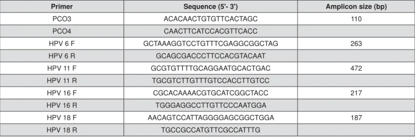
the absence of PCR inhibitors, a 110 bp segment of
primers6. Sequences of primers are displayed in Figure
stained with Gel RedTM (New England BioLabs; Ipswich,
MA, USA) at 100 V and 10 mA for 1 hour. Results were
visualized by UV transillumination.
Detection of HPV sequences was performed by
nested PCR using two sets of consensus primers, MY09/
MY11 and GP5+/GP6+, which amplify a 450 bp fragment
and an internal fragment of 150 bp, respectively, of the highly conserved L1 HPV gene (9.15). PCR reactions
were performed in Veriti® Thermal Cycler (Applied
Biosystems; Foster City, CA, USA) and contained 10 PL PCR Buffer Go Taq Green (Promega; Madison, WI, USA), 0.6 mL of each primer (10 mM), Milli-Q water
(Milipore; Billerica, MA, USA) and 2 PL of each sample PL. First, the 450 bp fragment was amplified using the following PCR conditions:
initial denaturation for 5 minutes at 95°C; followed by
36 cycles of 1 minute at 94°C, 1 minute at 50°C and
region was performed with 2 PL of the amplicon and
PCR conditions as follows: initial hold of 5 minutes at
95°C; 45 seconds at 95°C, 45 seconds at 47.7°C and
of 7 minutes at 72°C. Negative controls (sterile water)
and HPV-16 positive controls extracted from HeLa cells
were used in all reactions.
Genotyping was performed using multiplex PCR with
(Figure 1) using the same mix conditions as described
above. Thermo cycling parameters used were: initial denaturation for 1 minute at 94°C; followed by 37 cycles
of 1 minute at 94°C, 30 seconds at 53.5°C and 1 minute
Statistical analysis
Comparisons of HPV frequency related to lesions type
were made using Fisher test using the GraphPad Prism
5.0 software (GraphPad; La Jolla, CA, USA), considering
Results
Sociodemographic characteristics of the participants are displayed in Table 1. The majority of patients were
(90.4%). Regarding educational status and health behaviors, 52.4% of patients were literate and 52.4%
declared to regularly consume alcohol or tobacco.
The lesions were located throughout the hard palates
(28.6%), lips (19.0%) and dorsal and ventral surfaces
of the tongue (28.6%). HPV prevalence in collected
samples was 81.0%. From this total, 35.3% of the HPV
could not be genotyped with the techniques employed in
and HPV-16 (5.9%). Multiple-type infections (HPV-6
and 18) were presented in 23.5% of patients with HPV.
Histopathologically, lesions were categorized as
hyperplasia (4.8%), squamous papillomas (14.3%),
squamous cell carcinoma (28.6%) or epithelial dysplasia
As per histolopathological diagnosis, HPV was detected
in 2/3 of papilloma cases, in 5/6 of carcinomas, in one
case of hyperplasia and in 9/11 of cases with dysplasia. The statistical analysis between HPV genotype and
histopathological diagnosis was performed and no
association was found (p>0.165). HPV detection and
genotyping according to the histopathological diagnosis of the lesions is shown in Table 2.
Primer Sequence (5'- 3') Amplicon size (bp)
PCO3 ACACAACTGTGTTCACTAGC 110
PCO4 CAACTTCATCCACGTTCACC
HPV 6 F GCTAAAGGTCCTGTTTCGAGGCGGCTAG 263 HPV 6 R GCAGCGACCCTTCCACGTACAAT
HPV 11 F GCGTGTTTTGCAGGAATGCACTGAC 472 HPV 11 R TGCGTCTTGTTTGTCCACCTTGTCC
HPV 16 F CGCACAAAACGTGCATCGGCTACC 217 HPV 16 R TGGGAGGCCTTGTTCCCAATGGA
HPV 18 F AACAGTCCATTAGGGGAGCGGCTGGA 187 HPV 18 R TGCCGCCATGTTCGCCATTTG
Discussion
Apart from a lot of debate regarding the role of
HPV as etiological agent in oral cancer development
and worsening of the lesions, the mechanism of HPV transmission to oral cavity has also not been fully
elucidated. Oral-genital contact has been considered
the main route of HPV transmission to oral cavity7,13,
although other theories include perinatal transmission, self-contamination and mouth-to-mouth transmission5.
Although HPV has been detected in specimens of oral
dysplasia and cancer, the prevalence of HPV and its
role in the pathogenesis of dysplasia and cancer is still unclear24.
The present study detected HPV-DNA in 81.0%
of clinically detectable lesions in oral mucosa, thus
indicating a probable role HPV could play in oral lesions.
Morbini, et al.15
and dysplasia in Italy, and also Jordan, et al.10 (2012)
who detected HPV in 78.3% of 233 samples with SCC
in the United States.
However, our data differs from other studies
performed in Brazil that report HPV rates ranging from 0% to 30% in oral lesions1,14,22. In São Paulo state, Rivero
and Nunes22 (2006) could not identify HPV in any of the
40 samples with SCC. Using in sit u hybridization, Acay,
et al.1 (2008) detected HPV in 24% of samples with oral
study by Miyahara, et al.14 (2011) reported that 33.7%
of 83 samples with SSC were positive for presence of
HPV. Regarding the Northeast region of the country, few studies have evaluated viral presence in oral lesions so
Characteristics N (%) HPV occurrence
Gender
Female 15 (71.4%) 12 (80%)
Male 06 (28.6%) 05 (83.33%)
Ethnic group
White 02 (9.5%) 02 (100%)
1RQZKLWH 19 (90.4%) 15 (78.9%)
Education
Literate 11 (52,4%) 09 (81.8%)
Nonliterate 10 (47.6%) 08 (80%)
Age
Up to 50 06 (28.6%) 04 (66.6%)
50 to 65 09 (42.9%) 07 (77.7%)
65 to 83 06 (28.6%) 06 (100%)
6PRNLQJ6WDWXV
Smoker 7 (33.3%) 06 (85.7%)
Nonsmoker 14 (66.6%) 10 (71.4%)
Alcohol Use
Yes 6 (28.6%) 03 (50%)
No 15 (71.4%) 14 (93.3%)
Table 1-6RFLRGHPRJUDSKLFGDWDRIWKHSDWLHQWVZLWKRUDOPXFRVDOHVLRQVXVHGLQWKLVVWXG\
Type of Lesion HPV occurrence HPV-6 HPV-16 HPV-6/18 Not genotyped
Hyperplasia 1/1 1
Papilloma 2/3 1 1
SCC 5/6 3 2
Dysplasia 9/11 2 4 3
Total 17/21 6 1 4 6
SCC: Squamous cell carcinoma
far. In a study carried out in Pernambuco state, Vidal,
et al.29 (2004) detected HPV in 27.5% of samples with
in Sergipe state.
The high HPV index found in our study could
be related to demographic issues inherent in our population, in which 90.4% of patients declared
of Tsao, et al.28 (2016), according to which the only
patients and a higher prevalence of HPV in oral swabs.
An interesting aspect of HPV prevalence in oral
within the same country. For instance, in two studies
performed in Italy that used similar techniques, Termine,
et al.27 (2012) reported prevalence of 25.3% HPV cases
while Morbini, et al.15 (2012) described a rate of 87.5%.
It has been suggested that geographical variability
21. Moreover, distinct
techniques used may also account for the difference in
the rates reported in different studies, which interferes with comparisons6,21, and variability in evaluated lesions
types.
Although the patients with HPV lesions are generally
these data are still controversial. Our data, similar
to the data of several authors, show no association
between these factors and HPV status20,24
difference was observed in the frequency of HPV infection between men and women, as also observed
in the study by Tatar, et al.26 (2015). This is also in
accordance with other studies that suggest that in some
cultures both genders are exposed in a similar manner
18.
In order to enhance HPV detection we used the
nested-PCR technique that uses more than one pair of primers. Thus, we were able to detect the virus even in
very low concentrations. High quality DNA is required for
this technique to reach its optimal conditions. Acay, et
al.1 (2008) could not obtain the required amount of DNA
that this may be the explanation for the low prevalence
of HPV reported in studies with premalignant and
malignant lesions using nested-PCR in that specimen type. Another advantage of our study is uniformity of
material, sampling and processing procedures.
The presence of HPV reported in the literature is
independent of malignant grading14,27. Presently, we
evaluated malignant (SCC), premalignant (dysplasia)
and benign (hyperplasia and papilloma) lesions and
reported HPV infection in all types of lesions.
signature of HPV infections14,29. According to Hajdu, et
al.8 (2006), HPV-DNA is present in 75% of lesions with
study we found 71.4% of HPV prevalence in samples
For genotyping, primers pertaining to the four most
cancer were utilized4,12,13. HPV-6 was the most frequent
genotyped HPV samples. Bharti, et al.3 (2013) in their
study showed the presence of this type in six out of nine
oral lesions. Along with HPV-16, HPV-6 is one of most
frequently reported types in oral cancer. Kristoffersen,
et al.12
lesions and Sugiyama, et al.25 (2003) detected HPV-16
in 61% and 35% of samples with dysplasia and SCC,
respectively, and HPV-18 in only two samples with SCC
(3.5%). Here we report prevalence of 23.5% for HPV-18 and 5.9% for HPV-16 in HPV-positive samples. We
were not able to genotype 35.3% of our HPV-positive
samples probably because these patients were infected
with types different than what we tested for. Similar
17
(2013) who could not genotype half of their HPV-positive
samples.
Comparative studies concerning HPV infection sites have been performed around the world. Presence of
HPV-DNA has been detected in healthy oral mucosa7
as in all types of oral lesions11. However, there are few
studies so far, especially in the Northeast region, to evaluate HPV oral infection. The main limitation of our
study was the small sample size, which prevented a
more robust statistical analysis. Another limitation was
could help understand the route of viral transmission.
The HPV vaccination is currently available in several
countries, including Brazil, where the quadrivalent vaccine is distributed by the government to female
preadolescents aged 11 to 13 since March 2014.
Although the importance of vaccination in reducing
genital infections is well documented, the effect of the
been addressed yet30. It is possible that the rate of
oral cancer in vaccinated women will be reduced in the
the role of HPV in this disease3.
Conclusions
Similarly to other studies, we report high occurrence
of HPV in oral lesion. The results are of special
importance, since they are one of the few from
comprehension of HPV distribution in Sergipe and in Brazil as a whole. Moreover, even though HPV seems
to play an important role in oral carcinogenesis, studies
that evaluate the association of HPV with normal oral
mucosa are crucial to improve the comprehension of the role of HPV in oral lesions.
This study was supported by research grant from
FAPITEC/SE - Fundação de Apoio à Pesquisa e Inovação Tecnilógica do Estado do Sergipe (grant MS/CNPq/
FAPITEC/SE/SES 02/2013 - PPSUS Sergipe).
References
1- Acay R, Rezende N, Fontes A, Aburad A, Nunes F, Souza S. Human
in sit u
2008;23:271-4.
2- Andrade, JO, Santos CA, Oliveira MC. Associated factors with oral
cancer: a study of case control in a population of the Brazil's Northeast. Rev Bras Epidem. 2015;18(4):894-905.
3- Bharti AH, Chotaliya K, Marfatia YS. An update on oral human
papillomavirus infection. Indian J Sex Transm Dis. 2013;34:77-82.
2016;38:68-72.
5- Campisi G, Panzarella V, Giuliani M, Lajolo C, Di Fede O, Falaschini
S, et al. Human papillomavirus: its identity and controversial role in oral oncogenesis, premalignant and malignant lesions. Int J Oncol.
2007;30:813-23.
6- Feller L, Wood NH, Khammissa RA, Lemmer J. Human
papillomavirus-mediated carcinogenesis and HPV-associated oral and oropharyngeal squamous cell carcinoma. Part 2: Human papillomavirus associated oral
and oropharyngeal squamous cell carcinoma. Head Face Med. 2010;6:15.
Prevalence of oral HPV infection in the United States, 2009-2010. JAMA. 2012;307:693-703.
Ann Clin Lab Sci. 2006;36:485-7.
9- Instituto Nacional de Câncer José Alencar Gomes da Silva – INCA. Estimativa 2016: incidência de câncer no Brasil. Rio de Janeiro: INCA;
2016.
M, et al. Validation of methods for oropharyngeal cancer HPV status determination in US cooperative group trials. Am J Surg Pathol.
2012;36:945-54.
Human papillomavirus DNA in oral squamous cell carcinomas and normal oral mucosa. Acta Virol. 2003;47:11-6.
Solheim T, et al. Human papillomavirus subtypes in oral lesion compared
to healthy oral mucosa. J Clin Virol. 2012;53:364-6.
13- Lajer CB, von Buchwald CV. The role of human papillomavirus in
14- Miyahara GI, Simonato LE, Mattar NJ, Camilo DJ Jr, Biasoli ER.
PCR in oral and oropharynx squamous cell carcinoma biopsies. Mem
Inst Oswaldo Cruz. 2011;106:166-9.
15- Morbini P, Dal Bello B, Alberizzi P, Mannarini L, Mevio N, Bertino G,
et al. Exfoliated cells of the oral mucosa for HPV typing by SPF10 in head
16- Mosele JC, Barancelli M, Oliveira da Silva S, De Carli JP, Trentin
of the HPV in squamous cell carcinoma of the oral cavity. Rev Odonto.
2009;17:7-12.
papillomavirus in the lesions of the oral mucosa according to topography. Plos One. 2013;8:e69736.
18- Nasher AT, Al-Hebshi NN, Al-Moayad EE, Suleiman AM. Viral infection
Yemen: a case-control study. Oral Surg Oral Med Oral Pathol Oral Radiol. 2014;118:566-72.
19- Ndiaye C, Alemany L, Diop Y, Ndiaye N, Diémé MJ, Tous S, et al. The role of human papillomavirus in head and cancer in Senegal. Infect
Agents Cancer. 2013;8:14.
20- Petito G, Carneiro MA, Santos SH, Silva AM, Alencar RC, Gontijo AP,
et al. Human papillomavirus in oral cavity and oropharynx carcinomas in the central region of Brazil. Braz J Otorhinolaryngol. 2016 Apr 9. doi:
10.1016/j.bjorl.2016.01.004.
21- Quintero K, Giraldo GA, Uribe ML, Lopez C, Alvarez E, et al. Human
papillomavirus types in cases of squamous cell carcinoma of head and
22- Rivero ER, Nunes FD. HPV in oral squamous cell carcinomas of a
23- Simonato LE, Miyahara GI. The role of human papillomavirus in oral carcinogenesis. Rev Bras Cancerol. 2007;53:471-6.
human papilloma viruses play any role in oral squamous cell carcinoma
in North Indians? Asian Pac J Cancer Prev. 2015;16:7077-84.
T. Detection of human papillomavírus-16 and HPV-18 DNA in normal, dysplastic, and malignant oral epithelium. Oral Surg Oral Med Oral Pathol
Oral Radiol Endod. 2003;95:594-600.
26- Tatár TZ, Kis A, Szabó É, Czompa L, Boda R, Tar I, et al. Prevalence
of human papillomaviruses in the healthy oral mucosa of women with high-grade squamous intraepithelial lesion and of their partners as
compared to healthy controls. J Oral Pathol Med. 2015;44:722-7. 27- Termine N, Giovannelli L, Rodolico V, Matranga D, Pannone G, Campisi
G. Biopsy vs. brushing: comparison of two sampling methods for the detection of HPV-DNA in squamous cell carcinoma of the oral cavity.
Oral Oncol. 2012;48:870-5.
FC, et al. Concordance of oral HPV prevalence between patients with oropharyngeal cancer and their partners. Infect Agent Cancer.
2016;11:21.
29- Vidal KL, Caldas AF Jr, Mello RJ, Brandão VR, Rocha GI, Taromaru E.
HPV detection in oral carcinomas. J Bras Patol Med Lab. 2004;40:21-6.