©
201
5
Nature
America,
Inc.
All rights reserved.
DMD is a devastating childhood genetic muscular disorder that is manifested by progressive debilitating muscle weakness and wast-ing, and ultimately by death in the second or third decade of life1.
DMD is characterized by the absence of the 3,600-residue rod-shaped dystrophin protein, encoded by the DMD gene in humans, which is expressed in differentiated myofibers and connects the myofiber cytoskeleton to the extracellular matrix through the dystrophin-asso-ciated glycoprotein complex (DGC)2,3. In the absence of dystrophin,
myofibers are extremely susceptible to injury, which leads to multiple cycles of degeneration and regeneration that, in turn, lead to elevated inflammation, fibrosis and eventual progressive loss of muscle mass and function4.
Previous studies have shown that impairment of the DGC by myofiber-specific deletion of Dag1, the gene encoding the laminin-binding DGC component dystroglycan, does not mimic the robust muscle degeneration observed in the dystrophin-null mdx mouse model (which contain the Dmdmdx allele; we hereafter refer to these
animals as mdx mice), suggesting that myofiber fragility is not the only mechanism involved in muscle degeneration in patients with DMD5. It has been suggested that human DMD progression is
exac-erbated by reduced function of muscle stem cells owing to exhaustion caused by telomere shortening6,7. However, in human and mouse
dystrophic skeletal muscles, satellite cell numbers are elevated, even in advanced stages of dystrophy, suggesting that the depletion of
satellite cells is not the primary cause for failed regeneration8–10.
Notably, the proportion of progenitors entering the differentiation program and expressing the muscle-specific transcription factor myo-genin (Myog) is unusually low in DMD muscle8. Together, these data
suggest a hypothesis in which the homeostasis between stem cells and committed progenitors within the satellite cell compartment is perturbed in dystrophin-deficient muscle.
A recent study has indicated that the polarity protein microtubule-associating protein (MAP)/microtubule affinity–regulating kinase 2 (Mark2; also known as partitioning-defective 1b, Par1b) binds to the R8–R9 spectrin-like repeats of dystrophin in differentiated myofibers11. Mark2 has also been shown to be required for the
baso-lateral formation of a functional DGC in epithelial cells12. Notably,
Par1 (the homolog of Mark2 in Drosophila) has an essential role in regulating asymmetric divisions of stem cells by establishing cellular polarity through direct phosphorylation of Par3 (or Pard3 in mam-mals), thereby redistributing the PAR complex—which is composed of the cell polarity–regulating proteins Par3, Par6 and atypical protein kinase C (aPKC)—to the side of the cell opposite to that at which Par1 is localized13. This asymmetric localization of Par1 and the PAR
complex during asymmetric stem cell divisions is important for the unequal segregation of cellular factors that determine the fates of individual daughter cells. For example, in dividing neuroblasts in Drosophila, the PAR complex accumulates in the apical cell surface
1Sprott Center for Stem Cell Research, Ottawa Hospital Research Institute, Regenerative Medicine Program, Ottawa, Ontario, Canada. 2Department of Cellular and Molecular Medicine, Faculty of Medicine, University of Ottawa, Ottawa, Ontario, Canada. 3Present addresses: Fritz-Lipmann Institute for Age Research, Jena, Germany (J.v.M.); Nestle Institute of Health Sciences, Swiss Federal Institute of Technology Campus, Lausanne, Switzerland (C.F.B.). 4These authors contributed equally to this work. Correspondence should be addressed to M.A.R. (mrudnicki@ohri.ca).
Received 1 May; accepted 13 October; published online 16 November 2015; doi:10.1038/nm.3990
Dystrophin expression in muscle stem cells regulates their
polarity and asymmetric division
Nicolas A Dumont
1,2,4, Yu Xin Wang
1,2,4, Julia von Maltzahn
1–3, Alessandra Pasut
1,2, C Florian Bentzinger
1–3,
Caroline E Brun
1,2& Michael A Rudnicki
1,2©
201
5
Nature
America,
Inc.
All rights reserved.
and relocalizes the Notch receptor antagonist Numb to the basal cell surface, which gives rise to the differentiated ganglion daughter cell14.
In intestinal stem cells, however, integrin bound to the basement membrane segregates the PAR complex to the apical daughter cell that becomes the differentiated cell15. Consistent with the hypothesis that
the PAR complex regulates satellite cell polarity, Pard3 knockdown in satellite cells results in loss of asymmetric divisions and reduced capacity to form myogenic progenitors16.
Here we demonstrate that dystrophin is expressed in activated satel-lite cells, in which it regulates polarity establishment by interacting with Mark2. Dystrophin-deficient satellite cells show impaired polar-ity establishment, loss of apicobasal asymmetric division and a higher proportion of abnormal division, leading to reduced generation of myogenic progenitors and impaired muscle regeneration.
RESULTS
Dystrophin is expressed in satellite cells
Dystrophin is not expressed in myoblasts cultured in vitro, and con-sequently it was generally not considered to be expressed in satellite cells17,18. However, our RNA-seq and microarray analysis of
pro-spectively isolated satellite cells from mouse muscle revealed that DGC-encoding genes such as Dmd and Dag1, which are transcribed at low levels in myoblasts and upregulated in differentiated myotubes, are highly transcribed in satellite cells (Fig. 1a and Supplementary Fig. 1a,b)19. Clustering analysis of expression changes in DGC protein–
encoding genes also revealed that distinct components of the DGC complex, such as E-sarcoglycan, B2-syntrophin and B-dystrobrevin (encoded by Sgce, Stnb2 and Dtnb, respectively), are exclusively expressed in satellite cells and not in myotubes (Fig. 1a and Supplementary Fig. 1c). Validation by qPCR confirmed that Dmd and Dag1 mRNA levels are elevated by 475% and 250%, respectively, in prospectively isolated satellite cells as compared to the levels found in in vitro–differentiated myotubes (Fig. 1b,c and Supplementary Fig. 1d).
In sections from normal muscle, dystrophin protein expression in satellite cells is not easily discernible from dystrophin expression in the myofiber because of their close juxtaposition. Therefore, we isolated satellite cells by FACS from cardiotoxin-injured muscles
(2 d after injury), and we cytospun and immunostained the sorted sat-ellite cells for dystrophin and the paired-box transcription factor Pax7 (a satellite cell marker). We observed dystrophin protein expression in satellite cells from wild-type (WT) but not mdx mice (Fig. 1d). To examine the dystrophin expression pattern during satellite cell activa-tion, we isolated myofibers from the extensor digitorum longus (EDL) muscle of WT mice and cultured them ex vivo for 0, 12, 24 and 36 h. We found that a high level of dystrophin protein is expressed 24 h after satellite cell activation and that dystrophin is polarized on one side of the cell by 36 h (Fig. 1e). Moreover, we immunostained myofibers that were cultured for 72 h, a time point at which satellite cells have proliferated extensively, with antibodies to the N or C terminus of dystrophin and found that dystrophin could be visualized with both antibodies in a subset of WT satellite cells, whereas a small subset of mdx satellite cells were stained only with the antibody to the C termi-nus of dystrophin (which was only observed at the 72 h time point and was absent at previous time points) (Supplementary Fig. 1e).
Dystrophin regulates generation of myogenic progenitors
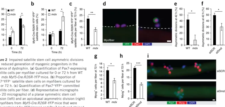
We next examined the developmental program of WT versus dys-trophin-deficient satellite cells after activation in myofiber cultures (Fig. 2 and Supplementary Fig. 2). We observed that the number of Pax7-expressing satellite cells per myofiber was 75% higher in freshly isolated myofibers (i.e., at time 0) from mdx mice relative to WT mice (Fig. 2a). However, after 72 h of culture the number of satellite cells in myofibers from WT mice increased by about 3.4-fold, whereas the number of satellite cells only increased by 1.4-fold in myofibers from mdx mice (Fig. 2a).
Studies from different laboratories have demonstrated that popu-lations of satellite cells are heterogeneous, with a subpopulation of satellite cells that can self-renew and another that is more prone to differentiate10,20–22. By using transgenic mice carrying the Myf5-Cre
and R26R-YFP alleles (in which Myf5 expression leads to permanent yellow fluorescent protein (YFP) expression), we previously showed that about 10% of satellite cells, which never expressed Myf5-Cre (i.e., Pax7+YFP− satellite stem cells), are able to self-renew and extensively contribute to the satellite cell pool after transplantation into host skel-etal muscle20. By contrast, satellite cells that did express Myf5-Cre
Myob lasts
D2 DMD5 DM
Dmd Dag1 Sntb1 Myh2 Myog Sgcb Sgca Dtna Sgcg Snta1 Mki67 Myod1 Pax7 Dtnb Sgce Sntb2 Sgcd
Fold change (log2)
30 25 20 15 10 5 0
*** ***
R
elativ
e
Dmd
e
xpr
ession
Myob lasts
Myotubes Satellit
e cells Satellit
e cells
6.0 5.0 4.0 3.0 2.0 1.0 0
R
elativ
e
Dag1
ex
pr
essio
n
*** ***
Myob lasts
Myotubes Satellit
e cells
WT
mdx
Dmd Pax7 DAPI
0 h 12 h
24 h 36 h
Myofiber
Dmd Pax7 DAPI
a
b
d
c
e
–3 –2 –1 1 2 3
Median
Figure 1 Dystrophin expression in satellite cells. (a) Microarray heat map representing myogenic genes and genes from the DGC from prospectively isolated satellite cells, proliferating myoblasts cultured in vitro, and 2- and 5-d-differentiated myotubes. Signal intensities represent the average of n = 3 microarrays for myoblasts and myotubes and n = 1 microarray for satellite cells obtained from pooled freshly isolated satellite cells of nine mice. (b,c) Quantitative real-time PCR for Dmd (b) and Dag1 (c) expression in satellite cells, myoblasts and myotubes. Error bars represent means o s.e.m. ***P < 0.005; Student’s t-test. (d) Representative micrographs (n > 20 micrographs per condition) of
immunostaining for Pax7 (red) and the N terminus of Dmd (green) and DAPI staining for nuclei (blue) of satellite cells that were isolated using FACS from cardiotoxin-injured WT (top) and mdx (bottom) mice 2 d after injury (n = 3 mice per group). (e) Representative
©
201
5
Nature
America,
Inc.
All rights reserved.
during development (i.e., Pax7+YFP+ satellite cells) are committed
to undergo differentiation and do not efficiently contribute to the satellite cell pool after transplantation20. Here we found that the
pro-portion of YFP− satellite stem cells was about 10% in freshly isolated myofibers (i.e., at time 0) from both WT and mdx mice (Fig. 2b). Although this proportion remained relatively stable after 72 h of cul-ture in myofibers from WT mice, it increased by 2.5-fold in myofibers from mdx mice (Fig. 2b). Accordingly, the generation of YFP+
com-mitted satellite cells per fiber after 72 h of culture was substantially lower in myofibers isolated from mdx mice as compared to those from WT mice (Fig. 2c).
Changes in the proportions of YFP− satellite stem cells could be explained by their ability to undergo apicobasal asymmetric cell divi-sion to generate one YFP− and one YFP+ daughter cell (Fig. 2d)20.
Therefore, we examined satellite stem cells immediately after the first round of cell division in isolated myofibers that were cultured for 42 h, and we observed an 80% reduction in the proportion of asymmetric satellite stem cell divisions in myofibers from mdx mice as compared to those from WT mice (Fig. 2e and Supplementary Fig. 2a). We also observed an unusually low proportion of asymmetric cell divisions in 2-week-old mdx mice, which precedes the major regenerative and inflammatory phase that is observed in mdx mice at 4–5 weeks of age (Supplementary Fig. 2b,c). By performing knockdown experiments on myofibers from WT mice using a siRNA that specifically targets Dmd (siDmd), we confirmed that the low proportion of asymmetric division is a direct consequence of the loss of dystrophin expression (Fig. 2f and Supplementary Fig. 2d–f). Consistent with this hypoth-esis, we observed a reduced proportion of asymmetric divisions after specifically deleting the dystrophin-binding transmembrane protein Dag1 in satellite cells from mice carrying loxP-flanked (‘floxed’) alle-les of Dag1 and the satellite cell–specific Pax7-CreER driver of Cre
recombinase (Pax7-CreER:Dag1fl/fl) as compared to Dag1fl/fl mice
without Cre recombinase activity (Supplementary Fig. 2g,h)5,20,23.
Notably, we did not observe any change in the proportion of asym-metric divisions using myofibers from adult A-sarcoglycan–deficient (Sgca−/−) mice, in which DGC assembly is not dysregulated, as com-pared to littermate control mice (Sgca+/−) (Supplementary Fig. 2i).
To examine the consequences of reduced asymmetric division on the generation of myogenic progenitors, we cultured myofibers for 72 h and enumerated the number of differentiating cells on the basis of immunostaining for Myog. Notably, we observed a 36% reduction in the number of Myog-expressing cells in myofibers isolated from mdx mice as compared to those from WT mice (Fig. 2g). Similarly, we observed a reduction of 52% in the number of Myog-expressing cells in myofibers isolated from WT mice that were treated with siDmd as compared to those treated with siSCR (Fig. 2h,i).
Dystrophin regulates polarity by interacting with Mark2
Using cultured myofibers, we detected that dystrophin expression is polarized (i.e., asymmetrically distributed to one side of the cell) in activated satellite cells that are about to divide after 36 h of culture (Fig. 3a,b and Supplementary Video 1). We performed an in situ prox-imity ligation assay (PLA) on isolated myofibers to investigate whether Mark2 associates with dystrophin in satellite cells as it does in myofibers (Fig. 3c–f and Supplementary Fig. 3)11,24. PLA detection of
endog-enous Mark2 and dystrophin generated a signal in activated satellite cells in cultured EDL myofibers and in prospectively isolated satellite cells from injured WT mice, whereas the signal was absent in satellite cells from mdx mice (Fig. 3c and Supplementary Fig. 3e,f). We also found that Mark2 interacts with Dag1; however, we could not detect a signal for interaction of Pard3 with dystrophin or Dag1 (Fig. 3d–f and Supplementary Fig. 3c,d)12. Notably, PLA also revealed that
35 30 25 20 15 10 5 0
WT
mdx
0 72
Time (h)
No. of satellite cells per fiber
35 30 25 20 15 10 5 0
0 72
Time (h) WT
mdx
Myf5-Cre:R26R-YFP
YFP
–
satellite stem cells (%)
30 25
20
15
10
5
0
WT mdx
**
Myf5-Cre:R26R-YFP
YFP
+
satellite cells per fiber at 72 h (%)
60
50
40
30
20
10
0
WT mdx
*
Myofiber
Asymmetric divisions at 42 h (%)
60
50
40
30
20
10
0
siSCR siDmd *
Asymmetric divisions at 42 h (%)
18 16 14 12 10 8 6 4 2 0
WT mdx
*
Myog
+ cells per fiber at 72 h
25
20
15
10
5
0 ***
siSCR siDmd
Myog
+ cells per fiber at 72 h
siSCR
siDmd
YFP Pax7 DAPI
Pax7 Myog DAPI
* *
*
a
b
c
d
e
f
g
h
i
Figure 2 Impaired satellite stem cell asymmetric divisions and reduced generation of myogenic progenitors in the absence of dystrophin. (a) Quantification of Pax7-expressing satellite cells per myofiber cultured for 0 or 72 h from WT and mdx Myf5-Cre:R26R-YFP mice. (b) Proportion of Pax7+YFP− satellite stem cells on myofibers cultured for 0 h or 72 h. (c) Quantification of Pax7+YFP+ committed satellite cells per fiber. (d) Representative micrographs (n > 20 micrographs) of a planar symmetric stem cell division (left) and an apicobasal asymmetric division (right) in myofibers from Myf5-Cre:R26R-YFP mice that were immunostained for YFP (green) and Pax7 (red) and stained
©
201
5
Nature
America,
Inc.
All rights reserved.
dystrophin and Dag1 interact with A7-integrin (Itga7), another lam-inin-binding receptor that has an important role in polarity, although this interaction is temporally restricted and is not exclusive (Fig. 3a and Supplementary Fig. 3a,b)15.
To further interrogate whether the PAR proteins are involved in satellite cell polarity, we analyzed the expression patterns of Mark2 and Pard3 by immunostaining satellite cells on myofibers that were cultured for 36 h, a time point before the first cell division occurs. We observed the polarized localization of Mark2 or Pard3 in about half of the satellite cells from WT mice (Fig. 3g–j). By contrast, we observed that the majority of dystrophin-deficient satellite cells expressed low levels of Mark2, whereas Pard3 was localized around the cell periph-ery in a nonpolarized manner (Fig. 3g–i). These findings are diagram-matically summarized in Figure 3j.
PAR proteins regulate satellite cell asymmetric division
We isolated EDL myofibers from Myf5-Cre:R26R-YFP mice and cul-tured them for 42 h, a time point immediately after the first cell divi-sion, to measure the asymmetric inheritance of the different polarity effectors in relation to the myogenic fate of the daughter cells. In asymmetric cell pairs (i.e., YFP+-YFP− cell pairs), we observed that expression of dystrophin and Mark2 was retained in YFP− satellite stem cells, but that it was rarely detectable in YFP+ daughter cells
(Fig. 4a and Supplementary Table 1). By contrast, we observed that Pard3 was segregated to the YFP+ satellite cells and was rarely
detect-able in YFP− daughter cells (Fig. 4a and Supplementary Table 1). Contrary to what we observed in asymmetric cell pairs, dystrophin,
Mark2 and Pard3 were equally distributed in both YFP− daughter cells after symmetric division (Fig. 4b and Supplementary Table 1). Asymmetric segregation of dystrophin was also observed in EDL myofibers that were cultured for 72 h (a time point at which satellite cells undergo multiple rounds of division), in which staining for dys-trophin was only observed in a subset of satellite cells (Supplementary Figs. 1e and 4a).
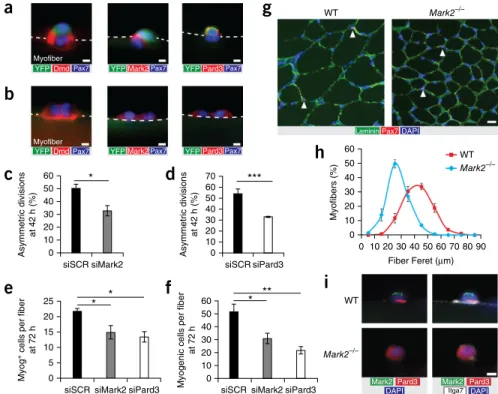
To investigate the specific function of PAR proteins on satellite cell–fate decision, we performed siRNA knockdowns for Mark2 (using siMark2) or Pard3 (using siPard3) in myofibers isolated from Myf5-Cre:R26R-YFP mice and cultured them for 42 h (Supplementary Fig. 4b–e). We found that myofibers treated with siMark2 or siP-ard3 resulted in an ~35% reduction in the proportion of asymmetric divisions as compared to siSCR-treated myofibers cultured for 42 h (Fig. 4c,d and Supplementary Fig. 4f). Moreover, we observed that treatment of freshly isolated myofibers with siMark2 and siPard3 resulted in a reduction of 30% and 38%, respectively, in the number of Myog-expressing cells and an ~50% decrease in the number of total myogenic (Pax7+ or Myog+) cells than in siSCR-treated
myofib-ers after 72 h of culture (Fig. 4e,f). Double knockdown with both siMark2 and siDmd did not exacerbate the reduction in the number of Myog-expressing cells as compared to knockdown with siMark2 alone (Supplementary Fig. 4g,h).
To further analyze Mark2 function, we investigated the muscle phenotype of mice with null alleles of Mark2 (Mark2−/− mice)25. We
observed that the myofiber size of muscle from Mark2−/− mice was strikingly smaller than those from WT mice (Fig. 4g,h). Moreover,
)'(($&
!$#
"! mdx
"!
mdx
"!
Dmd
Mark2
Pard3 Pard3
mdx "! mdx
)'(( '%$* %$*
)'(( '%$* %$*
)'(($&
)'(($&
"! mdx "!
mdx
a
b
c
d
e
f
h
i
j
g
Figure 3 Dystrophin regulates PAR polarity protein localization. (a) Representative micrographs (of n = 20 micrographs) showing immunostaining for the C terminus of Dmd (green), Dag1 (red) and Itga7 (cyan), and DAPI staining for the nuclei (blue) of cultured myofibers from WT mice at 36 h. (b) Quantification of Dmd expression (as measured by immunostaining of the rod domain) and Dmd localization in satellite cells from cultured myofibers of WT mice at 0, 12, 24 and 36 h (n = 3 mice; ~50 cells per mouse). Only undivided cells were quantified. (c,d) Representative micrographs (of n = 10 micrographs per condition) of PLA for Dmd (Dy4/6D3 clone) and Mark2 (red) (c) or for Dmd and Pard3 (red) (d), with immunostaining for Itga7 (green) and DAPI staining for nuclei (blue), on cultured myofibers from WT (left) and mdx (right) mice at 36 h (n = 3 mice per group). (e,f) Representative micrographs (n = 10 micrographs per condition) of PLA for Dag1 and Mark2 (red) (e) or for Dag1 and Pard3 (red) (f), with immunostaining for Itga7 (green) and DAPI staining for nuclei (blue) on cultured myofibers from WT (left) and mdx (right) mice at 36 h (n = 3 mice per group). (g) Representative micrographs (of n = 10 micrographs per condition) showing examples of polarity protein distribution after immunostaining for Mark2 (green), Pard3 (red) and Itga7 (white) and staining with DAPI for nuclei (blue) of cultured myofibers from WT (top) and mdx (bottom) mice at 36 h (n = 3 mice per group).
©
201
5
Nature
America,
Inc.
All rights reserved.
we detected asymmetric distribution of polarity proteins in satellite cells from WT but not Mark2−/− mice, in which the absence of Mark2 expression resulted in nonpolarized localization of Pard3 (Fig. 4i). Nonetheless, polarization of dystrophin was still observed in satellite cells from Mark2−/− mice (Supplementary Fig. 4i).
Impaired mitotic spindle orientation
Centrosomes specify the alignment of the mitotic spindle (i.e., api-cobasal versus planar orientations)26. Therefore, to determine the
axis of cell division, we immunostained mitotic centrosomes with an antibody specific to the phosphorylated forms of Aurora kinases (p-Aurk)27. In pro-metaphase, metaphase and anaphase satellite cells,
immunostaining for p-Aurk detects two centrosomes per cell that are localized at opposite cell poles28. In telophase and cytokinesis, p-Aurk
dissociates from the centrosome and accumulates at the midbody (Fig. 5a)28.
In myofibers from mdx mice, we observed an unexpectedly high number of abnormal mitotic divisions, both in YFP− and YFP+
sat-ellite cells, as compared to myofibers from WT mice (Fig. 5b). We characterized abnormal divisions as any event that did not corre-spond to the classical staining for p-Aurk—for instance, abnormally high numbers of centrosomes or abnormal kinetics of p-Aurk stain-ing, such as persistence of the midbody from previous cell divisions. Paradoxically, we also observed a higher proportion of mitotic cells in myofibers from mdx mice at 36 h, although the number of success-ful cell divisions was lower at 72 h as compared to myofibers from WT mice (Figs. 2a and 5c). Notably, the high proportion of abnor-mal divisions in dystrophin-deficient satellite cells corresponded to the specific loss of apicobasal divisions (Fig. 5d and Supplementary Fig. 5a,b). Taken together, our experiments suggest that dystrophin
expression is required to establish the apicobasal mitotic axis required for asymmetric cell division (Fig. 5e). Similar to what we observed in dystrophin-deficient satellite cells, cultured myofibers from Mark2−/− mice had a higher proportion of satellite cells with an abnormal p-Aurk staining pattern in addition to a specific reduction in apicoba-sal divisions, as compared to those from WT mice (Fig. 5f,g).
DGC-deficient satellite cells display impaired regeneration
To investigate the progression of the myogenic program in dystrophin-deficient satellite cells in vivo, we cardiotoxin-injured muscles from WT and mdx mice carrying the Myf5-Cre and R26R-YFP alleles, and we analyzed satellite cells by flow cytometry 3 d after injury (Supplementary Fig. 6a). We assessed the activa-tion state of the satellite cells using side scatter analysis to measure internal cell complexity, and we assessed cell cycle status by Hoechst 33342 staining to measure DNA content. In muscles from WT mice, cardiotoxin injury induced satellite cells to activate, exit quiescence (G0; quadrant Q4 of the FACS plot) and become proliferative (S to G2/M; quadrant Q2 of the FACS plot) (Fig. 6a–d). In the contralateral muscle from the mdx mice, the proportion of activated satellite cells was higher than in the contralateral muscle from WT mice, probably reflecting ongoing repair, which was consistent with the presence of cells expressing Myog or the myogenic regulatory factor Myod1 in these samples (Fig. 6a–d and Supplementary Fig. 6b–d). As com-pared to injured muscle from WT mice, cardiotoxin injury did not provoke a strong proliferative response of satellite cells in muscles of mdx mice, with a high proportion of myogenic cells that remained in G1 (quadrant Q1 of the FACS plot) (Fig. 6a–d).
Muscles from WT mice respond to injury through the production of YFP+ committed satellite cells, and we observed a lower proportion of
1+ !
- * - *
* 3!
* 3! 3!
3! 3!
- 3!
*
* 3!
!
01)*200
1,
+))..0/
1!,
! " #
1µ
(' ('
Mark244
('
Mark244
Mark244
0
0&$% 0 - 0 * 0&$% 0 *
0&$%
01)*200
1,
0
-a
b
c
d
0&$%
+
)..0/ 1!,
0 - 0 *
e
f
g
h
i
Figure 4 PAR polarity proteins are requiredfor muscle stem cell asymmetric divisions. (a,b) Representative micrographs of myofibers from Myf5-Cre:R26R-YFP mice cultured for 42 h and immunostained for YFP (green), Pax7 (blue), and the rod domain of Dmd (red; left), Mark2 (red; middle) or Pard3 (red; right) in asymmetric cell pairs (YFP+-YFP− pairs) (a) and symmetric YFP− cell pairs (YFP−-YFP− pairs) (b) (n = 3 mice; >12 micrographs per condition). (c,d) Quantification of the number of asymmetric divisions relative to the total number of satellite stem cell divisions in cultured myofibers from Myf5-Cre:R26R-YFP mice after knockdown of Mark2 (siMark2; n = 5 mice per group; 40 myofibers per mouse) (c) or Pard3 (siPard3; n = 4 mice per group; 40 myofibers per mouse) (d), as compared to that from myofibers treated with scramble siRNA (siSCR). (e,f) Quantification of Myog-expressing cells per fiber (e) and total myogenic cells (Pax7- or Myog-expressing cells) per fiber (f) from WT myofibers that were treated with siMark2, siPard3 or siSCR and cultured for 72 h (n = 3 mice per group; 30 myofibers per mouse). (g) Representative micrographs (n > 10 per condition) of tibialis anterior (TA) muscle sections from WT (left) or Mark2−/− (right) mice
©
201
5
Nature
America,
Inc.
All rights reserved.
YFP− satellite stem cells in these muscles 3 d after injury than in unin-jured muscles (11% and 22%, respectively) (Fig. 6a,e). However, the proportion of YFP− satellite stem cells remained unchanged between the contralateral and injured muscles of mdx mice (28% versus 31%, respectively) (Fig. 6a,e). This resulted in fewer Myog-expressing cells in the injured muscles of mdx mice as compared to those of WT mice (Supplementary Fig. 6c,d).
A previous study showed that myofiber-specific deletion of Dag1 in mice carrying the muscle creatine kinase (MCK) driver of Cre recombinase (MCK-Cre:Dag1fl/fl) results in a relatively mild
regenera-tion deficit as compared to MORE-Cre:Dag1fl/− mice, which express Cre from the Meox2 locus, leading to global recombinase activity (as Dag1 is deleted in both satellite cells and myofibers)5. Therefore, we
analyzed muscle regeneration in Pax7-CreER:Dag1fl/fl mice to assess
the regenerative capacity of muscle stem cells that specifically lack the DGC. We observed a marked delay in muscle regeneration, a lower number of satellite cells and lower caliber of regenerated myofib-ers in tamoxifen-treated Pax7-CreER:Dag1fl/fl mice as compared to
tamoxifen-treated Dag1fl/fl littermate controls (Fig. 6f–h).
DISCUSSION
Our findings show that Dmd and Dag1 are expressed at the RNA level in prospectively isolated quiescent satellite cells, consistent with microarray data from other laboratories29,30. Furthermore, we show
that high levels of dystrophin protein are expressed in satellite cells 24 h after their activation, which is consistent with the time predicted to translate full-length Dmd mRNA31. Immunostaining with
antibod-ies to the N terminus, C terminus and rod domain of dystrophin
suggest that full-length dystrophin is expressed in WT satellite cells, whereas shorter isoforms of dystrophin (for example, the Dp71 iso-form that does not interact with Mark2), with transcription start sites after the point mutation in the Dmd allele of mdx mice, may be expressed in satellite cells from mdx mice. Our experiments identify an essential role for dystrophin in regulating the establishment of PAR-mediated polarity in satellite cells. In the absence of dystrophin, the polarity effector Mark2 is dysregulated, leading to the failure of Pard3 to be polarized on the pole of the cell that will give rise to the YFP+ committed progenitor daughter cell (summarized in Fig. 3j
and Supplementary Fig. 7). Polarity deficits observed in Mark2−/− mice phenocopy the behavior of dystrophin-deficient satellite cells, supporting the notion that Mark2 is a critical effector of the dys-trophin-deficient satellite cell phenotype.
Impaired polarity leads to the dysregulation of mitotic spindle ori-entation and a lower proportion of apicobasal divisions. These results are consistent with the role of Mark2 in epithelial cells in establishing apicobasal polarity32. The biased loss of apicobasal divisions
sug-gests that alternative mechanisms exist in satellite cells to determine proper mitotic spindle assembly. Establishment of the apicobasal axis is known to promote asymmetric distribution of cell-fate determi-nants and to lead to asymmetric cell division in various types of stem cells20,33. By using WT and mdx Myf5-Cre:R26R-YFP transgenic mice,
we showed that asymmetric divisions are markedly reduced in the absence of dystrophin and that the generation of YFP+ myogenic
pro-genitors is impaired both in vitro and in vivo. Moreover, we observed a high proportion of aberrant divisions in satellite cells from mdx and Mark2−/− mice. Consistent with our results, Par1 has been identified Pro-metaphase Metaphase
WT
WT
Mitotic cells (%)
mdx
mdx Telophase
p-Aurk
p-Aurk p-Aurk
Pax7 DAPI
Dmd Dmd Itga7
p-Aurk p-Aurk DAPI
p-Aurk Pax7 DAPI
WT
Mark2–/–
Itga7 p-Aurk DAPI Cytokinesis
10 **
8
6
4
2
0
mdx WT
Abnormal
Planar
Apicobasal
***
***
Mitotic divisions (%)
0 20 40 60 80 100
Mitotic divisions (%)
Mark2–/– WT
Abnormal
Planar
Apicobasal
**
***
0 20 40 60 80 100
a
b
c
g
f
e
d
© 201 5 Nature America, Inc.
All rights reserved.
in Drosophila S2 cells as a gene required for centrosome clustering, and knockdown of Par1 leads to centrosome multipolarity34. Notably,
abnormal divisions and dystrophin-expressing cells are not exclu-sive to YFP− cells but are also observed in a subset of YFP+ satellite
cells. These results suggest that dystrophin has a role in other types of asymmetric divisions that have been reported in the progenitor stages of myogenesis35.
The loss of polarity in dystrophin-deficient satellite cells ultimately affects the kinetics of cell proliferation. Alterations to the phospho-rylation pattern of Aurora kinases are a sign of errors in the mitotic process. Overexpression of Aurora induces abnormal spindle forma-tion and causes cells to bypass the spindle checkpoint, leading them to undergo apoptosis or senescence36. Notably, a recent study showed
that, similarly to what we observed in mdx satellite cells, satellite cells deficient in spindle assembly checkpoint effectors display cell-cycle arrest in G1 and resist differentiation37. The presence of higher
numbers of centrosomes could be hypothesized to trigger mitotic catastrophe followed by cell cycle arrest and cellular senescence38.
Moreover, persistence of p-Aurk activity in the midbody is a sign of DNA segregation errors that delay abscission and are involved in a cellular mechanism to prevent tetraploidization39. Therefore,
abnormal p-Aurk staining pattern could also signal the activation of corrective mechanisms that delay division in dystrophin-deficient satellite cells, which is consistent with our observation that there is a higher number of cells in M phase together with a lower number of successful divisions. Similar defects in satellite cell proliferation have also been observed in Largemyd mice, in which Dag1 glycosylation
is impaired40.
Previous studies documenting impaired muscle regeneration in mdx mice suggest that satellite cell function is perturbed9,41. However,
the contribution of satellite cell–autonomous defects to the impaired regeneration has not been experimentally addressed. Our results indicate that the failure of regenerative myogenesis to keep pace with disease progression in DMD is not due to muscle stem cell exhaus-tion, but rather is due to impaired polarity, leading to a deficit in cell division, a lack of asymmetric divisions and reduced generation of myogenic progenitors (Supplementary Fig. 7). Consistent with this hypothesis, recent results indicate that the granulocyte–colony stimulating factor receptor (G-CSFR) is asymmetrically segregated in activated satellite cells where it plays a role in myogenic progression, and G-CSF treatment improves muscle regeneration in mdx mice42.
Our findings explain why the regenerative capacity of muscles from mdx mice or from DMD patients is impaired despite high numbers of satellite cells9,41. Moreover, although the total number of satellite cells
remains elevated in aged dystrophic muscles, their relative number gradually declines during aging, which is explained by our observa-tions that dystrophin-deficient satellite cells display mitotic errors and a slower division rate.
Different isoforms of dystrophin are expressed in various tissues and a generalized polarity deficit in DMD patients may explain other aspects of the disease, such as neurological deficits43,44. Notably,
defects in asymmetric stem cell division are associated with tum-origenesis45, and it was recently reported that dystrophin acts as a
tumor suppressor gene in rhabdomyosarcoma, consistent with its role in regulating muscle stem cell polarity via its interaction with Mark2 (ref. 46).
100 ***
** *** 80
Quiescent
satellite cells (%
) 60 40 20 0 WT mdx
Contra Injured Contra Injured
Q1 1.59 105 104 103 102 101 100 105 104 103 102 101 100 WT Contralateral leg WT CTX-injured leg mdx CTX-injured leg mdx Contralateral leg Q2 0.50 Q4 90.7 Hoechst SSC Q3 7.19 Q1 4.09 Q2 95.5 Q4 0.087 Q3 0.28 Q1 14.8 Q2 27.2 Q4 51.4 Q3 6.61 Q1 71.4 Q2 26.0 Q4 2.27 Q3 0.32 Q5 1.47 Q6 1.22 Q8 19.4 Q7 77.9 Q5 5.02 Q6 94.6 Q8 0.15 Q7 0.22 Q5 13.5 Q6 28.5 Q8 18.6 Q7 39.4 Q5 27.7 Q6 70.8 Q8 0.49 Q7 1.05 SSC YFP
a
b
** * Activatedsatellite cells in G1 (%
) 100 80 60 40 20 0 WT mdx
Contra Injured Contra Injured
c
WT mdx
Contra Injured Contra Injured
40 20 0 Myf5-Cre:R26R-YFP YFP –
satellite stem cells (%
)
*
*
WT mdx
Contra Injured Contra Injured
Activated
satellite cells in S–G2M (%
) 100 *** 80 60 40 20 0 *
d
e
Pax7 -CreER : Dag1 fl/flPax7
-CreER
:
Dag1 fl/fl Dag1
fl/fl
Dag1 fl/fl
100 25
20
Pax7
+ cells per
fi
eld
Relative
fi
ber Feret (%)
15 10 5 0 80 60 40 20 0 *** **
g
h
Pax7-CreER:Dag1fl/fl Dag1fl/fl
Analysis 7 d 5 d
Tamoxifen CTX
f
Figure 6 Dystrophin-deficient satellite cells have reduced ability to generate myogenic progenitors in regenerating muscle. (a–e) Representative flow cytometry plots (top, distribution of myogenic cells by side scatter (SSC, y axis) and DNA content (x axis)
on the basis of Hoechst 33342 staining; bottom, distribution of myogenic cells by SSC (y axis) and YFP expression (x axis)) (a) and quantification of the proportions of quiescent (SSC-low, DNA-low) (b), activated G1 (SSC-high, DNA-low) (c) and proliferating S–G2M (SSC-high, DNA-high) (d) myogenic cells, and of YFP− satellite stem cells (e) from the cardiotoxin-injured (CTX) or contralateral (Contra) muscles of WT and mdx Myf5-Cre:R26R-YFP mice (n = 3 mice per condition). (f) Representative micrographs (n > 10 micrographs per condition) from H&E-stained TA muscle sections from tamoxifen-treated Dag1fl/fl and Pax7-CreER:Dag1fl/fl mice 7 d after CTX injury (n = 3 mice per group). Scale bar, 20 Mm. (g,h) Relative fiber Feret diameter (g) and satellite cell density (Pax7-expressing cells) (h) from TA muscle sections of tamoxifen-treated
Dag1fl/fl and Pax7-CreER:Dag1fl/fl mice 7 d after CTX injury (n = 3 mice per condition). Throughout,
©
201
5
Nature
America,
Inc.
All rights reserved.
Overall, our findings demonstrate that the muscle-wasting process observed in individuals with DMD is more complex than anticipated and reveal that in addition to muscle fragility, DMD is also a mus-cle stem cell disease. These results have important implications for therapeutic interventions such as gene therapy or exon skipping47.
For instance, adeno-associated virus (AAV) mini-dystrophin vectors have been shown to partially rescue dystrophin expression in myofib-ers of mdx mice; however, the current mini-dystrophin vectors do not contain the R8–R9 spectrin-like repeats to which Mark2 binds48.
Gene therapies targeting satellite cells potentially have long-term efficiency owing to satellite cell self-renewal. Therefore, in addition to differentiated myofibers, muscle stem cells should be considered as a therapeutic target for restoring muscle function in individuals with DMD.
METHODS
Methods and any associated references are available in the online version of the paper.
Accession codes. Gene Expression Omnibus: all microarray data are available in the series GSE59272.
Note: Any Supplementary Information and Source Data files are available in the
online version of the paper.
ACKNOWLEDGMENTS
We thank J. Dilworth and L. Megeney for careful reading of the manuscript. We also thank J. Ritchie for animal husbandry, and J. Fernandes and P. Oleynik of the flow cytometry facility of the StemCore laboratories for technical assistance. N.A.D. is supported by a Postdoctoral Fellowship from the Canadian Institutes of Health Research (CIHR); Y.X.W. is supported by fellowships from the Queen Elizabeth II Graduate Scholarships in Science and Technology and the CIHR; J.v.M. was supported by a grant from the Deutsche Forschungsgemeinschaft; C.F.B. was supported by a grant from the Swiss National Science Foundation; C.E.B. is supported by a Postdoctoral Fellowship from the Ontario Institute for Regenerative Medicine; and M.A.R. holds the Canada Research Chair in Molecular Genetics. These studies were carried out with support from grants to M.A.R. from the US National Institutes for Health (grant no. RO1AR044031), the CIHR (grant no. MOP-12080 and MOP-81288), the E-Rare-2 program from the CIHR and Muscular Dystrophy Canada (grant no. ERA-132935), the Muscular Dystrophy Association, the Stem Cell Network and the Ministry of Research and Innovation (MRI), Government of Ontario (grant no. ORF-RE05-084).
AUTHOR CONTRIBUTIONS
N.A.D. and Y.X.W. designed and carried out experiments, analyzed results and wrote the manuscript. J.v.M. designed and conducted experiments and analyzed results. A.P., C.F.B. and C.E.B. conducted experiments. M.A.R. designed experiments, analyzed results, wrote the manuscript and provided financial support.
COMPETING FINANCIAL INTERESTS
The authors declare no competing financial interests.
Reprints and permissions information is available online at http://www.nature.com/
reprints/index.html.
1. Anderson, M.S. & Kunkel, L.M. The molecular and biochemical basis of Duchenne muscular dystrophy. Trends Biochem. Sci.17, 289–292 (1992).
2. Cohn, R.D. & Campbell, K.P. Molecular basis of muscular dystrophies. Muscle Nerve
23, 1456–1471 (2000).
3. Koenig, M. et al. Complete cloning of the Duchenne muscular dystrophy (DMD) cDNA and preliminary genomic organization of the DMD gene in normal and affected individuals. Cell50, 509–517 (1987).
4. Serrano, A.L. et al. Cellular and molecular mechanisms regulating fibrosis in skeletal muscle repair and disease. Curr. Top. Dev. Biol.96, 167–201 (2011).
5. Cohn, R.D. et al. Disruption of Dag1 in differentiated skeletal muscle reveals a role for dystroglycan in muscle regeneration. Cell110, 639–648 (2002).
6. Sacco, A. et al. Short telomeres and stem cell exhaustion model Duchenne muscular dystrophy in mdx/mTR mice. Cell143, 1059–1071 (2010).
7. Webster, C. & Blau, H.M. Accelerated age-related decline in replicative life span of Duchenne muscular dystrophy myoblasts: implications for cell and gene therapy.
Somat. Cell Mol. Genet.16, 557–565 (1990).
8. Kottlors, M. & Kirschner, J. Elevated satellite cell number in Duchenne muscular dystrophy. Cell Tissue Res.340, 541–548 (2010).
9. Reimann, J., Irintchev, A. & Wernig, A. Regenerative capacity and the number of satellite cells in soleus muscles of normal and mdx mice. Neuromuscul. Disord.
10, 276–282 (2000).
10. Chakkalakal, J.V. et al. Early forming label-retaining muscle stem cells require p27kip1
for maintenance of the primitive state. Development141, 1649–1659 (2014). 11. Yamashita, K. et al. The eighth and ninth tandem spectrin-like repeats of utrophin
cooperatively form a functional unit to interact with polarity-regulating kinase PAR-1b. Biochem. Biophys. Res. Commun.391, 812–817 (2010).
12. Masuda-Hirata, M. et al. Intracellular polarity protein PAR-1 regulates extracellular laminin assembly by regulating the dystroglycan complex. Genes Cells14, 835–850 (2009).
13. Neumüller, R.A. & Knoblich, J.A. Dividing cellular asymmetry: asymmetric cell division and its implications for stem cells and cancer. Genes Dev.23, 2675–2699 (2009).
14. Knoblich, J.A. Asymmetric cell division: recent developments and their implications for tumor biology. Nat. Rev. Mol. Cell Biol.11, 849–860 (2010).
15. Goulas, S., Conder, R. & Knoblich, J.A. The Par complex and integrins direct asymmetric cell division in adult intestinal stem cells. Cell Stem Cell11, 529–540 (2012).
16. Troy, A. et al. Coordination of satellite cell activation and self-renewal by Par-complex–dependent asymmetric activation of p38-A/B MAPK. Cell Stem Cell11, 541–553 (2012).
17. Miranda, A.F. et al. Immunocytochemical study of dystrophin in muscle cultures from patients with Duchenne muscular dystrophy and unaffected control patients.
Am. J. Pathol.132, 410–416 (1988).
18. Huard, J., Labrecque, C., Dansereau, G., Robitaille, L. & Tremblay, J.P. Dystrophin expression in myotubes formed by the fusion of normal and dystrophic myoblasts.
Muscle Nerve14, 178–182 (1991).
19. Bentzinger, C.F. et al. Fibronectin regulates Wnt7a signaling and satellite cell expansion. Cell Stem Cell12, 75–87 (2013).
20. Kuang, S., Kuroda, K., Le Grand, F. & Rudnicki, M.A. Asymmetric self-renewal and commitment of satellite stem cells in muscle. Cell 129, 999–1010 (2007).
21. Rocheteau, P., Gayraud-Morel, B., Siegl-Cachedenier, I., Blasco, M. & Tajbakhsh, S. A subpopulation of adult skeletal muscle stem cells retains all template DNA strands after cell division. Cell148, 112–125 (2012).
22. Ono, Y. et al. Slow-dividing satellite cells retain long-term self-renewal ability in adult muscle. J. Cell Sci.125, 1309–1317 (2012).
23. Nishijo, K. et al. Biomarker system for studying muscle, stem cells and cancer
in vivo. FASEB J.23, 2681–2690 (2009).
24. Fredriksson, S. et al. Protein detection using proximity-dependent DNA ligation assays. Nat. Biotechnol.20, 473–477 (2002).
25. Hurov, J.B. et al. Immune system dysfunction and autoimmune disease in mice lacking Emk (Par-1) protein kinase. Mol. Cell. Biol.21, 3206–3219 (2001). 26. Lu, M.S. & Johnston, C.A. Molecular pathways regulating mitotic spindle orientation
in animal cells. Development140, 1843–1856 (2013).
27. Wang, G., Jiang, Q. & Zhang, C. The role of mitotic kinases in coupling the centrosome cycle with the assembly of the mitotic spindle. J. Cell Sci. 127, 4111–4122 (2014).
28. Carmena, M. & Earnshaw, W.C. The cellular geography of Aurora kinases. Nat. Rev. Mol. Cell Biol.4, 842–854 (2003).
29. Fukada, S. et al. Molecular signature of quiescent satellite cells in adult skeletal muscle. Stem Cells25, 2448–2459 (2007).
30. Liu, L. et al. Chromatin modifications as determinants of muscle stem cell quiescence and chronological aging. Cell Rep.4, 189–204 (2013).
31. Tennyson, C.N., Klamut, H.J. & Worton, R.G. The human dystrophin gene requires 16 hours to be transcribed and is cotranscriptionally spliced. Nat. Genet. 9, 184–190 (1995).
32. Lewandowski, K.T. & Piwnica-Worms, H. Phosphorylation of the E3 ubiquitin ligase RNF41 by the kinase Par-1b is required for epithelial cell polarity. J. Cell Sci.127, 315–327 (2014).
33. Knoblich, J.A. Mechanisms of asymmetric stem cell division. Cell132, 583–597 (2008).
34. Kwon, M. et al. Mechanisms to suppress multipolar divisions in cancer cells with extra centrosomes. Genes Dev.22, 2189–2203 (2008).
35. Yennek, S., Burute, M., Théry, M. & Tajbakhsh, S. Cell adhesion geometry regulates nonrandom DNA segregation and asymmetric cell fates in mouse skeletal muscle stem cells. Cell Rep.7, 961–970 (2014).
36. Marumoto, T., Zhang, D. & Saya, H. Aurora-A—a guardian of poles. Nat. Rev. Cancer
5, 42–50 (2005).
37. Kollu, S., Abou-Khalil, R., Shen, C. & Brack, A.S. The spindle assembly checkpoint safeguards genomic integrity of skeletal muscle satellite cells. Stem Cell Reports
4, 1061–1074 (2015).
38. Galluzzi, L. et al. Molecular definitions of cell death subroutines: recommendations of the Nomenclature Committee on Cell Death 2012. Cell Death Differ. 19, 107–120 (2012).
39. Steigemann, P. et al. Aurora B–mediated abscission checkpoint protects against tetraploidization. Cell136, 473–484 (2009).
40. Ross, J. et al. Defects in glycosylation impair satellite stem cell function and niche composition in the muscles of the dystrophic Largemyd mouse. Stem Cells30,
©
201
5
Nature
America,
Inc.
All rights reserved.
41. Irintchev, A., Zweyer, M. & Wernig, A. Impaired functional and structural recovery after muscle injury in dystrophic mdx mice. Neuromuscul. Disord.7, 117–125 (1997). 42. Hayashiji, N. et al. G-CSF supports long-term muscle regeneration in mouse models
of muscular dystrophy. Nat. Commun.6, 6745 (2015).
43. Giliberto, F., Ferreiro, V., Dalamon, V. & Szijan, I. Dystrophin deletions and cognitive impairment in Duchenne/Becker muscular dystrophy. Neurol. Res.26, 83–87 (2004). 44. De Stefano, M.E., Leone, L., Lombardi, L. & Paggi, P. Lack of dystrophin leads to
the selective loss of superior cervical ganglion neurons projecting to muscular targets in genetically dystrophic mdx mice. Neurobiol. Dis.20, 929–942 (2005).
45. Morrison, S.J. & Kimble, J. Asymmetric and symmetric stem-cell divisions in development and cancer. Nature441, 1068–1074 (2006).
46. Wang, Y. et al. Dystrophin is a tumor suppressor in human cancers with myogenic programs. Nat. Genet.46, 601–606 (2014).
47. Long, C. et al. Prevention of muscular dystrophy in mice by CRISPR/Cas9–mediated editing of germline DNA. Science345, 1184–1188 (2014).
©
201
5
Nature
America,
Inc.
All rights reserved.
Dag1fl/fl, Pax7-CreER, R26R-YFP, Mark2−/−, Myf5-Cre and Myf5-LacZ23,25,49–52.
We performed all experiments in accordance with the University of Ottawa guidelines for animal handling and animal care as determined by the University of Ottawa Animal Care Committee, which is based on the guidelines of the Canadian Council on Animal Care. If not stated differently, we used 2- to 3-month-old mice for all experiments. Male or female mice were used and always gender-matched for each specific experiment. We delivered tamoxifen as described previously53. We performed cardiotoxin injections
(Latoxan, 50 Ml of 10 MM solution in saline) under general anesthesia intra-muscularly (i.m.) through the skin in the right tibialis anterior (TA) muscle (and gastrocnemius muscle for flow cytometry experiments) 5 d after the last tamoxifen treatment.
Gene expression analysis. Microarray analysis of prospectively isolated satellite cells was performed previously19. Briefly, we obtained cells from 6-week-old
BALB/c mice. We cultured primary myoblasts in Ham’s F10 nutrient mixture medium (Wisent) containing 20% FBS (Wisent) and 5 ng/ml basic fibroblast growth factor (bFGF; Cedarlane), and we differentiated myotubes in Dulbecco’s modified Eagle medium (DMEM) with Ham’s F10 nutrient mixture (Wisent) containing 5% horse serum (HS; Hyclone). We isolated total RNA from freshly FACS-isolated quiescent satellite cells that were pooled from nine mice, or in triplicates from established mouse primary myoblasts and differentiated myo-tubes using the RNeasy mini kit (Qiagen). The purity of RNA was analyzed by a Bioanalyzer (Agilent Technologies). We only used samples with an RNA integrity number (RIN) >9.0 for subsequent labeling and hybridization with Mouse Gene 1.0 ST Arrays (Affymetrix). Expression data was processed using Gene Expression Consol (Affymetrix). We deposited all microarray data in the Gene Expression Omnibus database in the series GSE59272. We performed expression clustering analysis on normalized gene expression fold change with respect to the median expression value using Cluster 3.0 (http://bonsai.hgc. jp/~mdehoon/software/cluster/software.htm).
RNA-seq. We prospectively isolated satellite cells by FACS from 4-week-old mice that were WT or knockouts for Pax7, as described previously54. A minimum
of four mice were pooled together for each experimental condition. RNA-seq libraries were constructed using the Nugen Ovation system from pre- amplifed RNA. We performed single-end RNA sequencing on a Solexa GIIx sequencer (Illumina).
Real-time PCR. We isolated total RNA with a commercial kit (NucleoSpin RNA II, Macherey-Nagel). We carried out reverse transcription using a mix-ture of oligo(dT) and random hexamer primers (iScript cDNA Synthesis Kit, Bio-Rad). We performed real-time PCR analysis (SSoFast EvaGreen Supermix, Bio-Rad) using SYBR Green and the CFX384 real-time PCR detection sys-tem (Bio-Rad). We used Gapdh and Tbp expression for normalization and analyzed the results by the Bio-Rad CFX Manager software. We used the following primers: Dmd forward: 5`-CAACAACTCCTTCCCTAGTT-3`,
Dmd reverse: 5`-GCTCTGCCCAAATCATCT-3`; Dag1 forward: 5` -CTCATTTCGAGTGAGCATTCC-3`, Dag1 reverse: 5`-ACTGTGTGGG TCCCAGTGTAG-3`; Sgca forward: 5`-ACACAGCGCAGTCCCTATAAC-3`,
Sgca reverse: 5`-CCAGGAACTCAGCTTGGTATG-3`; Mark2 forward: 5`-GTCC GCAGGAACCTGAATGA-3`, Mark2 reverse: 5`-CCCGAAACTCCTCCTT GTCC-3`; Pard3 forward: 5`-CATGATCCAGCTCATTGTGG-3`, Pard3 reverse: 5`-CGTTCTCGGTCATCCAGTTC-3`; Pax7 forward: 5`-GACGA CGAGGAAGGAGACAA-3`, Pax7 reverse: 5`
-ACATCTGAGCCCTCATCCAG-3`; Myod1 forward: 5`-GGCTACGACACCGCCTACTA-3`, Myod1 reverse:
5`-GTGGAGATGCGCTCCACTAT-3`; Gapdh forward: 5`-CCCAGA AGACTGTGGATGG-3`, Gapdh reverse: 5` -ACACATTGGGGGTAGGAACA-3`; Tbp forward: 5`-AGAACAATCCAGACTAGCAGCA-3`, Tbp reverse: 5` -GGGAACTTCACATCACAGCTC-3`.
EDL fiber culture and siRNA transfection. We performed myofiber culture as described earlier55. Briefly, we carefully dissected EDL muscles and incubated
the muscles in DMEM (Gibco) containing 0.2% collagenase I (Sigma) for 45 min.
FBS (Wisent), 1% chick embryo extract (MP Biomedicals) and 2.5 ng/ml bFGF (Cedarlane). We transfected satellite cells on myofibers using Lipofectamine RNAimax (Life technologies) with validated Smartpool siRNAs for Mark2,
Pard3 or Dmd, or a scrambled (SCR) control (Dharmacon). To ensure maximal
efficiency, we performed two transfections at 4 h and 16 h after isolation of the myofibers, as described earlier19.
Immunostaining and antibodies. For immunostaining, we first fixed EDL myofibers for 5 min in 2% paraformaldehyde (PFA) and permeabilized them for 15 min in 0.1% TritonX-100, 0.1 M glycine in PBS. We then blocked the permeabilized fibers in 5% HS in PBS for 1–2 h. We applied primary antibod-ies in blocking solution for 2 h at room temperature or at 4 °C overnight. We subsequently washed the samples with PBS and stained them with appropriate fluorescently labeled secondary antibodies (Alexa Fluor 488, 546, or 647) for 1 h at room temperature. After washing with PBS, we stained the fibers with DAPI for 5 min and mounted the samples with Permafluor (Fisher). We used the following antibodies: mouse anti-Pax7 (Developmental Studies Hybridoma Bank (DSHB)), chicken anti-GFP (cat. no. ab13970, Abcam), mouse anti–Dmd C terminus (MANDRA1, clone 7A10, DSHB), rabbit anti–Dmd C terminus (cat. no. ab15277, Abcam), mouse anti–Dmd rod domain (clone Dy4/6D3, cat. no. VP-D508, vector labs), mouse anti–Dmd rod domain (clone Dy4/ 6D3, cat. no. DYS1-CE-S, Leica microsystems), mouse anti–Dmd N termi-nus (MANEX1011B, clone 1C7, DSHB), mouse anti-Dag1 (cat. no. ab49515, Abcam), rabbit anti-Dag1 (cat. no. D1945, Sigma), rabbit anti-Mark2 (cat. no. 9118S, Cell signaling technology), rabbit anti–p-Mark2 AF647-conjugated (cat. no. bs-5742R, Bioss), rabbit anti-Pard3 (cat. no. 07-330, Millipore), rab-bit anti–p-Aurk (cat. no. 2914S, Cell Signaling technology), rat anti-Itga7 AF647-conjugated (clone R2F2, cat. no. 67-0010-10, AbLab), mouse anti-Itga7 (cat. no. K0046-3, Cedarlane), rat anti-laminin (cat. no. L0663, Sigma), rabbit anti-Myod1 (C-20, cat. no. sc-304, Santa Cruz) and rabbit anti-Myog (M225, cat. no. sc-576, Santa Cruz). We performed proximity ligation assays (PLA) using Duolink (Sigma) PLA probes for mouse and rabbit according to the manufacturer’s instructions, as described earlier19. We captured images of
immunostainings on an Axio Observer.Z1 microscope equipped with a LSM510 META confocal laser scanner and a plan-Apochromat 63×/1.40 Oil differen-tial interference contrast (DIC) M27 objective or an Axioplan 2 microscope equipped with a plan-Neofluar 40×/1.30 Oil DIC and a plan-Neofluar 100×/1.30 Oil DIC objective. We analyzed images with Axiovision, Zen and FIJI software, and we measured myofiber Feret size as described previously56.
Characterization of satellite-cell divisions. We fixed single myofibers after 36 h of in vitro culture, as described above. Satellite cells were identified by Pax7 expression. Pax7 staining becomes cytoplasmic in mitotic satellite cells after the dissociation of the nuclear envelope but is still discernible. We identified mitotic satellite cells by positive p-Aurk staining, which labels cells from pro-metaphase to cytokinesis (Fig. 5a). We quantified abnormal division as any mitotic satellite cells with p-Aurk staining patterns that were not observed in WT cells, includ-ing monopolar, multipolar (>2) and abscission defects (Fig. 5b). We manually counted mitotic orientations according to the angle between the mitotic spindle and the tangential plane of the satellite cell’s attachment point to the myofiber (Supplementary Fig. 5a). Generally, apicobasal divisions were clearly oriented 60°–90° away from the myofiber whereas planar divisions were oriented 0°–30° along the myofiber (Supplementary Fig. 5b). Note that telophase cells that have undergone apicobasal divisions could become slanted or be pushed over during the staining process; therefore, we assessed mitotic orientations of these telophase cells on the basis of the position of the midbody that was found in the daughter cell that is attached to the myofiber57. We projected 3D z-stack images
by maximum intensity using Fiji software (http://fiji.sc/Fiji).
©
201
5
Nature
America,
Inc.
All rights reserved.
10 cycles of 5 s at o 360 r.p.m. for trituration, and 12 min at −30 r.p.m. for secondary digestion. We filtered mononuclear cells through a 50-Mm nylon filter, washed the cells with FACS buffer (5% FBS with 1 mM EDTA in PBS) and stained them with phycoerythrin (PE)-conjugated anti-Sca-1 (clone D7, cat. no. 553108, BD Biosciences), anti-CD45 (clone 30-F11, cat. no. 12-0451-83, BD Biosciences), anti-CD31 (clone 390, cat. no. 12-0311-81, BD Biosciences), anti-CD11b (clone M1/70, cat. no. 12-0112-81, BD Biosciences), APC-conju-gated anti-Itga7 (clone R2F2, cat. no. 67-0010-10, AbLab) and Hoechst 33342. We gated satellite cells based on forward scatter (FSC) and side scatter (SSC) profiles, followed by negative lineage selection in PE for removal of Sca-1–, CD45-, CD31- and CD11b-positive cells, and positive lineage selection in APC for Itga7. We further purified this population by eliminating autofluorescent cells by gating APC against APC-Cy7 (Supplementary Fig. 6a)54. We established
the gates from the cytometric profiles of Itga7+YFP+ satellite cells from resting
and injured muscles of WT and mdx Myf5-Cre:R26R-YFP mice. The FSC and SSC gates resulting from this strategy also agree with our observation that acti-vated satellite cells isolated from Pax7-ZsGreen mice have higher SSC signal. We sorted satellite cells from Pax7-ZsGreen mice on the basis of ZsGreen expression. We cytospun prospectively isolated cells onto slides immediately after isolation with the Cytospin 4 (Thermo Scientific) at 500 r.p.m. for 10 min, and analyzed them for purity and expression of myogenic markers by immunofluorescence staining. All flow cytometry and FACS analyses were performed at StemCore Laboratories on a MoFlo XDP (Beckman Coulter) equipped with five excitation lasers (355 nm, 405 nm, 488 nm, 561 nm and 640 nm).
Statistical analysis. We performed experiments in at least biological tripli-cates, unless otherwise stated. We replicated the data presented in the main figures at least twice in the laboratory. We did not exclude any animals from an
ment allowed it. We did not perform any power calculation analysis. Unless otherwise stated, data displayed normal variance. We performed a two-tailed Student’s t-test to determine statistical significance. We used Wilcoxon rank-sum test for data that were not normally distributed. Error bars denote s.e.m. The level of significance is indicated as follows: *P < 0.05, **P < 0.01, ***P < 0.005.
49. Wang, J. et al. Dilated cardiomyopathy and atrioventricular conduction blocks induced by heart-specific inactivation of mitochondrial DNA gene expression.
Nat. Genet.21, 133–137 (1999).
50. Tajbakhsh, S. et al. Gene targeting the myf-5 locus with nlacZ reveals expression of this myogenic factor in mature skeletal muscle fibers as well as early embryonic muscle. Dev. Dyn.206, 291–300 (1996).
51. Tallquist, M.D., Weismann, K.E., Hellström, M. & Soriano, P. Early myotome specification regulates PDGFA expression and axial skeleton development.
Development127, 5059–5070 (2000).
52. Srinivas, S. et al.Cre reporter strains produced by targeted insertion of EYFP and
ECFP into the ROSA26 locus. BMC Dev. Biol.1, 4 (2001).
53. von Maltzahn, J., Jones, A.E., Parks, R.J. & Rudnicki, M.A. Pax7 is critical for the normal function of satellite cells in adult skeletal muscle. Proc. Natl. Acad. Sci. USA110, 16474–16479 (2013).
54. Pasut, A., Oleynik, P. & Rudnicki, M.A. Isolation of muscle stem cells by fluorescence-activated cell sorting cytometry. Methods Mol. Biol.798, 53–64 (2012). 55. Pasut, A., Jones, A.E. & Rudnicki, M.A. Isolation and culture of individual myofibers
and their satellite cells from adult skeletal muscle. J. Vis. Exp. 73, 50074 (2013).
56. Briguet, A., Courdier-Fruh, I., Foster, M., Meier, T. & Magyar, J.P. Histological parameters for the quantitative assessment of muscular dystrophy in the mdx mouse.
Neuromuscul. Disord.14, 675–682 (2004).