UNIVERSIDADE DA BEIRA INTERIOR
Ciências
Regulation of Aβ-scavengers in the choroid plexus
of Alzheimer´s disease patients.
André Pedro Oliveira
Dissertação para obtenção do Grau de Mestre em
Bioquímica
(2º ciclo de estudos)
Orientador: Prof. Dra. Cecília Santos
Coorientadoras: Dra. Joana Figueiró Silva e Dra. Eva Carro, Instituto de
Investigación Hospital 12 de Octubre, Madrid
iii
Acknowledgments
I would like to thank all the people involved in the course of this work, Prof. Cecilia Santos for the opportunity to study abroad, Eva Carro for her hospitality, and also thanks to the Neuroscience group of Instituto de Investigación 12 de Octubre.
A special thanks addressed to Joana Figueiró, a person that was always present since the very beginning of this work, for her personableness, support, advice, endless patience, for never doubting my capacities and, more importantly, for teaching me everything I put on this work. A person whose door was always open whenever I ran into a trouble spot or had a question about my work. Her willingness to give her time so generously has been very much appreciated. This dissertation is dedicated to her. Thank you Joanita.
Finally, I must express my very profound gratitude to my parents for providing me with unfailing support and continuous encouragement throughout my years of study and through the process of researching and writing this thesis. This accomplishment would not have been possible without them. Thank you.
v
Abstract
Alzheimer’s disease (AD) is a neurodegenerative disorder described as a progressive cognitive impairment and represents the most frequent cause of dementia in the elderly. AD is considered to be a multifactorial disease, which is characterized by accumulation of extracellular amyloid-β peptide (Aβ) into soluble plaques in the brain, associated with the aggregation of tau protein into intracellular neurofibrillary tangles leading to neuronal loss. Recently, it has been demonstrated that the choroid plexus (CP) has a role counteracting in the disease progression, specifically, by producing Aβ-scavengers and performing Aβ clearance. Insulin-degrading enzyme (IDE), angiotensin-converting enzyme (ACE), gelsolin (GLS), neprilysin (NEP), transthyretin (TTR) and metallothionein-2A (MT2A), are categorized Aβ-scavengers and perform the Aβ clearance. Memory formation and cognitive functions are deeply influenced by sleep which is controlled by the circadian clock. The circadian rhythm comprises the human internal clock system, characterized by the suprachiasmatic nucleus (SCN). SCN represents the core of the circadian system, responsible for oscillations with a periodicity of approximately 24h in sleep-wake cycle, body temperature, feeding, rest-activity behavior, hormonal levels, among others. The regulation of the circadian rhythms depends on a set of clock genes, including circadian locomoter output cycles kaput (CLOCK), brain and muscle Arnt-like protein-1 (BMAL1) or aryl hydrocarbon receptor nuclear translocator-like (ARNTL), three PERIOD genes (PER1, PER2, and PER3) and two plant CRYPTOCHROME gene homologues (CRY1 and CRY2). During sleep there is an increase of Aβ clearance in brain and also in CSF of healthy individuals, which contrasts with the diminished Aβ clearance in AD brains. The hypothesis of this work study is that the circadian rhythm regulates the expression of Aβ-scavengers in the CP of AD patients. Firstly, primers were designed for IDE, NEP, ACE, GLS, TTR, MT2A, CLOCK, BMAL1, PER2, PER3 and CRY2 genes, and respectively optimizations of their RT-qPCR reactions were also carried out with success. Finally, an analysis of mRNA expression levels of TTR, GLS and NEP genes was performed on the CP of AD patients along the stages of the disease by real-time quantitative PCR (RT-qPCR). TTR and GLS mRNA levels were significantly upregulated in Braak stage II, when compared to age-matched controls. In the following Braak stages, the levels returned normal levels as observed in age-matched controls. NEP mRNA levels displayed the same tendency but it did not reach statistical significance.
Keywords
Choroid plexus, circadian rhythm, Aβ-scavengers, amyloid β, Insulin-degrading enzyme, angiotensin-converting enzyme, gelsolin, neprilysin, transthyretin, metallothionein-2A, clock genes, Alzheimer´s disease
vii
Resumo alargado
A doença de Alzheimer (DA) é uma doença neurodegenerativa descrita como um declínio cognitivo progressivo e representa a causa mais comum de demência na faixa etária mais avançada. A DA é considerada como uma doença multifatorial, caracterizada pela acumulação excessiva do péptido β-amilóide (Aβ) extracelular em placas solúveis em várias regiões do cérebro, associado com a agregação da proteína tau em emaranhados neurofibrilares intracelulares que levam a uma significativa perda neuronal. A acumulação de Aβ é considerado o processo mais patogénico da doença. Este é gerado pela clivagem da proteína precursora amilóide (PPA) pelas β- e γ-secretases que podem gerar três principais isoformas distintas: Aβ 38, Aβ 40 e Aβ 42. A isoforma Aβ 40 representa a mais solúvel e a Aβ 1-42 a mais neurotóxica e mais propensa a agregar-se. Recentemente, foi demonstrado que o plexo coroide (PC) desempenha um papel importante na progressão da doença. O PC é uma estrutura altamente vascularizada localizada nos ventrículos cerebrais. Está envolvida numa panóplia de funções no cérebro, incluindo a secreção de líquido cefalorraquidiano, síntese e secreção de numerosas substâncias bioativas, algumas delas direta- ou indiretamente envolvidas na eliminação do péptido Aβ. Este processo envolve proteínas e enzimas que são capazes de metabolizar e/ou eliminar o péptido no cérebro (Aβ-scavengers), diminuindo assim a sua quantidade. Dentro das numerosas substancias bioativas secretadas pelo PC, destacam-se algumas que são relevantes para a eliminação de Aβ do cérebro. Neste grupo incluem-se a enzima degradadora de insulina (EDI), a enzima conversora de angiotensina (ECA), a gelsolina (GLS), a neprilisina (NEP), a transtiretina (TTR) e a metalotioneína-2A (MT2A). Todas estas proteínas interagem com o péptido Aβ, por mecanismos distintos, podendo encontrar-se sobre- ou sub-expressas no cérebro devido à doença de Alzheimer. Uma das patologias mais comuns da doença de Alzheimer são as perturbações no sono e no ritmo circadiano, ambos bastante relacionados. O ritmo circadiano é estabelecido por um sistema relógio interno, que é um componente fundamental da fisiologia dos mamíferos, localizado no núcleo supraquiasmático (NSQ). O NSQ representa o centro do ritmo circadiano, responsável por oscilações com uma periodicidade de, aproximadamente, 24 h no ciclo dormir-acordar, manutenção da temperatura corporal, fome, comportamento repouso-atividade, níveis hormonais, entre outros. A regulação dos ritmos circadianos encontra-se sob a responsabilidade de um conjunto de genes relógio, incluindo circadian locomotor output cycles kaput (CLOCK), proteína-1 tipo-Arnt de cérebro e músculo (BMAL1), três genes PERIOD (PER1, PER2 e PER3) e dois genes homólogos CRYPTOCHROME (CRY1 e CRY2). Estes genes são capazes de controlar aproximadamente 10% de todos os genes expressos e podem ser considerados ubíquos. Quase todos os tecidos periféricos, incluindo regiões do cérebro fora do NSQ, contêm relógios circadianos autónomos, incluindo o PC. O mau funcionamento do relógio circadiano possui uma grande influência nos ciclos de sono/vigília e pensa-se que é capaz de
viii gerar mudanças que influenciam a formação de memórias e funções cognitivas. Durante a fase do sono existe um aumento da eliminação do péptido Aβ no cérebro e também no líquido cefalorraquidiano de indivíduos saudáveis, o que contrasta com o baixo poder de eliminação de Aβ no cérebro de pacientes com DA.
O objetivo deste estudo é verificar se a expressão de Aβ-scavengers sofre alterações em plexos coroide de pacientes com DA por PCR quantitativa em tempo real e optmizar as condições de reação de PCR quantitativa em tempo real para os genes do ritmo circadiano com vista a proceder à sua análise também no PC de doentes com AD. Os pacientes foram divididos entre os estágios de Braak (I-VI) que são utilizados para caracterizar o estágio da doença pela quantidade e localização dos emaranhados neurofibrilares da proteína tau no cérebro de um individuo com DA.
A hipótese colocada neste trabalho seria verificar o ritmo circadiano dos Aβ-scavengers no PC de pacientes com DA. Em primeiro lugar, foram desenhados primers para os genes IDE, NEP, ACE, GLS, TTR MT2A, CLOCK, BMAL1, PER2, PER3 e CRY2 usando um programa específico (PrimerBlast), seguindo os melhores ideais de termodinâmica. As otimizações da reação dos genes para RT-qPCR com vista a obter os melhores valores de eficiência e especificidade, foram realizadas com sucesso. Depois, foi realizada uma análise de expressão dos níveis de mRNA dos genes TTR, GLS e NEP ao longo dos estágios da doença por PCR quantitativa em tempo-real (RT-qPCR). Os resultados indicam uma sobre-regulaçao significativa dos niveis de mRNA de TTR e GLS no estágio II de Braak, em comparação com o grupo de controlo [TTR (p< 0.0001), GLS (p< 0.01). O mRNA do gene NEP não apresentou significância estatística nos seus níveis de expressão, apesar de se observar um ligeiro aumento da sua expressão nos estágio de Braak II, III e IV/V. Os genes TTR e GLS demonstraram diferenças significativas nos seus níveis de expressão em comparação com os restantes estágios de Braak, indicando uma sobre-regulação maioritária no estágio II de Braak, seguidos por uma sub-sobre-regulação da sua expressão nos estágios seguintes. Mais resultados são necessários para que seja possível verificar a existência de um padrão de regulação dos genes Aβ-scavengers. Para uma futura análise da expressão génica dos genes do ritmo circadiano seria necessário aumentar o número de pacientes com DA com uma maior correspondência do número de horas de morte/recolha dos plexos coroide nos diferentes estágios da doença.
Palavras-chave
Plexos coroide, ritmo circadiano, Aβ-scavengers, β amilóide, enzima degradadora de insulina, enzima conversora de angiotensina, gelsolina, neprilisina, transtiretina, metalotioneína-2A, genes relógio, doença de Alzheimer
xi
INDEX
Acknowledgments ... iii
Abstract... v
Keywords ... v
Resumo alargado ... vii
Palavras-chave ... viii
I. INTRODUCTION ... 1
1. Alzheimer´s disease ... 2
1.1. Pathology and risk factors ... 2
1.2. Clinical stages of AD ... 3
2. Amyloid-β processing in AD ... 3
2.1. Aβ peptide formation ... 3
2.2. Aβ clearance in brain ... 4
2.2.1. Aβ-scavengers ... 6 2.2.1.1. Insulin-degrading enzyme ... 6 2.2.1.2. Neprilysin ... 7 2.2.1.3. Angiotensin-converting enzyme ... 8 2.2.1.4. Gelsolin ... 9 2.2.1.5. Transthyretin ... 9 2.2.1.6. Metallothioneins ... 10 3. Circadian rhythm ... 12
3.1. Human circadian rhythm model, components and location ... 12
3.2. Circadian rhythm molecular mechanisms ... 13
3.3. Circadian rhythmicity, sleep disorders and AD ... 15
4. Choroid plexus... 17
4.1. Choroid plexus location and structure ... 17
4.2. Choroid plexus function ... 18
4.3. Choroid plexus role in Alzheimer’s disease ... 19
II. AIM ... 22
xii
2. RNA extraction ... 27
2.2. Total RNA extraction ... 27
3. Reverse-transcription – cDNA synthesis ... 28
4. Polymerase-chain reaction (PCR) ... 28
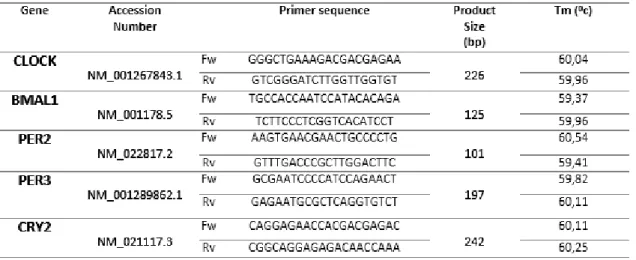
4.1. Primer design ... 28
4.2. Primer testing ... 32
4.2.1. Conventional polymerase-chain reaction (PCR) ... 32
4.3. Real-time quantitative PCR ... 32
4.3.1. Conditions optimization ... 32
4.3.1.1. Real-time qPCR steps ... 33
4.3.1.2. Real-time qPCR components and groundwork ... 33
4.3.2. Real-time qPCR analysis ... 34
4.3.2.1. Standard curve ... 34
4.3.2.2. Slope ... 34
4.3.2.3. Efficiency ... 34
4.3.2.4. Threshold cycle (Ct) ... 35
4.3.2.5. Melting curve (dissociation curve) ... 35
4.3.2.6. Relative quantification ... 37
4.3.2.7. Optimized real-time qPCR conditions ... 37
5. Data analysis ... 38 6. Statistical analysis ... 38 1. Primer testing ... 40 1.1. Aβ-scavengers ... 40 1.2. Circadian rhythm ... 42 2. RT-qPCR optimization ... 44
3. Real-time qPCR relative quantification ... 46
3.1. PGK1 housekeeping gene ... 46
3.3. Gelsolin (GLS) ... 48
3.4. Neprilysin (NEP)... 49
xiii
...
BIBLIOGRAPHY ... 541
I. INTRODUCTION
2
1. Alzheimer´s disease
Alzheimer’s disease (AD) is a neurodegenerative disorder described as a progressive cognitive impairment, and is a current and emergent public health crisis with only a few number of effective treatments available (Lucey et al., 2014). It is considered the most frequent cause of dementia in the elderly (Urrestarazu et al., 2016).
1.1. Pathology and risk factors
The main pathological change in AD consist of brain atrophy, most significantly in the hippocampal formation, temporal lobes and parietotemporal cortices, accompanied by cortical thinning, enlarged ventricles, and white matter abnormalities (Tarasoff-Conway et al., 2015). The major early step in the pathogenesis of AD is the deposition of extracellular amyloid-β peptide (Aβ) into insoluble plaques in the brain, also associated with the aggregation of tau protein into intracellular neurofibrillary tangles leading to neuronal loss, synaptic dysfunction and cognitive impairment. (Hardy et al., 2002; Jack et al., 2010; Lucey et al., 2014). Accumulation of amyloid-β peptide (Aβ) into parenchymal senile plaques (also known as neuritic plaques) or in the walls of cerebral capillaries and arteries (known as cerebral amyloid angiopathy), aggregation of hyperphosphorylated tau into intracellular neurofibrillary tangles (NFTs) and neuropils threads are all microscopic alterations described in AD pathology (Tarasoff-Conway et al., 2015).
One of the most reported abnormalities in AD is circadian rhythm disruption, which affects more than 80% of AD patients over 65 years and include disturbances in sleep-wake cycles and thermoregulation (Van Someren, 2000; Song et al., 2015).
AD includes two main types of the disease, early-onset AD (EOAD) and sporadic or late-onset AD (LOAD). EOAD represents a minority of AD patients, while LOAD goes up to over 95% of patients with AD (Tarasoff-Conway et al., 2015).
When characterized by autosomal dominant inheritance, EOAD is associated with mutations in the presenilin 1 (PSEN1), presenilin 2 (PSEN2) or amyloid precursor protein (APP) genes. The main overall risk factor for LOAD is ageing, although the evolution of cognitive impairment comes from genetics variants in the apolipoprotein E (APOE) ε4 allele. Environmental risk factors for LOAD, include cardiovascular disease and factors providing a risk of cardiovascular disease, such as diabetes mellitus and hypertension; head trauma and mental inactivity, and sleep impairment (Tarasoff-Conway et al., 2015).
The amyloid cascade theory proposes that Aβ is central to the pathogenesis of AD. In this concept, either over-production or decreased clearance of Aβ, will result in the formation of protofibrillar oligomers of Aβ that aggregate and deposit as plaques. The deposition of
3 plaques causes the activation of microglia and astrocytes and all together can cause synaptic and neuritic injury. Then dementia is followed as a consequence of neuronal injury (Hirko, Meyer et al. 2007). However, there are other pathological mechanisms in this cascade involving neurotoxic effects of amyloid peptides probably producing oxidative damage and apoptosis in brain cells, including CP epithelial cells (Perez-Garcia et al., 2009; Vargas et al., 2010; Krzyzanowska et al., 2012).
1.2. Clinical stages of AD
AD can be clinically divided in three distinct phases: i) the pre-symptomatic or preclinical phase, ii) the symptomatic predementia phase, often referred to as mild cognitive impairment (MCI), and iii) the dementia phase. The MCI applies to patients who have some evidence of cognitive impairment but have not progressed to dementia (Sperling and Johnson, 2013).
Braak and Braak, 1991 proposed a neuropathological staging to differentiate initial, intermediate, and advanced AD based on the spread of neurofibrillary tangles (NFTs) within the medial temporal lobe memory circuit. Braak stage 0 corresponds to absence of NFTs, stages I-II (or transentohinal stages) to entorhinal-perirhinal cortex NFTs, stages III-IV (or limbic stages) to NFTs additionally in hippocampus and stages V-VI (or isocortical stages) to NFTs distributed in wider neocortical areas. Stages I-II represent clinically silent periods of the disease, the preclinical phase. Stages III-VI corresponds to clinically incipient AD, and stages V-VI correspond to fully developed AD (Braak and Braak, 1991; Mufson et al., 2016).
2. Amyloid-β processing in AD
2.1.
Aβ peptide formation
The Aβ peptide is mostly formed in the brain by neurons upon the cleavage of the amyloid precursor protein (APP) by β- and γ-secretases (Figure 1) in the endoplasmic reticulum, Golgi apparatus, and endosomal-lysosomal pathway (Gonzalez-Marrero et al., 2015). It is cleaved into numerous isoforms of different amino acid lengths (Strooper et al., 2010; Lucey and Bateman 2014). The C-terminus of Aβ plays a key role in amyloid aggregation (Chauhan et al., 1999). To be more exact, γ-secretase cuts the C-terminal end of the Aβ peptide followed by cleavage of β-secretase to produce 3 major isoforms, which are Aβ1-38, Aβ1-40 and Aβ1-42. The
4 Figure 1 - The amyloid precursor protein (APP) is cleaved by the enzymes β-secretase and γ-secretase
into amyloid-β (Aβ) peptides (scissor symbols represent the cleavage sites; adapted from Peter et al., 2012).
and Bateman, 2014) and were identified in amyloid deposits in AD brain (Chauhan et al., 1999). On the contrary, if α-secretase cleaves APP inside the Aβ domain, it prevents the formation of Aβ in normal APP metabolism (Gonzalez-Marrero et al., 2015).
The Aβ peptide exists in solution in a monomeric α-helical conformation. Aβ fibril formation is a multi-step process which is headed by oligomerization and aggregation of Aβ, and comprises conformational change of the peptide from α-helical to cross β-pleated sheet secondary conformation (Ray et al., 2000). The Aβ circulates in biological fluids at low nanomolar levels in a soluble form. Aβ1-40 is considered to be one of the major species of soluble Aβ in body
fluids and Aβ1-42, in turn is the main component of brain fibrillar deposits (Pérez et al., 2000).
The Aβ1-40 is produced at higher concentrations while Aβ1-42 is more likely to aggregate due to
its hydrophobic features, and is more neurotoxic (Jarrett et al., 1993; Pérez et al., 2000; Lucey and Bateman, 2014). Aβ1-42 neurotoxicity is correlated to its higher β-sheet structure in
comparison to Aβ1-40, being Aβ1-42 more fibrillogenic and cytotoxic than shorter Aβs. The
peptide can be detectable in the cerebrospinal fluid (CSF) and plasma of normal individuals and patients with AD (Chauhan et al., 1999). Both neuritic and diffuse plaques contain predominantly Aβ1-42 and the cerebrovascular amyloid contains predominantly Aβ1-40. In the Aβ
soluble form it binds to several circulatory proteins, which will prevent Aβ fibrillization or, alternatively, promote its polymerization (Qiao et al., 2004).
2.2. Aβ clearance in the brain
There are various pathways for the elimination of unwanted proteins in the brain, proteasomal and lysosomal degradation stand out as the major ones. Cells deploy numerous constitutive and regulated proteolytic activities that maintain proteostasis, all of them to work on regulating the biogenesis, trafficking, and degradation of proteins to maintain their steady-state levels. Evidences suggest that deficient clearance of Aβ rather than increased production of the peptide contributes to its accumulation in LOAD (Baranello et al., 2015). In healthy individuals, the production and clearance are both fast, being the clearance slightly fast than the production, approximately 7,6% and 8,3%,respectively, of the total volume of Aβ produced per hour. With this information it is possible to note that even a minor change in the production or clearance of Aβ would soon cause anomalous accumulation in AD (Miners et
5 al., 2014). Despite the fact that only a few proteolytic pathways have been associated with degradation of Aβ, there are quite a few enzymes with a wide specificity that seems to degrade Aβ (Baranello et al., 2015).
Clearance through the blood-brain barrier (BBB) is suggested as the main pathway of Aβ clearance, where Aβ is removed across the BBB via transcytosis into the vascular lumen. This mechanism is thought to involve several transporter proteins and cell-surface receptors. Proteolytic degradation of Aβ is considered the second most important pathway for Aβ removal (Bohm et al., 2015). Proteolytic cleavage in vitro of the Aβ generates fragments less neurotoxic and less prone to aggregation, and therefore, predicted peptide to be more easily cleared from the brain (Miners et al., 2014). The ability of the monomeric Aβ peptide to assemble into soluble oligomers and then into more stable fibrils due to its tendency to aggregate is considered to be the third in the list of important pathways of Aβ clearance (Bohm et al., 2015). Interestingly, experiments from large post-mortem human brain and in vitro studies have shown that the level and activity of many Aβ-degrading proteases are increased in post-mortem brain tissue and upregulated by Aβ which proposes the idea that the increases are secondary to Aβ accumulation, probably demonstrating physiological responses to the rise in concentration of the Aβ peptide (Miners et al., 2014). It is common sense that a healthy lifestyle enhances quality of life of the individual, and exercise has been demonstrated to benefit Aβ clearance in mouse models of AD. Furthermore, physical activity showed to be linked with delayed Aβ deposition in preclinical AD, whereas cognitively normal sedentary AD mouse models appeared to have an increased risk for cerebral amyloid deposition (Gallina et al., 2015).
In the last decade and half, several studies focused on possible aberrations of Aβ clearance in late-onset sporadic AD, and described age- and disease-associated disruptions in the many clearance pathways, such as reductions in Aβ-degrading protease activities (Miners et al., 2014). Along with aging, Aβ aggregates become extremely difficult to be eliminated compared to soluble types, which explains why they are only visible many years previously to the beginning of other pathogenic events (Gallina et al., 2015). The rare autosomal dominant familial AD is mainly due to an overproduction of Aβ, or promoting Aβ protofibril formation, on the other hand, the late-onset sporadic AD is more common and is highly probable to be caused, in part, by diminished clearance of the Aβ peptide from the CNS. Therefore, the deregulation of Aβ clearance pathways in brain and at choroid plexus - cerebrospinal fluid (CP-CSF) seems to be a central disease event in some AD cases (Gonzalez-Marrero et al., 2015).
There is a group of proteins named Aβ-scavengers, which can be produced by CP, characterized for being able to degrade and/or eliminate the Aβ peptide from several brain regions in which they play a crucial role in the prevention of AD pathology (Carro et al., 2002; Zlokovic, 2004; Vargas et al., 2010).
6
2.2.1. Aβ-scavengers
These proteins mainly comprise the degrading protease activity group. These Aβ-scavengers include insulin-degrading enzyme, neprilysin, angiotensin-converting enzyme, gelsolin, transthyretin and metallothioneins (Chauhuan et al., 1999; Manso et al., 2011; Jochemsen et al., 2015; Alshehri et al., 2015; Hubin et al. 2016;).
2.2.1.1. Insulin-degrading enzyme
Insulin degrading enzyme (IDE) is a 110 kD zinc-dependent metallopeptidase which can be located in the peroxisomes, cytosol, and at the cell surface; it was also detected in human CSF. IDE is able to cleave a variety of small peptides, including insulin, glucagon, and insulin-like growth factors I and II, and this enzyme also converts β-endorphin to γ-endorphin (Pérez et al., 2000; Baranello et al., 2015; Kochkina et al., 2015; Tang, 2016;). This enzyme is expressed in all tissues, and its levels can be controlled by various signals such as cellular stress, glucagon and free fatty acids (Tang, 2016).
Insulin-degrading enzyme (IDE), along with neprilysin (NEP), are considered to be the primary Aβ-cleaving enzymes (Hubin et al., 2016). IDE is capable of cleaving Aβ intra- and extracellularly and may prove valuable as a target to upregulate Aβ clearance in impaired AD patients (Baranello et al., 2015). In addition, IDE actively regulates extracellular levels of Aβ and has been reported to be proficient in degrading monomeric Aβ, although, on the other hand there are few studies regarding IDE effect on Aβ aggregates such as oligomers, and fibrils. In a more specific manner, reports have shown that IDE is capable of degrading Aβ1-40
into fragments that do not form aggregates upon prolonged incubation and to equally cleave Aβ1-42 and inhibit the formation of oligomers when acts in an early aggregation formation
process of the oligomers, although IDE effect on Aβ1-42 is lower when compared to Aβ1-40 which
reflects the higher aggregation propensity of Aβ1-42. Further experiments demonstrated that
IDE exclusively degrades monomeric Aβ due to its small catalytic chamber in comparison with the oligomeric and fibrillar forms of Aβ which are too large to fit in the chamber itself. Regarding this data, IDE presence appears to induce a disruption in monomer-aggregate equilibrium, directing the Aβ assembly away from toxic oligomer formation. The fragments generated by IDE upon cleavage of Aβ are aggregation prone but not toxic (Hubin et al. 2016). Curiously, IDE is regulated by Aβ1-40 and Aβ1-42 via a feedback mechanism, which proposes that
cells seem to regulate IDE expression to eliminate these toxic peptides (Baranello et al., 2015). Upregulation of IDE expression in the brain of AD transgenic mice has been shown to diminish the amyloid burden and cognitive decline (Kochkina et al., 2015). In agreement, when the IDE gene was selectively deleted in mouse models, these presented key phenotypic characteristics of AD, including chronic elevation of cerebral Aβ. An inverse relationship between IDE expression and age has also been reported, what proposes that its loss of activity
7 may be crucial in the development of AD pathology (Baranello et al., 2015). Indeed, decrease of IDE activity was shown to be linked to AD pathology (Kochkina et al., 2015).
2.2.1.2. Neprilysin
Neprilysin (NEP; aka neutral endopeptidase 24.11 or enkephalinase) is a membrane bound-zinc metallopeptidase synthesized in the Golgi and transported to the cell surface, where it anchors into the extracellular space (Baranello et al., 2015; Li et al., 2015). NEP is included in the group of vasoendopeptidases formed by endothelin-converting enzyme and angiotensin-converting enzyme, the main drug targets for the control of hypertension (Baranello et al., 2015). In fact, NEP cleaves several other substrates due to its wide range of specificity, being the substance P the most efficiently hydrolyzed substrate. Other substrates include enkephalins, atrial natriuretic peptide, tachykinins, bradykinin, adrenomedullin, members of the vasoactive intestinal peptide family, glucagon, thymopentin, and of utmost importance in pathophysiological terms, the Aβ peptide (Nalivaeva et al., 2012).
This enzyme is abundant in the kidney but its content in other organs, including the brain, is much lower. In the brain, NEP appears to have mostly neuronal localization especially in the striatonigral pathway, even though it is expressed by activated astrocytes and microglia. NEP was also found in the hippocampus and cortical regions. NEP role in neuronal function is highlighted by its pre- and postsynaptic localization in the nervous system (Nalivaeva et al., 2012). Analysis of the enzyme sequence reveals a type 2 integral-membrane protein structure that starts with a short cytoplasmic domain and a single transmembrane domain that serves as a signal and anchor, followed by a long extracellular domain that set up its catalytic active domain (Baranello et al., 2015).
Along with IDE, NEP is considered to be a major Aβ-degrading enzyme with the ability to degrade both monomeric and oligomeric forms of Aβ1-40 and Aβ1-42 (Li et al., 2015; Hubin et
al., 2016). Several studies have shown that NEP mRNA, protein and activity levels decline with age in the hippocampus and cortex of rodents and humans, and also in the AD brain where senile plaques formation and Aβ aggregation was present (Nalivaeva et al., 2012; Baranello et al., 2015; Li et al., 2015). These data suggested that diminished NEP activity during aging might contribute to the development of AD by promoting Aβ accumulation. Previous studies have shown NEP inactivation and downregulation during aging and the early stages of AD. Cell culture and in vivo experiments revealed an inverse correlation between NEP and Aβ levels in AD (Li et al., 2015). Nevertheless, although NEP levels decrease with age and with AD pathology seen in the neuronal cells, it has been demonstrated that NEP is upregulated in reactive astrocytes surrounding amyloid plaques in an AD mouse model (Nalivaeva et al., 2012; Li et al., 2015; Baranello et al., 2015). Age-related decrease of NEP capability to degrade Aβ might be due to enzyme oxidation or conformational inactivation, for example, by amyloid peptide (Nalivaeva et al., 2012). Numerous studies demonstrated
8 that NEP gene therapy in order to increase its levels in AD mice models can decrease Aβ plaque formation and preserve brain function (Li et al., 2015).
2.2.1.3. Angiotensin-converting enzyme
Angiotensin-converting enzyme (ACE; aka dipeptidyl carboxypeptidase-1 or DCP-1) is a membrane-bound ectoenzyme, zinc peptidase (Baranello et al., 2015; Larmuth et al., 2016). ACE plays a crucial role in blood pressure and body fluid homeostasis by converting angiotensin I to angiotensin II, and degrading bradykinin (a potent vasodilator). It is mainly secreted in the lung and kidney by cells of the inner layer of blood vessels (Baranello et al., 2015). It is known to cleave a variety of peptides, including Aβ peptide and therefore its role in AD. It has been evidenced that ACE is able to inhibit Aβ toxicity in cultured cells, by blocking and cleaving the aggregation of Aβ (Baranello et al., 2015). Recent studies provided evidence of both endoproteolytic and classical dicarboxypeptidase activities of ACE on Aβ peptides (Larmuth et al. 2016).
There are two conflicting hypothesis regarding the correlation between ACE and AD. In order to understand this statement, there are several facts to be considered. Hypertension has been shown to be an important contributor to dementia, including AD, and ACE is a key enzyme in the renin-angiotensin system, which is one of the mechanisms that regulate blood pressure. The amyloid hypothesis proposes that high ACE activity decreases the accumulation of Aβ peptide, therefore decreasing the risk of AD. On the contrary, the vascular hypothesis suggests that high ACE-activity increases the risk of AD by increasing blood pressure, consequently leading to cerebral small-vessel disease. Although, Jochemsen and colleagues found a weak and non-significant association between higher ACE activity and more cerebral small-vessel disease, they also suggested that ACE activity would be more indicative and relevant to Aβ degradation and brain atrophy (Jochemsen et al., 2015). High levels of ACE protein on the CSF could have a beneficial effect on brain atrophy caused by the Aβ burden. Indeed, ACE activity degrades Aβ peptide and contributes to less accumulation of Aβ into senile plaques in the brain and thereby, reducing the extent of brain atrophy (Jochemsen et al., 2015). In fact, other studies have shown that the level and activity of ACE seems to be increased in AD brains (Baranello et al., 2015).
Several studies suggest that ACE is not a direct physiological regulator of steady-state Aβ concentrations in the brain, despite ACE’s ability to cleave Aβ in vitro, in vivo data point out that it does not appear to regulate cerebral amyloidosis (Baranello et al., 2015). Further investigation is needed regarding the role and importance of ACE in the AD pathology.
9
2.2.1.4. Gelsolin
Gelsolin (GSN) is a highly conserved 89 kD protein whose main function is to sustain the cytoplasmic architecture by regulating the actin filament assembly, and therefore, capping and severing the actin filaments. It is mainly localized in the cell’s cytosol and in the plasma, and CSF as secretory protein (Chauhuan et al., 1999; Ray et al., 2000; Hirko et al. 2007;). The intracellular functions of GSN are regulated by calcium and phosphoinositides (Chauhuan et al., 1999). GSN was also reported to participate in other activities, including modulation of calcium channel and NMDA receptor, apoptosis, and in tumor suppression (Ray et al., 2000). Extracellular GSN functions are not known and studies reported that gelsolin binds to the monomeric form of Aβ, and forms an Aβ-gelsolin complex. It was proposed as main function of extracellular gelsolin in the plasma and CSF, to sequester and maintain Aβ in the soluble form (Chauhuan et al., 1999). Furthermore, Ray and colleagues reported that plasma GSN binds to Aβ1-40 and Aβ1-42, and forms a sodium dodecyl sulfate (SDS)-stable complex, which
inhibits the fibrillization of soluble Aβ1-40 and Aβ1-42 in plasma and CSF. GSN also defibrillizes
the performed Aβ fibrils in a time-dependent manner, which suggests that GSN plays a vital role in the regulation of Aβ fibrillogenesis (Ray et al., 2000). The binding of Aβ to GSN is saturable and contains two Aβ binding sites. Kinetics of Aβ suggest that Aβ binding to GSN fits better with two binding sites rather than single binding site. Binding of Aβ to GSN is dependent on the concentrations of both GSN and Aβ, which is increased with higher concentrations of Aβ (Chauhuan et al., 1999). In addition, it was observed that inhibition of GSN gene expression in an AD mouse model was associated with increased apoptotic response in the presence of Aβ, demonstrating that interfering with GSN leads to a sensitization to proapoptotic stimuli (Qiao et al., 2004).
Despite GSN having the ability to bind Aβ, it was not found in detectable amounts in the amyloid plaques of AD patients (Chauhuan et al., 1999; Ray et al., 2000). Authors suggest that GSN is a major Aβ-sequestering protein in the plasma and CSF where it prevents Aβ from fibrillization and maintains it in the soluble form (Ray et al., 2000).
2.2.1.5. Transthyretin
Transthyretin (TTR; aka prealbumin) is a homotetrameric protein with a molecular mass of approximately 55 kDa. TTR has an extensive β-structure, with each monomer being composed of eight β-strands arranged in an antiparallel configuration. This protein is very stable under physiological conditions both in vivo and in vitro. The TTR tetramer contains the two thyroid hormones-binding sites and therefore, its main function is to bind and distribute thyroid hormones (THs) (Alshehri et al., 2015). TTR transports thyroxine (T4) and retinol, by its association with retinol binding protein (RBP), apolipoprotein A-I, neuropeptide Y, and a panoply of exogenous disrupting chemicals and drugs, in CSF and plasma (Alshehri et al.,
10 2015; Ciccone et al., 2016). TTR is mainly synthesized in the liver and in the choroid plexus (Ciccone et al., 2016; Alemi et al., 2016). Regarding the liver, TTR is secreted into the bloodstream and distributes the thyroid hormones around the body, whereas TTR synthesized in the choroid plexus is involved in the movement of thyroxine from the blood into the cerebrospinal fluid and distribution of THs in the brain (Alshehri et al., 2015).
TTR was described as the major Aβ binding protein in the CSF by Schwarzmen and colleagues. These authors proposed that TTR was able to inhibit aggregation and toxicity of Aβ, suggesting that when TTR fails to sequester Aβ, amyloid formation take place (Schwarzmen et al., 1996). In fact, new roles for TTR have been suggested such as protection against neurodegeneration, involvement in behavior, memory and learning, and also promotion of neurodegeneration. It has been recently discovered that TTR may play a wide role in neurobiological function due to the high number of chemicals that can bind to TTR, therefore, interfering with its functions in the brain. TTR is associated with a growing list of neurological diseases including AD, depression, anxiety, schizophrenia, familial amyloid polyneuropathy and familial cardiomyopathy (Alshehri et al., 2015; Ciccone et al., 2016). There are some theories concerning the TTR mechanism associated to the prevention of AD: (i) TTR has a direct role in AD by sequestering Aβ and thus preventing amyloid formation; (ii) in late onset AD patients, diminished TTR in the CSF results in an increase in the Aβ fiber formation; (iii) it was suggested, as an indirect role for TTR, that it prevents Aβ formation in the amyloidogenic pathway by inhibiting the cleavage of the CTF99 by γ-secretase (CTF99 is responsible for the release of the Aβ peptide); (iv) TTR facilitates the non-amyloidogenic pathway by increasing α-secretase to cleave APP, which results in the production of sAPPα (Alshehri et al., 2015). TTR levels have been shown to be decreased in both CSF and plasma in aging and in AD patients, which supports the idea of neuroprotection by TTR (Alemi et al., 2016). Curiously, authors demonstrated that TTR expression is increased in some neurons of the AD brain. The multiplicity of molecular mechanisms involving TTR interactions with Aβ peptides and their precursor seems to clarify why TTR expression is increased in some neurons of the AD brain (Li et al., 2016).
2.2.1.6. Metallothioneins
Metallothioneins (MTs) are the major endogenous zinc- and copper-binding proteins present in the brain and play a crucial role in maintaining metal homeostasis (Chung et al., 2010). MTs are high cysteine and metal content cadmium-binding proteins. MTs are subdivided into numerous families based on primary sequences. Mammals possess several isoforms, including MT-1, MT-2, MT-3, and MT-4. The first three isoforms are known to be synthesized in the central nervous system (CNS), composed of a single polypeptide chain, of which some of them are cysteine residues (Manso et al., 2011). The MT-2A isoform is the most expressed isoform of MTs and is described as a neuroprotective protein essential for brain repair and represents
11 the MTs (Chung et al., 2010). The Mt-2A gene is widely expressed and can be found throughout the brain and spinal cord, and is mainly expressed by the astrocytes, with lower levels of expression in ependymal cells, epithelial cells of choroid plexus, meningeal cells of the pia mater, and endothelial cells of blood vessels. MT-2 has been reported to be synthesized in neurons but in very low levels compared to astrocytes (Manso et al., 2011). MT-2A is a neuroprotective protein, via intracellular functions that include metal detoxification and quenching of oxidative free radicals (Chung et al., 2010). The MT-2A isoform amino acids typically bind seven divalent metal ions [Zn(II), Cd(II)] and up to 12 monovalent copper ions through thiolate bounds. These metals are separated into two metal-thiolate clusters holding three and four divalent metal ions or six monovalent metal ions each (Manso et al., 2011). MT-2A isoform is upregulated in neuroinflammatory environments, as can be caused by the neuropathology of AD. MT-2 expression levels are highly regulated by cytokine interleukine-6 (IL-6), which is a neuroinflammatory factor. This MT isoform has been suggested to have a role has antioxidant, anti-inflammatory, and antiapoptotic, in addition to its zinc and copper binding capabilities, underlying MT-2A neuroprotective activities (Manso et al., 2011).
MT-2A expression is significantly upregulated in regions where Aβ plaque burden is present in the pre-clinical and clinical AD brain, as well as in the brains of an AD mouse model (Chung et al., 2010). The upregulation of this protein in the surrounding area of the amyloid plaques will benefit the nearby cells, considering the antioxidant, anti-inflammatory and antiapoptotic assets of MT-2A. This MT is also a metal binding protein, a very important property in this case since Aβ plaques are enriched with zinc, copper, and iron, which are metals probably involved in the aggregation of Aβ (Manso et al., 2011). Studies demonstrated that Zn7-MT-2A, can prevent the Cu-mediated aggregation of Aβ 1-42 although Zn7-MT-2A is
incapable of de-aggregate either Cu-Aβ1-42 or Cu-Aβ1-40. Based on different data, the same
authors predicted that the zinc bound is required for the ability of MT-2A to block copper mediated Aβ1-40 and Aβ1-42 aggregation, due to the existence of a metal exchange of copper
and zinc between Zn7-MT-2A and Cu(II)Aβ. This observation might be explained by the MTs
ability of higher binding affinity metals (copper) to shift lower affinity metals (zinc). The same authors also observed that Zn7-MT-2A is able to protect against Cu(II)-Aβ induced
neuronal degeneration, again due to the metal exchange of copper and zinc (Chung et al., 2010; Manso et al., 2011).
12
3. Circadian rhythm
Circadian rhythms are defined as oscillations with a periodicity of approximately 24h and constitute a fundamental component of mammalian physiology. These internal circadian clock systems have the function of coordinating to external synchronizing cues (Zeitgebers), such as the light-dark cycle (Thome et al., 2011; Musiek et al., 2015). The circadian system commands the 24h rhythmicity in sleep-wake cycle, body temperature, feeding, rest-activity behavior, hormonal levels, among other biological processes of the organism (Videnovic et al., 2014).
3.1. Human circadian rhythm model, components and
location
The suprachiasmatic nucleus (SCN) characterizes the core of the circadian system, being the main component and represents the master clock of the circadian system (Cassone et al., 1988; Videnovic et al., 2014). The SCN can be divided in two parts: a core and shell subnuclei. The subnucleus has distinct neurochemical properties, containing γ-aminobutiric acid (GABA) which is the main neurotransmitter in almost all neurons of the SCN. Neurons present in the SCN core secrete vasoactive intestinal polypeptide (VIP), whereas neurons in the shell secrete arginine vasopressin (Dardente et al., 2007; Golombek et al., 2010; Videnovic et al., 2014; Musiek et al., 2015). The arginine vasopressin works in a complementary manner to impart stability and flexibility to the circadian timing system (Saper, 2013; Wang et al., 2015). The role of the VIP neurons of the SCN core is to obtain direct light input from the retina and to deliver intranuclear projections to the rest of the SCN itself (Wang et al., 2015).
The circadian timekeeping is arranged by complex molecular loops and is composed by three distinct components: a pacemaker (SCN), afferent pathways (for light and other stimuli that synchronize the pacemaker to external cues) and efferent output rhythms regulated by the SCN (Videnovic et al., 2014). The SCN will signal via multiple mechanisms, including the autonomic nervous system and hormone release, to synchronize cell-autonomous peripheral clocks in the body (Weaver, 1998; Musiek et al., 2015). The SCN must be able to afford a steady, robust circadian output while remaining adaptable to differences between endogenous and exogenous cycles that arise from seasonal changes in the time of sunrise and endogenous circadian periods not exactly equal to 24h in length (Wang et al., 2015).
One of the most apparent example of a circadian process is the sleep-wake cycle, which occurs with a consistent 24h rhythm and can be shifted according to environmental cues (Musiek et al., 2015). Circadian rhythmicity, can also be observed in body temperature, and
13 secretion of numerous hormones, red blood cells production and other physiological parameters (Volicer et al., 2012). The rhythmic clock gene expression has been described, in humans, in peripheral tissues such as skin, oral mucosa, colonocytes, bone marrow, heart, and white blood cells but there’s very few information on human brain oscillators (Cermakian et al., 2011). It is thought that the one of the main reasons for the global disruption of the body´s circadian activity is directly linked with decreased electrical, hormonal and gene-expression activity of SCN cells which is associated with aging (Costa et al., 2013). The circadian clock controls many pathways involved in neurodegeneration, such as autophagy, metabolism, reactive oxygen species (ROS) homeostasis and oxidative stress response and DNA damage repair (Costa et al., 2013).
The SCN is able to preserve these 24h rhythms in physiological functions through a complex molecular based mechanism (Videnovic et al., 2014).
3.2. Circadian rhythm molecular mechanisms
The regulation of the circadian rhythms depends on a set of clock genes, including circadian locomoter output cycles kaput (CLOCK), brain and muscle Arnt-like protein-1 (BMAL1) or aryl hydrocarbon receptor nuclear translocator-like (ARNTL), three PERIOD genes (PER1, PER2, and PER3) and two plant CRYPTOCHROME gene homologues (CRY1 and CRY2). These genes have the very important task of controlling a considerable percentage of the genome: approximately 10% of all expressed genes estimated to be regulated by the clock genes. These genes can be considered ubiquitous, as almost all peripheral tissues, including brain regions outside the SCN, contain autonomous circadian clocks. These clocks are most likely synchronized by the SCN but they also can be influenced by their own distinct circadian synchronizers (Videnovic et al., 2014).
The maintenance of the circadian rhythms in the SCN in most cells is made by a cellular clock transcriptional machinery involving the bHLH/PAS transcription factors ARNTL and CLOCK. These two heterodimerize and drive transcription of several genes which includes their own negative feedback repressors PER and CRY genes; these last ones repress BMAL1/CLOCK-mediated transcription (Mohawk et al., 2012; Musiek et al., 2015). This feedback loop functions as a regulator and can be divided into positive and negative limbs. The positive limb comprises the CLOCK and ARNTL, two PAS helix-loop-helix transcription factors; they form heterodimers and bind to E-box enhancer element, thus, are able to regulate the transcription of both PER and CRY genes. The negative limb comprises PER and CRY proteins, and its main function is to attenuate activation of their own genes by CLOCK/BMAL1, consequently producing the negative feedback (Figure 2; Abe et al., 2002; Kunieda et al., 2006; Marpegan et al., 2011; Musiek et al., 2015). More specifically, the intracellular level of CLOCK remains the same throughout the 24 h period. There is a peak of ARNTL expression at the beginning of the day that promote the formation of ARNTL–CLOCK heterodimers, which
14 will activate transcription of PER and CRY. As PER accumulates in the cytoplasm, it becomes phosphorylated and degraded by ubiquitylation. CRY accumulates in the cytoplasm late in the subjective day, and translocates to the nucleus to inhibit ARNTL–CLOCK-mediated transcription. At night, the PER–CRY complex is degraded, and the cycle starts again. This feedback loop ensures a high level of ARNTL and low levels of PER and CRY at the beginning of a new circadian day (Figure 2) (Videnovic et al., 2014).
An important feature of CLOCK genes is their close interrelation with external, environmental factors because very few other gene families demonstrate such strong environment affiliation and interdependency. Consequently, they are highly subjective to exogenous factors (such as stress) and endogenous vulnerability factors, such as inherited genetic variants, eventually leading to the clinical phenomenology of AD (Thome et al., 2011).
One aspect to keep in mind is that it is extremely difficult to monitor CLOCK genes, either one monitors the genes themselves or their RNA or protein products. Regarding the gene level, it is important to know exactly when the material was collected during the day. As it is known the genes are under the influence of circadian rhythms, a sample collected during the day will be different from the one collected during the night and vice-versa. Finding a matched control is also difficult. It is also important to eliminate false positives and false negatives during background subtraction and data manipulation, which can be difficult to address (Thome et al. 2011).
15 Figure 2 – Molecular mechanism of the circadian system. The proteins encoded by the core set of clock
genes—PER, CLOCK, BMAL1/ARNTL and CRY—cooperate to create a self-sustaining negative transcription–translation feedback loop (adapted from Videnovic et al., 2014).
3.3. Circadian rhythmicity, sleep disturbances and AD
One of the most common features in AD are sleep and circadian rhythm disturbances, and it has been reported that up to 45% of patients may have sleep problems. Excessive awakenings (23%), excessive daytime sleepiness (10%), early morning awakening (11%), and napping for more than 1 hour during the day (14%) makes the most of the major disturbances. These features can be present early in the progression of the disease, despite the fact that they tend to be correlated with the severity of the cognitive decline (Urrestarazu and Iriarte, 2016). Circadian clock malfunction has a major influence in sleep and it is thought to make changes that influence memory formation and cognitive functions, which are sleep-dependent processes (Costa et al., 2013; Musiek et al., 2015). Indeed, sleep has a very important role in restoring the functions of the brain and it is involved with memory retention (Born and Wilhelm 2012; Lucey and Bateman, 2014). It has been observed that in extreme cases, a complete day/night sleep pattern reversal can occur (Urrestarazu and Iriarte 2016). Circadian clock disruptions also leads to reduced amplitudes and phase delay of circadian variation in core body temperature and activity. As a result of these circadian alterations, patients show increases in cognitive decay/dementia, number of daytime naps, wandering, nocturnal insomnia, misperception and disorientation, and decreases in motor control and metabolism (Costa et al., 2013; Videnovic et al., 2014). The changes in body temperature rhythms have been evidenced to differ between genders in older subjects, as alterations have their amplitude reduced in older males, but not in older females (Costa et al., 2013).
Studies monitoring the sleep-wake cycle in cognitively normal individuals aged between 45-75 years revealed decreased sleep efficiency and increased nap frequency in individuals with amyloid deposition in comparison with those of the same age deprived of amyloid deposition, determined by CSF Aβ42 levels (Ju et al., 2013; Lucey and Bateman, 2014). It was shown that individuals cognitively normal shared a statistically significant difference in sleep parameters when separated by amyloid deposition status which supports the hypothesis that sleep disturbances are associated with AD pathology and precede the onset of cognitive impairment (Lucey and Bateman, 2014). In addition, in animal models the levels of soluble Aβ are higher in the ISF of APP transgenic mice during wakefulness and lower during sleep. These mice have a normal sleep cycle when younger and before amyloid deposition. Then, when the Aβ plaque formation in APP transgenic mice begins, a loss of the Aβ diurnal pattern is observed and also an obvious disrupted sleep-wake cycle with increased wakefulness and sleep during the light phase when the mice should be sleeping. These findings regarding sleep alterations in transgenic mice indicate that the irregularities in sleep are due to Aβ aggregation in the brain (Roh et al., 2012; Lucey and Bateman, 2014). Also, sleep deprivation of mice for 21 days
16 increased ISF Aβ concentrations and accelerated Aβ deposition into plaques. (Kang et al., 2009; Lucey and Bateman, 2014). In addition, other authors evidenced that the levels of soluble Aβ in the extracellular ISF of the hippocampus in mice are dynamically and positively related with minutes awake per hour and negatively related with time asleep (Kang et al., 2009; Roh et al., 2014; Ju and Holtzman, 2013; Ooms et al., 2014; Roh et al., 2014). Two different authors demonstrated a 12 and a 15% increase in the sleep time during the dark phase that was related with more than 50% reduction in the progression of Aβ pathology in the brain (Yan et al., 2009; Roh et al., 2014). Observations with transgenic mice, when they were kept in standard 12h:12h light:dark conditions, the levels of Aβ were highest during the dark phase (period of mice activity) and lowest during the light phase (period of mice sleep; Musiek et al., 2015). These studies are in agreement with others where it has been reported that neurodegenerative disorders, such as DA, disrupt the circadian clock and these internal time alterations are very likely to be one of the primordial signs of the disease (Costa et al., 2013).
Regarding the molecular basis of the circadian rhythm, the deletion of the master clock gene BMAL1 annuls all circadian function which leads to a total loss of day-night rhythmicity of sleep (Bunger et al., 2000; Laposky et al., 2005; Musiek et al., 2015). Sleep deprivation can modify the expression of clock genes and also the DNA binding patterns of BMAL1/CLOCK heterodimers, consequently changing clock function (Mongrain et al., 2011). Recent studies suggest the mechanism of Aβ induced circadian rhythm disruption is by enhancing the degradation of BMAL1, leading to disruption of PER2 expression at both protein and mRNA levels (Song et al., 2015). Aβ destabilizes BMAL1 and CBP proteins by sumoylation, a post-translational modification (Cardone et al., 2005; Song et al., 2015). Sumoylation is present during oxidative-stress and Aβ peptide is able to activate the enzymes that lead to sumoylation of BMAL1 and, therefore, to its degradation and thereby the circadian rhythmicity (Song et al., 2015). Numerous studies in humans have suggested that circadian dysfunction in AD lies on the disruption and degeneration of the SCN (Harper et al., 2008; Farajnia et al., 2012; Musiek et al., 2015).
Sleep can limit Aβ production as well as regulate the clearance of Aβ from the brain. Sleep actually induces significant increases in the volume of extracellular fluid in the brain and promotes the convective bulk flow of metabolites and proteins out of the brain, a process controlled by glia (Xie et al., 2013; Musiek et al., 2015).
17
4. Choroid plexus
4.1. Choroid plexus location and structure
The choroid plexus (CP) is a highly vascularized tissue located within each ventricle of the brain, the lateral, third and fourth ventricles. Structurally, the CP is made of the telencephalic CP in each lateral ventricle, the diencephelic CP of the third ventricle and the hindbrain CP (Figure 3A; Liddelow, 2005; Lun et al., 2015).
The endothelium capillaries of the CP are fenestrated, and are connected by thin membranous diaphragms permeable to small molecules and water, which enables the rapid delivery of water from the blood to epithelial cells for CSF production. The solutes seem to cross from the blood into the stromal space by diffusion across endothelial fenestrae or by vesicular transport (Lun et al., 2015). The structure of CP mirrors consists on a monolayer of cuboidal cells that surrounds a stromal core of capillaries and connective tissue. The blood-CSF barrier (Bblood-CSFB) is formed by the adjacent CP epithelial cells joined together by tight junctions. This barrier prevents paracellular free passage of molecules from the systemic circulation into the CSF. The tight junctions, along with adherens junctions, guarantee the apico-basal polarity of membrane proteins, such as transporters, which play a key role for normal epithelial cell function (Damkier et al., 2013; Lun et al., 2015). The BBB, one of the main brain defense barriers, entails the tight junctions-containing endothelial cells of the brain capillaries, and the BCSFB is formed by the epithelial cell monolayer of the CP, located in the brain ventricles. The BBB entails the tight junctions-containing endothelial cells of the brain capillaries, and the BCSFB is formed by the epithelial cell monolayer of the CP, located in the brain ventricles (Demeestere et al., 2015). This epithelial cell monolayer locates in a basal lamina surrounding and enclosing a central stroma with dendritic cells, fibroblasts and macrophages (Marques et al., 2015). Attached to the apical side of CP epithelial cells, are present the epiplexus cells. These cells possess macrophage activity and examine the CSF filled brain ventricles and there are also present, supraependymal cells. These cells, in conjunct, play a key role in defensing the brain from infections, after bacteria transverse the CP (Figure 3B; Marques et al., 2015).
18 Figure 3 – Location of the CPs in the human brain. A) The CPs are present in the two lateral, third and
fourth ventricles (red bands). B) Every plexuses contain fenestrated vessels, with a single layer of closely opposite CP epithelial cells, joined by tight junctions which forms the blood-cerebrospinal fluid barrier (BCSFB; adapted from Liddelow, 2015). Abbreviations: BV, blood vessel; CPEC, choroid plexus epithelial cell; CSF, cerebrospinal fluid; LV, lateral ventricle; ST, stroma/basement membrane; TJ, tight junction; 3V, third ventricle.
4.2. Choroid plexus function
The CP perform a multitude of functions such as synthesis, secretion, active transport and selective reabsorption of deleterious substances (Lun et al., 2015). CP main function is to produce CSF in the vertebrate brain and to maintain the extracellular environment of the brain by monitoring the chemical exchange between the CSF and the brain tissue (Krzyzanowska et al., 2012; Lun et al., 2015). This monitoring involves the surveying of both chemical and immunological status of the extracellular fluid and the removal of toxic substances as well as important roles in the regenerative processes following traumatic events. The CP is also capable of producing various peptides which can have beneficial and neuroprotective properties, as well as monitor and maintain the biochemical and cellular homeostasis of the central nervous system (CNS) (Krzyzanowska et al., 2012).
Approximately two thirds of the CSF is produced and secreted by CP, the remaining part is produced by other brain regions, including the ependymal cells of the ventricular surface and those cells lining the subarachnoid space (Liddelow, 2005). The production of CSF by the CP
19 occurs by passive filtration of fluid across the extremely permeable capillary endothelium and regulated secretion across the CP endothelium (Demeestere et al., 2015). The CP-CSF system is the upmost system for the development and maintenance of the CNS. Several studies had showed the importance of a proper CP function to the formation and integrity of the CNS. Lack of CSF causes an impaired brain growth, due to the right CSF pressure during normal brain development. On the other hand, an excess of CSF, caused by overproduction, obstructed flow or even limited CSF reabsorption, can cause hydrocephalus (excess accumulation of CSF within the brain ventricles) (Lun et al., 2015).
4.3. Choroid plexus role in Alzheimer’s disease
The CP is highly sensible to the many external factors and goes through structural and physiological changes during aging and disease states. Harmful changes in the CP’s anatomy, function and also CSF turnover have been found in several neurological diseases, including AD (Alvira-Botero and Carro, 2010). The CP plays a key role in the most basic features of neural function, such as Aβ clearance by expressing several Aβ-scavengers (as mentioned previously in section 2.2) (Carro et al., 2002; Zlokovic, 2004; Vargas et al., 2010). It was shown that Aβ 1-42, the most neurotoxic form of the Aβ peptides, modifies BBB integrity and causes a
significant increase in NO production which is implicated in the AD pathogenesis (Zhang et al., 2002; Keil et al., 2004; Teng and Tang, 2005; Vargas et al., 2010). Oxidative stress, and more specifically, oxidative protein damage is present in the early onset of the disease. It may affect protein interactions, protein folding and protein kinase activity, abnormal functions of endothelial and vascular smooth muscle cells, and impaired HDL cholesterol metabolism, therefore having important implications in the deterioration of the CP’s functions (Perez-Garcia et al., 2009; Krzyzanowska et al., 2012).
In AD the CP develops irregularities similar to those related with aging, however very enhanced. Indeed, CP ability to secrete Aβ-carrier and scavenger proteins and to express important receptors that scavenge Aβ peptides is decreased with age and is compromised in several models of AD (Zlokovic et al., 1996; Krzyzanowska et al., 2015).
Accumulation of amyloid-like inclusions, also known as Biondi ring tangles in CP epithelial cells, might affect vital function of the CP in AD cases (Wen et al., 1999; Vargas et al., 2010). Those alterations such as epithelial atrophy of the CP (Serot et al., 2000), which is significantly accentuated in AD, may alter the choroid plexus main functions, including synthesis, secretion, and transport of proteins to other molecules. This is considered by many authors not to be enough to cause amyloid deposits in the majority of AD cases but plays a key role in the initiation and development of AD pathology. Additionally, authors hypothesize that disturbed CP functions may precede several AD hallmarks, such as amyloid brain accumulation. The altered CP function could be one of the primary pathogenic events in
late-20 onset AD (Krzyzanowska et al., 2012). In the LOAD stage there are numerous CP significant modifications such as epithelial atrophy, fibrosis, and thickened basement membranes which proposes a disruption on CP functions (Bolos et al., 2014).
Aβ peptide also accumulates in the CP of autopsied AD patients, and suggested a direct relationship between Aβ deposits at CP epithelium and the development of a functional and structural disruption of the organ (Dietrich et al., 2008; Krzyzanowska et al., 2012). Vargas and colleagues also revealed the presence of Aβ accumulation in CP of APP/PS1 transgenic mice and AD patients, and demonstrated its association with transcytosis impairment, increased nitric oxide (NO) production and cell death mediated by mitochondrial dysfunction. They showed that Aβ deposits were responsible for mitochondrial dysfunction in CP and also a direct effect of Aβ accumulation on cell death induction in CP (Vargas et al., 2010). Aβ can be found in CP epithelial cells from non-demented subjects, although the proportion of epithelial cells containing Aβ accumulation increases considerably in CP from AD patients (Krzyzanowska et al., 2012). The decreased activity of the enzymes involved in oxidative phosphorylation and mitochondrial activity might help to disrupt the protein synthesis in the CP. The impairment of the functional activity of the CP is associated with the decrease in protein secretion and renewal of the CSF, which can also be involved in the onset and progression of AD (Liu et al., 2013; Krzyzanowska et al., 2015).
Interestingly, recent studies have shown that Aβ has the ability to stimulate neural progenitor cells within the CP through prompting endogenous growth factor actions. Aβ peptide can induce neurogenesis, however many of these newly born neurons die shortly after differentiation which suggests that the neurons do mature but undergo cell death. Nevertheless, further results suggest that amyloidogenic environment tends to down-regulate neurogenesis (Bolos et al., 2013).
Due to the variety of the CP’s functions, even a slight modification in the CP can promote extensive effects. Modifications in the CP’s anatomy and physiology are linked with ageing and many others central nervous system diseases, comprising AD and are parallel with abnormal brain activity (Serot et al., 2003; Vargas et al., 2010).
21 Figure 4 – Proposed sequence of pathological events in Alzheimer’s disease – Several pathogenic
processes contribute to the dysfunction of the choroid plexus which results in a disrupted Aβ processing. These processes plus the accumulation of Aβ contributes to the development of the pathogenic progression (Adapted from Krzyzanowska et al., 2015).
22
II. AIM
23 The work hypothesis in this study was that the circadian rhythm and AD progression regulate the expression of Aβ-scavengers in CP of AD patients. For that purpose, the global aim was to analyze the expression of Aβ-scavengers in the CP of AD patients. This current work has the following specific objectives:
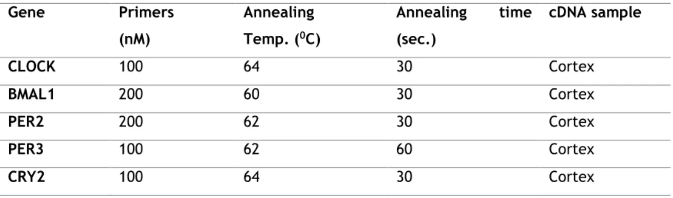
1. Design primers specific for IDE, NEP, ACE, GLS, TTR and MT2A, CLOCK, BMAL1, PER2, PER3 and CRY2 genes;
2. Optimize RT-qPCR reactions for the Aβ-scavengers and circadian rhythm genes; 3. Analyze relative mRNA levels of expression of TTR, GLS and NEP genes in CP of AD
24
III. MATERIALS AND METHODS
25
1. Samples
Choroid plexus tissue was obtained from Department of Patahology and Neuropathology (Xerencia de Xestión Integrada de Vigo-SERGAS, Spain) and Neuropathology Brain Bank (HUB-ICO-IDIBELL Biobank), and Banco de Tejidos. All samples were obtained following the ethical guidelines both of Spanish legislation on this matter and of the local ethics committee. The post-mortem interval between death and tissue processing was between 3 and 14 hours. Neuropathological diagnosis of AD was based on the Braak classification, mentioned in the introduction. Samples are represented in table 1, above.
26
Table 1 – Patients samples characteristics, including age and Alzheimer’s disease Braak stage.
Number of sample AD Braak Stage Age Gender
1 Control 44 Male 2 Control 40 3 Control 52 4 Control 52 5 Control 80 6 Control 63 7 Control 28 8 AD I 64 Male 9 AD I 68 10 AD I 64 11 AD I 58 12 AD I 76 13 AD I 63 14 AD I 63 15 AD II 55 16 AD II 57 17 AD II 60 18 AD II 83 19 AD II 74 20 AD II 86 21 AD III 73 Male 22 AD III 81 23 AD III 77 24 AD III 82 25 AD V 82 26 AD IV 79 27 AD III 76 28 AD V 75