EPR and Mo
¨ ssbauer Spectroscopic Studies on Enoate Reductase*
(Received for publication, January 26, 1996, and in revised form, March 26, 1996)Jorge Caldeira‡§, Richard Feicht¶, Hiltturd White¶, Miguel Teixeira‡i, Jose´ J. G. Moura‡,
Helmut Simon¶, and Isabel Moura‡**
From the ‡Departamento de Quı´mica (and Centro de Quı´mica Fina e Biotecnologia), Faculdade de Cieˆncias e Tecnologia, Universidade Nova de Lisboa, 2825 Monte de Caparica, Portugal, the §Instituto Superior de Cieˆncias da Sau´ de Sul, 2825 Monte de Caparica, Portugal, the¶Lehrstuhl fu¨ r Organische Chemie und Biochemie, Technischen Universita¨t Mu¨ nchen, D-85747 Garching, Germany, and theiInstituto de Tecnologia Quı´mica e Biologica, Universidade Nova de Lisboa, Apt. 127, 2780 Oeiras, Portugal
Enoate reductase (EC 1.3.1.31) is a protein isolated from Clostridium tyrobutyricum that contains iron, la-bile sulfide, FAD, and FMN. The enzyme reduces thea,b carbon-carbon double bond of nonactivated 2-enoates and in a reversible way that of 2-enals at the expense of NADH or reduced methyl viologen. UV-visible and EPR potentiometric titrations detect a semiquinone species in redox intermediate states characterized by an isotro-pic EPR signal at g5 2.0 without contribution at 580 nm. EPR redox titration shows two widely spread mid-point redox potentials (2190 and 2350 mV at pH 7.0), and a nearly stoichiometric amount of this species is detected. The data suggest the semiquinone radical has an ani-onic nature. In the reduced form, the [Fe-S] moiety is characterized by a single rhombic EPR spectrum, ob-served in a wide range of temperatures (4.2– 60 K) with g values at 2.013, 1.943, and 1.860 (2180 mV at pH 7.0). The gmaxvalue is low when compared with what has been reported for other iron-sulfur clusters. Mo¨ssbauer stud-ies reveal the presence of a [4Fe-4S]12/11center. One of the subcomponents of the spectrum shows an unusually large value of quadrupole splitting (ferrous character) in both the oxidized and reduced states. Substrate bind-ing to the reduced enzyme induces subtle changes in the spectroscopic Mo¨ ssbauer parameters. The Mo¨ssbauer data together with known kinetic information suggest the involvement of this iron-sulfur center in the enzyme mechanism.
Enoate reductase (EC 1.3.1.31) (1– 6) isolated from
Clostrid-ium tyrobutyricum catalyzes the NADH or
methyl-viologen-de-pendent reduction of the a,b carbon-carbon double bond of nonactivated 2-enoates (7) and in a reversible way that of 2-enals (8). The enzyme appears to be a multimer of identical subunits. The total molecular mass is 940 kDa (subunit circa 73 kDa). Sedimentation equilibrium experiments, molecular mass data, and electron microscopy indicate that the native enzyme is composed of a tetramer of trimers. Per enzyme subunit 1 FAD, 0.6 FMN, 4 iron, and 4 labile sulfur atoms were found (9), and thus enoate reductase belongs to the rare class of flavoen-zymes containing both FAD and FMN. The involvement of iron-sulfur centers in electron transfer is well established (10), and they have also been shown to be associated with other important physiological nonredox functions, such as those of
aconitase and other dehydratases (11, 12), glutamine 5-phos-phoribosyl-1-pyrophosphate aminotransferase (13), endonucle-ase III (14), and iron-responsive element-binding protein (15–17).
In this work, we report the involvement of an iron-sulfur cen-ter in a new type of biochemical reaction. EPR and Mo¨ssbauer data are analyzed in the oxidized and NADH- and dithionite-reduced states, as well as in substrate bound forms in order to identify the type of iron-sulfur core involved. A tentative mech-anism is presented that involves a hydride transfer from a flavin group and a carbon-carbon double bond polarized by the presence of the iron-sulfur cluster, thus including enoate reductase in the group of iron-sulfur enzymes in which the metal center interacts with substrate molecules during catalysis.
MATERIALS AND METHODS
Growth of C. tyrobutyricum (DSM 1460)—Cells of C. tyrobutyricum DSM1460 were grown on57Fe- or56Fe-containing medium on a 50-liter
scale according to Bader and co-workers (1) with slight modifications (1–3). The medium contained 150 mg/liter (NH4)3SO4 instead of
(NH4)2PO4and additionally 32 mg Na2SO4z10 H2O. The source of iron
was about 65 mg of57Fe in the form of an57FeSO
4solution, i.e., the
concentration amounted to about 2 3 1025 M iron. The cells were
harvested by centrifugation after reaching the stationary phase 20 h after inoculation.
Purification and Sample Preparation—Enoate reductase was puri-fied under strict anaerobic conditions essentially according to Kuno et al. (9). Wet packed cells (78 g) were suspended in 234 ml of 50 mM
potassium phosphate buffer, pH 7.0, containing 10 mMsodium tiglate, 1 mMEDTA, 320 mg of lysozyme, and 32 mg of DNase. The pH of the suspension was adjusted to 7.2 by the addition of 2.5MNa2CO3. After
incubating the suspension for 40 min at 35 °C, it was centrifuged for 20 min at 270003 g at 0 °C. The careful control of the pH is crucial for obtaining the enoate reductase in the supernatant.
All chromatographic steps were performed with oxygen-free buffers containing 0.1 mMEDTA, 250 mMsaccharose, 0.02% sodium azide, and
0.02% mercaptoethanol. The supernatant was applied to the following columns in two portions.
It was first applied to a DEAE Sepharose CL-6B column (18.53 4.5 cm) equilibrated with 20 mMpotassium phosphate buffer (pH 7.0) and
then applied directly to a hydroxyapatite column (18 3 2.4 cm) as described previously (3).
Pure enoate reductase both in57Fe and in56Fe were concentrated in
phosphate buffer, pH 7.0, 0.25Msaccharose, and 1 mMcrotonate (need-ed to maintain the enzyme in the oxidiz(need-ed state). In the final steps the enzyme buffer was exchanged with a buffer containing only phosphate and saccharose in the concentrations mentioned above. Enoate reduc-tase in57Fe at the final concentration of about 1 m
Mwas used to prepare four samples: native, dithionite-reduced, NADH-reduced, and dithion-ite-reduced in the presence of 50 mMof cinnamate. The enzyme reduc-tions were performed under strict anaerobic condireduc-tions.
Spectroscopic/Potentiometric Measurements—Redox potentials of enoate reductase were determined by UV-visible absorbance and EPR potentiometric titrations. The protein solution in phosphate buffer, pH 7.0, was kept under anaerobic conditions by flushing the solution dur-ing the titration with purified Argon (with an Indicatdur-ing Oxygen Trap from Chemical Research Supplies). An optical redox cell and an EPR * The costs of publication of this article were defrayed in part by the
payment of page charges. This article must therefore be hereby marked “advertisement” in accordance with 18 U.S.C. Section 1734 solely to indicate this fact.
** To whom correspondence should be addressed. Tel.: 351-1-2948381; Fax: 351-1-2948550; E-mail: [email protected].
© 1996 by The American Society for Biochemistry and Molecular Biology, Inc. Printed in U.S.A.
18743
by guest on September 9, 2019
http://www.jbc.org/
potentiometric cell, slightly modified from the design of Dutton (18) was used for the UV-visible and EPR titrations. The potentials were meas-ured with a Crison 2002 potentiometer with a platinum and a Ag/AgCl electrode and are quoted relative to the normal standard hydrogen electrode and calibrated with quinhydrone at pH 7.0. The following redox mediators were present at a final concentration of 3.5mM: 1,4-naphtoquinone, methylene blue, triquat, phenosaphranine, benzylviolo-gen, methylviolobenzylviolo-gen, dichlorophenol-indophenol, benzoquinone, anthra-quinone-2-sulfonic acid, phenazinamethosulfate, dimethyltriquat, indigo tetrasulfonate, 2-hydroxy-1,4-naphtoquinone, 5-hydroxy-1,4-naphtoquinone, duroquinone, phenazil, and safranine.
Solution potentials were varied by adding appropriate volumes of deareated 30 mM sodium dithionite or 30 mMNADH as reductant. UV-visible spectra were recorded during titration on a UV-265FS Shi-madzu spectrometer. EPR redox titrations were performed in the same conditions as described for the UV-visible titrations, and the potentials were varied as described in Ref. 9. The protein solution was poised at different redox potentials, and 180-ml aliquots were transferred under argon to EPR tubes and frozen in liquid nitrogen for later measurement.
EPR spectra were made on a Bruker ER 200 spectrometer equipped with an Oxford continuous flow cryostat. The Mo¨ssbauer spectrometer was similar to the one described in Ref. 19. The zero velocity of the Mo¨ssbauer spectra is referred to the centroid of metallic iron spectra at room temperature.
RESULTS
UV-visible and EPR Redox Titrations
The UV-visible spectra of enoate reductase are largely dom-inated by the flavin cofactor in the 300 –550 nm region. UV-visible measurements coupled with potentiometric titrations as shown in Fig. 1 do not distinguish between the flavin and [Fe-S] absorbance contribution due to the similarity of the redox potentials of these prosthetic groups. Also no 580 – 600 nm spectral contribution was detected in the UV-visible spec-trum during redox titrations. The native form of enoate reduc-tase is EPR silent, and the iron-sulfur and the flavin moieties are both diamagnetic (not shown).
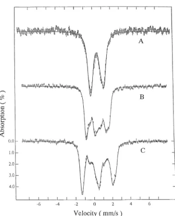
At 10 K, the EPR spectrum of the NADH-reduced state shows a rhombic signal with g values at 2.013, 1.943, and 1.860, which are assigned to an iron-sulfur center with a su-perimposed isotropic signal centered at g5 2.00, attributable to a flavin semiquinone (see Fig. 2C). At high temperature (120 K), the iron-sulfur center is too broad to be detected, and the EPR features are dominated by the flavin radical signal (Fig. 2B). The dithionite-reduced enzyme shows the same rhombic EPR signal as the NADH-reduced sample, but the isotropic signal is absent (Fig. 2A). The intensity of the rhombic EPR signals does not increase upon further reduction with dithion-ite: the spin quantification (relative to a CuEDTA standard)
gives 0.94 spin per monomer. The quantitation of the flavin signal is 0.9 spin per monomer, using as a standard
Desulfo-vibrio desulfuricans ATCC 27774 flavodoxin in the
semiqui-none state at 120 K (20). The temperature dependence of the rhombic signal shows that it can be detected up to 60 K and is better observed at 10 K, using a microwave power of 23.7mW. The line width of this rhombic species is increased in the
57Fe-enriched sample due to unresolved hyperfine interactions.
The optimal temperature to observe the isotropic signal at-tributable to the flavin is 120 K. A comparison of the EPR signals of the flavin radical in H2O and D2O shows a line width
reduction from 17 to 13 G (21). This observation is not in agreement with what was previously reported for anionic semiquinones where a decrease in the line width from 15 to 14 G was found, whereas in the neutral semiquinones the line width changes from 19 to 15 G due to the permutation of the flavin N-5 exchangeable proton. The span in redox potentials for this redox equilibrium (see below) is large enough (D 5 170 mV) to generate a nearly stoichiometric amount of the inter-mediate redox form. Then, in conclusion, despite the anoma-lous line width behavior, an anionic semiquinone form is pres-ent in this enzyme. Fig. 3 shows the relative intensity of the EPR spectrum of the [FeS] center recorded at 10 K versus the poised redox potential. The experimental data were fitted with a monoelectronic Nernst curve with a midpoint redox potential of2180 mV when dithionite is used as the reducing agent. When the protein is titrated with NADH, the midpoint poten-tial was found to be2167 mV (error bar 6 15 mV). The relative intensity of the semiquinone EPR spectrum recorded at 120 K follows a bell shaped curve, which was fitted using a simple sequential redox model (see Fig. 3) and assuming a maximum intensity of 0.9 spin, and the mid-point redox potentials deter-mined were2190/2177 mV for the quinone/semiquinone cou-ple (using either dithionite or NADH as reducing agent) and 2350 mV for the semiquinone/hydroquinone couple (using di-thionite). The redox titration performed in the presence of NADH, does not reach a redox potential value below2300 mV.
Mo¨ssbauer Spectroscopy Oxidized State
High Temperature Spectra—The Mo¨ssbauer spectra of the
na-tive enzyme enriched in57Fe were recorded at 80, 100, and 130 K.
FIG. 1. UV-visible spectra of enoate reductase at different
re-dox potentials. Spectra were obtained with a protein concentration of
17mMin phosphate buffer 0.7Mwith pH 7.0. at the following poised redox potentials: a,154; b, 149; c, 142; d, 136; e, -22; f, 248; g, 2105; h,2123; I, 2140; j, 2147; k, 2155; l, 2167; m, 2180; n, 2191; o, 2198
p,2275; q, 2291; r, 2338 mV. FIG. 2. EPR spectra of enoate reductase reduced with
dithio-nite measured at 10 K (A) and sample reduced with NADH recorded at 120 (B) and 10 K (C). Other experimental conditions are:
microwave frequency, 9.43 GHz; microwave power, 23.5mW, modula-tion amplitude, 1 millitesla; modulamodula-tion frequency, 100 KHz; receiver gain, 53 104
; protein concentration, 96mM.
by guest on September 9, 2019
http://www.jbc.org/
Fig. 4 shows the spectrum obtained at 130 K. An asymmetric quadrupole doublet is observed with different line widths for the positive velocity peak and the negative velocity peak.
The high temperature spectrum was fitted with subcompo-nents that have an average isomer shift (d) of 0.43 mm/s and an average quadrupole splitting (DEQ) varying from 1.30 to 1.33
mm/s depending on the recording temperature. These Mo¨ss-bauer parameters are characteristic of high spin iron in an oxidation state intermediate between the ferric and ferrous states, suggesting electron delocalization and tetrahedral coor-dinated by weak field ligands, such as sulfur atoms.
The analysis of these parameters and the temperature de-pendence of the quadrupole splitting (not shown) indicate that this cluster has some ferrous character in the native state, excluding the possibility of the presence of an oxidized [2Fe-2S] cluster, which contains only ferric atoms.
Low Temperature Spectra—The Mo¨ssbauer spectra of the
protein as isolated were recorded at 4.2 K in the presence of an applied magnetic field of 0.0, 4.0 or 8.0 tesla (Fig. 5). The spectra were simulated with four subcomponents, using an S5 0 spin Hamiltonian. The simulated spectra are shown as the
solid lines in Fig. 5, with the parameters summarized in Table
I. The good agreement between the theoretical and experimen-tal data indicates that all the iron atoms in the sample are in a diamagnetic environment in agreement with the EPR results. The diamagnetism of the center results from the antiferromag-netic coupling of the d electrons of the two ferric and two ferrous high spin iron atoms. The parameters shown in Table I are characteristic of a [4Fe-4S]12cluster, but the mean value of the quadrupole splitting, and in particular the mean value of the isomer shift, is high when compared with reported values for this class of cluster.
Reduced State
High Temperature Spectra—The Mo¨ssbauer spectrum of the
dithionite-reduced sample observed at high temperature (100 K) shows two resolved quadrupole doublets corresponding to the mixed valence and ferrous pairs (Fig. 6). In the ferrous pair, a small nonequivalence (broader line) is noticed in the peak detected at positive velocity. However, a deconvolution of these two subsites was not attempted due to the poor spectral reso-lution. For this reason, the data were fitted with two quadru-pole doublets in a 1:1 ratio. The mixed valence pair hasDEQ5 1.22 mm/s andd 5 0.50 mm/s. The ferrous pair in the dithio-nite-reduced sample has DEQ 5 2.32 mm/s and d 5 0.61(4)
mm/s. This value of quadrupole splitting is the highest re-ported for a [4Fe-4S]11center in biological systems (see Table II).
Low Temperature Spectra—The Mo¨ssbauer spectra recorded
at 4.2 K with applied magnetic fields of 0.095, 4.0, and 8.0 tesla are characteristic of a paramagnetic species with an S5 1/2 spin system (Fig. 7). The experimental data were fitted with an
S5 1/2 spin Hamiltonian:
FIG. 4. Mo¨ ssbauer spectrum of native enoate reductase re-corded at 130 K. The solid line corresponds to the least square fitting
of quadrupole doublets to the spectrum.
FIG. 5. Mo¨ ssbauer spectra of native enoate reductase [4Fe-4S]21cluster recorded at 4.2 K. The spectra were recorded with an
applied magnetic field of 0.0 (A), 4.0 (B), and 8.0 tesla (C), parallel to the g beam. The solid lines plotted over the experimental spectra represent the theoretical simulation of the [4Fe-4S]21cluster, with the
parame-ters reported in the text. FIG. 3. Relative intensity of the flavin (Ç) and FeS center (M)
EPR signals as function of measured redox potential (versus normal hydrogen electrode). Solid and dashed lines correspond to
the Nernst fit to the flavin and FeS center, respectively, using redox potentials given in the text.
by guest on September 9, 2019
http://www.jbc.org/
Hˆ 5 beSW z g˜ez HW 1 SW z A˜ z IW 1 eQV 4
F
Iz 22I~L 1 1! 3 1 h 3~Ix 22 I y 2!G
2 g˜ nbnHW z IW using the WMOSS analysis program from WEB Research Co. (Edina, MN). Two major sites were identified by the inner and outer movement of the spectral lines when the externally applied magnetic field is increased, corresponding to negative or positive hyperfine coupling constants (22). The site with positive coupling constants (ferrous component) was subdivided for analysis pro-poses into two subsites (a and b). The sites with negative coupling constants were labeled 2c. This model was adopted to improve the simulation, taking into account small nonequivalence among sites (already detected in the high temperature spectra). The subsite differentiation is more evident when small spectral changes are analyzed on cluster data obtained in the presence or the absence of substrate (see below).The parameters used in the data analysis of the reduced samples are presented in Table III. The DEQ value for the
ferrous pair in this cluster is higher than commonly found in ferredoxin type clusters. Site 2c hasd 5 0.53 mm/s and DEQ5
1.34 mm/s and negative hyperfine coupling constants (223.2, 228.4, and 224.0 tesla) and was therefore identified as the mixed valence (ferric/ferrous) pair. The ferrous site a hasDEQ
5 12.4 mm/s andd 5 0.65 mm/s, whereas ferrous site b has
DEQ5 -2.39 mm/s andd 5 0.61 mm/s.
Analysis of the Hyperfine Parameters—The simulation of the
low temperature spectra recorded in the presence of strong externally applied magnetic fields allow determination of the values of the 57Fe hyperfine coupling constants (Fig. 7). The
average value of the splitting constants of the mixed valence pair is found to be similar in most of the analyzed [4Fe-4S] clusters and ranges from217 to 226 tesla. Somewhat higher variability is found in the average splittings of the ferrous pair, which range from13 to 115 tesla. The intrinsic value of the coupling constant in a iron ion coordinated by an oxygen atom is higher than that with a sulfur ligand. However, as it has been shown (23–25), the magnitude of A is also dependent on the spin projection, and this parameter cannot be taken as absolutely conclusive of oxygen coordination. Fig. 8 shows the average values of the hyperfine coupling constant of the ferrous and ferric components in iron-sulfur clusters. Enoate reductase shows the second highest hyperfine coupling constants after aconitase.
Interaction of Reduced Enoate Reductase with Substrate—
Modifications were observed in the Mo¨ssbauer spectra of the reduced protein in the presence of substrate (Fig. 9). The peak at23.8 mm/s in the 8 tesla spectrum is sharper in the presence of substrates while some other minor modifications occur in other parts of the spectra (compare Figs. 7 and 9). A simulation of this spectrum was done and required some adjustments in TABLE I
Mo¨ssbauer simulation parameters of the oxidized sample at 4.2 K (averageDEQandd values are 1.359 and 0.4615 mm/s, respectively)
Site DEQ d G h mm/s I 1.63 0.49 0.29 0.40 II 1.62 0.49 0.29 0.45 III 1.29 0.45 0.30 0.7 IV 0.90 0.41 0.29 1.89
FIG. 6. Mo¨ ssbauer spectrum of dithionite-reduced enoate re-ductase recorded at 100 K. The solid line plotted over the
experi-mental spectrum is a least square fit of two quadrupole doublets to the spectrum (solid lines plotted above).
TABLE II
Mo¨ssbauer simulation parameters of enoate reductase at 100 K, reduced with dithionite, NADH and by dithionite in the presence
of 50 mMof cinnamate Sample DEQ d mm/s Dithionite-reduced 2.32 0.61 1.22 0.50 NADH-reduced 2.34 0.61 1.20 0.49
Dithionite-reduced in the presence of cinnamate
2.31 1.20
0.61 0.48
FIG. 7. Mo¨ ssbauer spectra of dithionite-reduced [4Fe-4S]11 cluster recorded at 4.2 K. The spectra were recorded with a magnetic
field of 0.095 (A), 4.0 (B), and 8.0 tesla (C) applied parallel to theg radiation. The solid lines above the spectra represent the components of the theoretical simulation according to the text, and the solid line over the spectra represents the sum of simulation components.
by guest on September 9, 2019
http://www.jbc.org/
the overall set of parameters as indicated in Table III. This table also presents the parameters used for the fitting of the Mo¨ssbauer spectra of the NADH-reduced sample. The changes are small and reflect subtle differences in the chemical envi-ronment of the cluster.
DISCUSSION
Comparison of 4.2 K Mo¨ssbauer Parameters of the11 and 12 Oxidation States—Iron-sulfur centers show a wide structural
variability in terms of metal stoichiometry and coordination. They may contain from one to six iron atoms, and the metal core can have only sulfur coordination, but other ligands can also be present substituting for cysteinyl residues. [2Fe-2S] clusters can have nitrogen containing ligands (histidine), and [4Fe-4S] clusters can have ligands containing oxygen atoms (aspartic acid) or even hydroxyl or water molecules (26). Re-cently, the x-ray structure of Desulfovibrio gigas hydrogenase showed a new type of tetranuclear cluster binding with one histidine and three cysteines in the coordination sphere (27).
The presence of one oxygen atom in a biological [4Fe-4S]12 cluster has been studied by Mo¨ssbauer spectroscopy in sub-strate-free form of aconitase: the site with no sulfur coordina-tion has adav5 0.46 mm/s and DEQav5 1.20 mm/s (25).
The amino acid sequence of enoate reductase was recently determined1and compared with other related enzymes whose
primary structures where available (29). A conserved pattern CX2CX2–3CX11–13C was detected. For enoate reductase, the
cysteine anticipated to bind the cluster (i.e., C364, C367, C371, and C384) are supported by x-ray structural data obtained on the related trimethylamine dehydrogenase (30). On the basis of this observation, the enoate reductase center should only in-volve cysteinyl coordination.
However, the spectral parameters here reported for oxidized enoate reductase (dav5 0.46 mm/s, and DEQav5 1.36 mm/s) also suggested a non-sulfur coordination at the cluster when compared with substrate-free aconitase. The coordination number, however, should not be higher than four, because a highly differentiated site is not observable. In the11 state, all [4Fe-4S]-containing proteins characterized so far exhibit dav
between 0.52 and 0.59 mm/s (hydrogenases have smaller re-ported values of 0.47/0.49) and DEQ between 1.15 and 1.75 mm/s (19, 31–33, 35–39).
The work performed in reduced aconitase bound to citrate and other substrates has shown a specific iron (Fea), which has
penta/octahedral coordination, which results in a value of12.5 mm/s for DEQ and 0.99 mm/s of isomer shift, whereas its
tetrahedral counterpart iron (Feb1) hasDEQ5 -2.5 mm/s andd
5 0.64 mm/s (25).
1
A. Bacher, personal communication.
TABLE III
Mo¨ssbauer fitting parameters of reduced samples at 4.2 K with an applied magnetic field of 0.095, 4.0, and 8.0 tesla
Site DEQ d Axx/gnbn Ayy/gn/bn Azz/gnbn h
mm/s Tesla
Reduced with dithionite Fea 12.39 (7) 0.64 (5) 6.7 20.5 4.2 0.0
Feb 22.38 (6) 0.61 (4) 5.6 23.4 16.2 21.0
Fe2c 11.42 (3) 0.53 (3) 223.2 228.4 224.0 0.8
Reduced with NADH Fea 12.39 (6) 0.59 (3) 3.1 21.9 7.2 0.2
Feb 22.41 (8) 0.59 (1) 4.5 23.4 15.2 20.8
Fe2c 1.39 (5) 0.55 (0) 222.7 229.0 224.5 0.9
Reduced with dithionite and cinnamate Fea 2.39 (5) 0.59 (3) 3.6 21.1 7.2 0.2
Feb 22.47 (9) 0.59 (1) 5.3 21.7 14.7 20.8
Fe2c 1.39 (6) 0.55 (0) 224.6 230.0 223.9 0.6
FIG. 8. Comparison of the average hyperfine coupling
con-stants of [4Fe-4S]11 clusters. The plotted data are the reported
averages of the x, y, and z hyperfine coupling constants of the mixed valence pairs and ferrous pairs of the [4Fe-4S]11clusters.
FIG. 9. Mo¨ ssbauer spectra of dithionite-reduced enoate reduc-tase in the presence of cinnamate recorded at 4.2 K. The spectra
were recorded with a magnetic field of 0.095 (A) and 8.0 tesla (B) applied parallel to theg radiation. The solid lines above the spectra represent the components of the theoretical simulation according to the text, and the solid line over the spectra represents the overall simulation.
by guest on September 9, 2019
http://www.jbc.org/
Enoate reductase exhibits the highest reported averageDEQ
(1.91 mm/s) and ad average value (0.58 mm/s) that is much more similar to the one found in APS center I (37). These results suggest also that enoate reductase should have a dif-ferent chemical environment, giving a stronger ferrous charac-ter to this cluscharac-ter component. The Mo¨ssbauer studies on acon-itase enable us to discard the possibility of having ferrous ions coordinated by five or six ligands; however, some distinction can be made relative to the ferredoxin type of clusters. The strong ferrous character (compared with ferredoxin [4Fe-4S]11 clusters) in the dithionite-reduced state corroborates the above mentioned character also found in the protein as isolated.
Mechanistic Implications of the Coordination of the Enoate Reductase [4Fe-4S] Cluster—The coordination of iron-sulfur
proteins by nitrogens or oxygen ligands is well documented in Rieske centers ([2Fe-2S], 2 Cys or 2 His), aconitase ([4Fe-4S], 3 Cys, 1 H2O, or substrate), Pyrococus furiosus ferredoxin
([4Fe-4S], 3 Cys, 1 Asp, or 1 H2O) and recently the monohistidine
coordinated cluster found in hydrogenase (27) ([4Fe-4S], 3 Cys or 1 His). Unfortunately, the Mo¨ssbauer studies on the these two last proteins provide little information, because in hydro-genase there is a spectral overlap with other clusters and P.
furiosus ferredoxin has a ground state spin mixture (S5 1/2
and S5 3/2).
The Mo¨ssbauer parameters determined for the [4Fe-4S] clus-ter of enoate reductase are closely related to the substrate-free aconitase but distinct from the four cysteinyl ligation [4Fe-4S] clusters. This could be an indication of non-cysteinyl coordina-tion at one iron site. Other plausible biological ligands can only be oxygen or nitrogen. The possibility of the direct coordination of nitrogen to the cluster was discarded by ESEEM and EN-DOR studies,2 leaving the possibility of oxygen coordination.
These techniques also exclude the possibility of water or hy-droxyl coordination, because the magnitudes of the observed1H
hyperfine coupling constants are much smaller than those ob-served in P. furiosus ferredoxin and do not show any significant isotopic (H2O/D2O) effect.
The sequence data together with the x-ray analysis of a related protein strongly suggest that a sufficient number of cysteines are available to coordinate the metal center. We suggest that the unusual properties of the core are due to structural constrains that impose a particular electronic delo-calization (ferrous character of subsites a and b) rather than non-sulfur coordination at these subsites.
Kinetic studies proposed that enoate reductase has Bi Bi Ping Pong type mechanism (7). In the first cycle the reducing agent (either NADH or reduced methyl viologen) will reduce the enzyme, making it competent to reduce the substrate (eno-ate or enal) double bond. Hydride transfer from NADH was proven by isotopic labeling to be stereospecific relative to the R hydrogen atom (7). The hydride transfer from NADH presum-ably involves the flavin cofactor.
The reduction of enoates with a halogen substituent at theb position lead to a interesting result, that the first reducing equivalents were used not to reduce the double bond but to eliminate HX from the enoate. Sedlmaier et al. (41) interpreted these results by proposing that a positive charge close to the carboxylate group will polarize the double bond in a way that favors the elimination reaction.
Based on the information from the Mo¨ssbauer data and the previous kinetic studies, we propose that the iron-sulfur center plays a role in the polarization of the substrate double bond, probably through second sphere coordination of the substrate
carboxylate group to the cluster. This idea is supported by theoretical studies of the enzymatic reactions, where the need for a strong polarizing agent is indicated (28, 40, 42). The strongly polarized double bond is then in a favorable condition for the hydride nucleophilic attack, completing the double bond reduction.
REFERENCES
1. Tischer, W., Bader, J., and Simon, H. (1979) Eur. J. Biochem. 97, 103–112 2. Giesel, H., and Simon, H. (1983) Arch. Microbiol. 135, 51–57
3. Simon, H. (1991) in Chemistry and Biochemistry of Flavoenzimes (Mu¨ ller, F., ed) Vol. II, pp. 317–328, CRC Press, Boca Raton, FL
4. Simon, H., Bader, J., Giinther, H., Neumann, S., and Thanos, J. (1985) Angew. Chem. Int. Ed. Engl. 24, 539 –553
5. Bu¨ hler, M. (1981) Untersuchungen zu Substrat-spezifita¨t, Kinetic und Mecha-nismus von Enoat-Reduktasen aus Clostridien Ph.D. thesis, Technischen Universita¨t, Munich, Germany
6. Bader, J., and Simon, H. (1983) FEMS Microbiol. Lett. 20, 171–175 7. Bu¨ hler, M., and Simon, H. (1982) Hoppe-Seyler’s Z. Physiol. Chem. 363,
609 – 625
8. Thanos, I., Deffner, A., and Simon, H. (1988) Biol. Chem. Hoppe-Seyler 369, 451– 460
9. Kuno, S., Bacher, A., and Simon, H. (1985) Biol. Chem. Hoppe-Seyler 366, 463– 472
10. Cammack, R. (1992) in Advances in Inorganic Chemistry - Iron Sulfur Proteins (Sykes, A. G., and Cammack, R., eds) Vol. 38, pp. 281–322, Academic Press, Inc.
11. Emptage, M. H. (1988) in Metal Clusters in Proteins, pp. 343–371, American Chemical Society, Washington, D. C.
12. Beinert, H., and Kennedy, M. C. (1989) Eur. J. Biochem. 186, 5–15 13. Switzer, R. L. (1989) Biofactors 2, 77– 86
14. Cunningham, R. P., Asahara, H., Bank, J. F., Scholes, C. P., Salerno, J. C., Surerus, K., Mu¨ nck, E., McCracken, J., Peisach, J., and Emptage, M. H. (1989) Biochemistry 28, 4450 – 4455
15. Cox, T. C., Bawden, M. J., Martin, A., and May, B. K. (1991) EMBO J. 10, 1891–1896
16. Rouault, T. A., Stout, C. D., Kaptain, S., Harford, J. B., and Klausner, R. D. (1991) Cell 64, 881– 883
17. Robbins, A. H., and Stout, C. D. (1989) Proc. Natl. Acad. Sci. U. S. A. 86, 3639 –3641
18. Dutton, P. L. (1971) Biochim. Biophys. Acta 226, 63– 80
19. Teixeira, M., Moura, I., Xavier, A. V., Moura, J. J. G., Le Gall, J., Der Vartanian, D. V., Peck, H. D., Jr., and Huynh, B. H. (1989) J. Biol. Chem.
264, 16435–16450
20. Caldeira, J., Palma, N., Lampreia, J., Regalla, M., Calvete, J., Schafer, W., Moura, I., LeGall, J., and Moura, J. J. G (1994) Eur. J. Biochem. 220, 987–995
21. Palmer, G., Mu¨ ller, F., and Massey, V. (1971) in Flavins and Flavoproteins (Kamin, H., ed) pp. 123–137, University Park Press, Baltimore
22. Christner, J. A., Janick, P. A., Siegel, L. M., and Mu¨nck, E. (1983) J. Biol. Chem. 258, 11157–11164
23. Kent, T. A., Dreyer, J.-L., Kennedy, M. C., Huynh, B. H., Emptage, M. H., Beinert, H., and Mu¨nck, E. (1982) Proc. Natl. Acad. Sci. U. S. A. 79, 1096 –1100
24. Kent, T. A., Emptage, M. H., Merkle, H., Kennedy, M. C., Beinert, H., and Mu¨ nck, E. (1985) J. Biol. Chem. 260, 6871– 6881
25. Emptage, M. H., Kent, T. A., Kennedy, M. C., Beinert, H., and Mu¨nck, E. (1983) Proc. Natl. Acad. Sci. U. S. A. 80, 4674 – 4678
26. Moura, J. J. G., Macedo, A. L., and Palma, P. N. (1994) in Methods in Enzymology, Inorganic Microbial Sulfur Metabolism (Peck, H. D., Jr., and LeGall, J., eds) Vol. 243, pp. 165–188, Academic Press, New York 27. Volbeda, A., Charon, M.-H., Piras, C., Hatchikian, E. C., Frey, M., and
Fan-tecilla-Camps, J. C. (1995) Nature 373, 580 –587
28. Flint, D. H., Emptage, M. H., Finnegan, M. G., Fu, W., and Johnson, M. K. (1993) J. Biol. Chem. 268, 14732–14742
29. Franklund, C. V., Baron, S. F., and Hylemon, P. B. (1993) J. Bacteriol 175, 3002–3012
30. Barber, M. J., Neame, P. J., Lim, L. W., White, S., and Mathws, F. S. (1992) J. Biol. Chem. 267, 6611– 6619
31. Moura, J. J. G., Moura, I., Kent, T. A., Lipscomb, J. D., Huynh, B. H., LeGall, J., Xavier, A. V., and Mu¨ nck, E. (1982) J. Biol. Chem. 257, 6259 – 6267 32. Middelton, P., Dickson, D. P. E., Jonhson, C. E., and Rush, J. D. (1978) Eur.
J. Biochem. 88, 135–141
33. Geary, P. J., and Dickson, D. P. E. (1981) Biochem. J. 195, 199 –203 34. Deleted in proof
35. Teixeira, M., Moura, I., Fauque, G., DerVartanian, D. V., LeGall, J., Peck, H. D., Jr., Moura, J. J. G., and Huynh, B. H. (1990) Eur. J. Biochem. 189, 381–386
36. Moura, I., LeGall, J., Lino, A. R., Peck, H. D., Jr., Fauque, G., Xavier, A. V., DerVartanian, D. V., Moura, J. J. G., and Huynh, B. H. (1990) J. Am. Chem. Soc. 110, 1075–1082
37. Lampreia, J., Moura, I., Teixeira, M., Peck, H. D., Jr., LeGall, J., Huynh, B. H., and Moura, J. J. G. (1990) Eur. J. Biochem. 188, 653– 664
38. Middelton, P., Dickson, P. E. D., Johnson, C. E., and Rush, J. D. (1980) Eur. J. Biochem. 104, 289 –296
39. Kent, T. A., Emptage, M. H., Merkle, H., Kennedy, M. C., Beinert, H., and Mu¨ nck, E. (1985) J. Biol. Chem. 260, 6871– 6881
40. Gerlt, J. A., and Gassman, P. G. (1992) J. Am. Chem. Soc. 114, 5929 –5934 41. Sedlmaier, H., Tischer, W., Rauschenbach, P., and Simon, H. (1979) FEBS
Lett. 100, 129 –132
42. Scherf, U., and Buckel, W. (1993) Eur. J. Biochem. 215, 421– 429
2G. Daeges, J. Caldeira, H. White, R. Feicht, J. J. G. Moura, H.
Simon, I. Moura, and J. Huttermann, submitted for publication.
by guest on September 9, 2019
http://www.jbc.org/
Simon and Isabel Moura
Jorge Caldeira, Richard Feicht, Hiltturd White, Miguel Teixeira, José J. G. Moura, Helmut
EPR and Mössbauer Spectroscopic Studies on Enoate Reductase
doi: 10.1074/jbc.271.31.18743
1996, 271:18743-18748.
J. Biol. Chem.
http://www.jbc.org/content/271/31/18743
Access the most updated version of this article at
Alerts:
When a correction for this article is posted
•
When this article is cited
•
to choose from all of JBC's e-mail alerts
Click here
http://www.jbc.org/content/271/31/18743.full.html#ref-list-1
This article cites 38 references, 12 of which can be accessed free at
by guest on September 9, 2019
http://www.jbc.org/