Circadian rhythms have broad implications for
understanding brain and behavior
Rae Silver1,2,3and Lance J. Kriegsfeld4,5 1
Department of Psychology, Barnard College, Columbia University, New York, NY, USA
2
Department of Psychology, Columbia University, Mail Code 5501, 1190 Amsterdam Avenue, New York, NY 10027, USA
3Department of Pathology and Cell Biology, Columbia University Health Sciences, New York, NY, USA 4
Department of Psychology, University of California, Berkeley, CA, USA
5Helen Wills Neuroscience Institute, University of California, Berkeley, CA, USA
Keywords: chronopharmacology, endocrine, feeding, mental health, obesity, sleep
Abstract
Circadian rhythms are generated by an endogenously organized timing system that drives daily rhythms in behavior, physiology and metabolism. In mammals, the suprachiasmatic nucleus (SCN) of the hypothalamus is the locus of a master circadian clock. The SCN is synchronized to environmental changes in the light:dark cycle by direct, monosynaptic innervation via the retino-hypo-thalamic tract. In turn, the SCN coordinates the rhythmic activities of innumerable subordinate clocks in virtually all bodily tissues and organs. The core molecular clockwork is composed of a transcriptional/post-translational feedback loop in which clock genes and their protein products periodically suppress their own transcription. This primary loop connects to downstream output genes by additional, interlocked transcriptional feedback loops to create tissue-specific ‘circadian transcriptomes’. Signals from peripheral tissues inform the SCN of the internal state of the organism and the brain’s master clock is modified accordingly. A consequence of this hierarchical, multilevel feedback system is that there are ubiquitous effects of circadian timing on genetic and metabolic responses throughout the body. This overview examines landmark studies in the history of the study of circadian timing system, and highlights our current understanding of the operation of circadian clocks with a focus on topics of interest to the neuroscience community.
Introduction
Early history
Daily changes in behavior and physiology have been known, most likely, since prehistoric times. Initially, it was believed that daily changes were not endogenously generated but were, instead, driven by external temporal cues. Early evidence for the endogenous nature of circadian rhythms came from a classic study by Jean-Jacques d’Ortous de Mairan (1729) in which he investigated the daily leaf motion in the heliotropic plant, Mimosa pudica (Somers, 1999). In addition to its best-known behavior, where the leaves of M. pudica rapidly fold inward when touched, the foliage of this plant also closes during the night and reopens during the day. To examine whether this rhythm was endogenous, de Mairan placed these plants into constant darkness and monitored leaf movements. Despite hav-ing been removed from the light:dark (LD) cycle, the plants in con-stant darkness continued to show daily leaf movement with a period close to 24 h.
Although the results of de Mairan’s work provided compelling evidence for endogenous daily rhythms, it was argued, quite
plausibly, that daily changes in temperature or unspecified geophysi-cal cues could be driving these oscillations. Although it was not their intention, research by Nathanial Kleitman and his colleague, Bruce Richardson, helped to provide further evidence for the endogenous nature of circadian rhythms (Kleitman, 1939). With their goal being to attempt to synchronize their sleep–wake cycle to a 28 h day, Kleit-man and Richardson spent over a month in Mammoth Cave, Ken-tucky, 150 feet below ground, where temperature and light were constant. The younger Richardson was capable of modifying his behavior to a 28 h day, whereas Kleitman was not, continuing to sleep on an approximately 24 h schedule. Kleitman noted daily rhythms in his body temperature, with peak efficiency occurring when body temperature was highest. Although inconclusive given the disparity between the two researchers, the fact that Kleitman’s behavior and temperature oscillated with a 24 h cycle in the face of 28 h time cues suggested the existence of an endogenous clock.
Definitions and criteria
In nature, rhythmic responses that oscillate with ultradian (< 24 h), infradian (> 24 h), circannual (~1 year) and circalunar (~29.5 days) periods are known, but the molecular, cellular, network and behav-ioral processes underlying these oscillations are understood only in the case of circadian rhythms. That said, several criteria must be met
Correspondence: Dr Rae Silver,2Department of Psychology, as above. E-mail: [email protected]
Received 15 January 2014, revised 14 March 2014, accepted 19 March 2014
in order to confirm that a particular variable is under endogenous cir-cadian control (as opposed to being driven by daily changes in the environment). First, circadian rhythms should persist when animals or tissues are removed from all daily temporal cues. This can be tested by housing animals in constant darkness or by examining tis-sues in culture. In addition, the response must persist for a minimum of two or more cycles. In general, the first 24 h interval following placement into constant conditions is not part of this assessment, as thisfirst cycle may be a consequence of the change in external condi-tions or temporary rhythm maintenance following removal from a driving stimulus. Thus, further confidence that a rhythm is endoge-nous is gained through observing additional cycles under such condi-tions. Finally, the measured response should be entrained (synchronized) to a daily temporal cue (e.g. the LD cycle) and resyn-chronized to this entraining agent following phase adjustments. Application of these criteria indicates that circadian rhythms are ubiquitous.
Many molecular, cellular, physiological or behavioral measures exhibit robust circadian rhythmicity. A dramatic example is seen in the circadian oscillation of the liver-enriched transcriptional activator protein, D-site of albumin promoter-binding protein (DBP), which is not detectable in liver nuclei in the morning hours. DBP levels rise during the afternoon and peak at about 20:00 h. During the night, the cellular DBP concentration again decreases below detectability (Wuarin & Schibler, 1990). Although somewhat more difficult to study, there are also circadian oscillations in gene expression in the brain, and these are region-specific. For example, rhythmicity in PER2 expression was described in 18 different brain regions, with clusters of peaks at different times of day (Harbour et al., 2013). Likewise, the transcriptional regulation of ~3–10% of genes in the brain and periphery show daily rhythms (Akhtar et al., 2002; Duf-field et al., 2002; Miller et al., 2007; Hughes et al., 2009). In this context, it is not surprising that there are pronounced daily rhythms in cognitive functioning, e.g. the ability to learn and recall in ani-mals held in an LD cycle or constant conditions (reviewed in Smarr et al., 2014).
Implications for the research
As there are significant circadian oscillations in many biological responses, it is important to control for time of day when collecting experimental data, as this can contribute significantly to response vari-ability. Direct assessment of circadian impact entails investigating a phenomenon across the day and night. Without consideration of circa-dian timing, one might fail to uncover the impact of experimental manipulation. Furthermore, exposure to light, even brief exposure, can lead to pronounced shifts in circadian phase. At night, light in ani-mal facilities, from windows on doors, leakage around door frames, or dim lights used for maintenance, can alter circadian rhythms of gene expression, shift feeding times, increase body mass, reduce glucose tolerance, alter melatonin rhythms and modulate oncogenicity (Minn-eman et al., 1974; Dauchy et al., 1999; Fonken et al., 2010; Butler et al., 2012). Such observations underscore the importance of taking into consideration the time of day and photic environment when con-ducting manipulations, tissue collection, or behavioral examinations.
Present goal
The foregoing background describes the phenomenology of circa-dian rhythms and the criteria used in delineating endogenous con-trolled processes. Today, it is clear that oscillations in functional state impact broad swaths of neuroscience research. Our goal in the
present article is to provide a broad overview of the circadian timing system for non-specialists and to underscore implications for circa-dian timing in the study of neuroscience and behavior. In addition, we highlight the significance of circadian timing particularly for researchers interested in feeding and metabolism, sleep biology, mental health, sex differences, and the pharmacological treatment of disease. Given the broad nature of this overview, our intention is to point readers to key considerations of circadian timing for research in the neurobiological basis of behavior, and to the recent literature, rather than exhaustively reviewing literature on more limited aspects of circadian rhythmicity.
Landmarks in the study of circadian rhythms
Modern history
Since the findings by de Mairan and Kleitman, numerous converg-ing lines of evidence support the endogenous nature of circadian timing. First, in constant conditions, the period of circadian rhythms is approximately, but not precisely, 24 h. Were rhythms driven by daily cues such as the LD cycle or geophysical signals, these cycles would be precisely 24 h. Second, unlike most biological processes, where increased temperatures hasten biochemical processes and decreased temperatures have the opposite effect, circadian rhythms are temperature compensated, such that the period of the rhythm is unaltered by temperature changes (Pittendrigh & Caldarola, 1973). This result rules out the possibility that daily changes in temperature are responsible for circadian rhythmicity, although some rhythms can be entrained to cycles in temperature. Together, thesefindings provide strong suggestive evidence for the endogenous regulation of circadian rhythms.
Suprachiasmatic nucleus as a brain master clock
The discovery of the suprachiasmatic nucleus (SCN) and its identifi-cation as a master brain clock launched the study of circadian rhythms into a fruitful era of mechanistic studies. In 1972, two labo-ratories showed that the destruction of a very discrete hypothalamic area, the SCN, led to the permanent loss of circadian rhythms (Moore & Eichler, 1972; Stephan & Zucker, 1972). Thefinding of a neural locus for circadian timekeeping provides definitive evidence for the endogenous control of circadian rhythmicity. In the two dec-ades following the characterization of the SCN as a master circadian clock, substantial support accumulated for the notion that, in mam-mals, this hypothalamic nucleus is an internal timekeeper, with a necessary role in circadian timing. The supporting evidence includes proof that, both in vivo (Inouye & Kawamura, 1979) and in vitro (Gillette & Prosser, 1988), the SCN is rhythmic when isolated from the rest of the brain. When transplanted from a fetal donor animal into an SCN-lesioned host, an SCN graft rescues rhythmicity and the restored behavior has the period of the donor rather than the host (Lehman et al., 1987; Ralph et al., 1990). In addition, the molecular mechanisms responsible for rhythm generation at the cel-lular level have been well characterized, and it has been shown that genetic mutations or knockout of essential clock genes leads to either arrhythmicity or gross deficits in circadian timekeeping of the SCN (Hastings et al., 2014; Partch et al., 2014).
The molecular clockwork
The SCN is comprised of about 20 000 cells that form a highly organized network to produce a coherent, tissue-level clock (Welsh
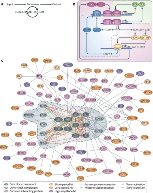
et al., 2010; Partch et al., 2014). At the cellular level, circadian rhythms are generated by interlocked transcriptional/translational feedback loops consisting of‘clock’ genes and their protein products (reviewed in Zhang & Kay, 2010; Hastings et al., 2014; Partch et al., 2014) (Fig. 1). In mammals, the core feedback loop consists of two transcriptional activators, CLOCK and BMAL1, and two transcriptional repressors, the PERIOD (PER) and CRYPTO-CHROME (CRY) proteins (Huang et al., 2012). In the morning, CLOCK and BMAL1 activate transcription of the Per (Per1, Per2 and Per3) and Cry (Cry1 and Cry2) genes by binding to the E-box (CACGTG) domain on their gene promoters. Over the course of the
day, the Per and Cry genes are translated into their respective pro-teins. PER and CRY proteins form heterodimers late in the day that translocate from the cytoplasm to the cell nucleus to inhibit CLOCK:BMAL1-mediated transcription. The timing of nuclear entry is balanced by regulatory kinases that phosphorylate the PER and CRY proteins, leading to their degradation (Lowrey et al., 2000; Shanware et al., 2011). REV-ERBa/ROR-binding elements (Preitner et al., 2002) act to regulate Bmal1 transcription via a sec-ondary feedback loop. The transcriptional retinoid-related orphan receptor (ROR) is a transcriptional activator of Bmal1, whereas REV-ERBa, an orphan nuclear receptor, negatively regulates Bmal1.
a
c
b
Fig. 1. A decade ago, our understanding of the pacemaker was limited to a single ‘core’ loop, in which the transcriptional activators circadian locomotor out-put cycles kaout-put (CLOCK) and brain and muscle ARNT-like 1 (BMAL1) dimerize and induce the expression of repressors PER and CRY, which feedback into the loop to inhibit their own expression. (b) During thefirst half of the past decade, a triple interlocking loops model was developed, including the PER–CRY loop, which is the primary loop, and the ROR–REV–ERB-associated loop and D-site of albumin promoter-binding protein (DBP)–E4 promoter-binding protein 4 (E4BP4)-associated loop. In addition to the primary loop, which was previously recognized as the core loop (a), transcriptional regulation of CLOCK and BMAL1 is controlled by ROR transcriptional activators and the dimeric REV–ERB repressors [by binding to REV response element (RRE)], the expression of which is governed by CLOCK–BMAL1 activity (through binding to E-box-containing DNA elements). Furthermore, the transcriptional activator DBP (the expression of which is controlled by an E-box) and E4BP4 (the expression of which is controlled by RRE) synergistically regulate the expression of D-box-con-taining genes, including PER. (c) Hundreds of clock modifiers have now been identified through a genome-wide screen, some of which directly or indirectly associate with known clock components. Genes that affect clock amplitude or period are shown in different colors. The dashed line circle indicates previously known clock genes as shown in the triple interlocking loops (b). Reproduced from Zhang & Kay (2010), with permission (Nature Publishing Group).
The same CLOCK:BMAL1 mechanism controlling Per and Cry gene transcription also controls transcription of REV-ERBa. This secondary feedback loop produces rhythmic expression of BMAL1, further stabilizing the clockwork. The clockwork at the cellular level is functionally similar across taxa, with interacting transcription/ translation feedback loops driving rhythms at the cellular level. Importantly, clock genes themselves are not conserved across higher taxa, but transcriptional feedback loops and post-transcriptional con-trols are common mechanisms for the generation of cell-based oscil-lation (reviewed in Harmer et al., 2001).
Circadian rhythms and temporal niche
Circadian oscillation is key to understanding how organisms are synchronized to their local environments, and species-typical adapta-tions to their temporal niches are markedly influenced by environ-mental LD cycles (reviewed in Hut et al., 2012). As noted above, in mammals, photic input from the retina entrains the SCN, but some-what surprisingly, the phases of SCN electrical, metabolic and molecular rhythms, relative to the light cycle, have the same day-time peaks in diurnally and nocturnally active species (reviewed in Smale et al., 2003). As an example, rhythms of Period gene expres-sion in the SCN peak at approximately the same time of day in diurnal as in nocturnal rodents, suggesting that the phase of clock gene expression in the SCN relative to the LD cycle is conserved across mammalian groups, and implying that the signaling cascade initiating daily activity lay beyond the SCN. This phenomenon has piqued the interest of investigators, especially because there is sig-nificant evidence that switching of temporal niches can occur (Mro-sovsky & Hattar, 2005; Gattermann et al., 2008). It appears that neural responses to light can mediate acute temporal-niche switch-ing. Thus, a switch from nocturnal to diurnal activity rhythms occurs in wild-type mice transferred from standard intensity to sco-topic levels of light in an LD cycle (Doyle et al., 2008). A similar switch from nocturnal to diurnal activity rhythms occurs in double-knockout mice, bearing little rod function, due to a lack of the inner-retinal photopigment melanopsin (OPN4) and of RPE65, a key protein used in retinal chromophore recycling. Interestingly, the rhythm of clock gene expression in the SCN is also reversed in these mice, suggesting that the nocturnal to diurnal switch is due to a change in the neural response to light upstream from the SCN. It is possible that a common mechanism produces phase reversals in Rpe65 / ;Opn4 / mice and in wild-type mice during dim LD cycles.
Ubiquity of circadian oscillators
The identification of ‘clock genes’ and the invention of reporter gene technology enabled the assessment of rhythmicity in cultured cells and tissues, such as SCN slice preparations. The technical developments and experimental findings based on assessing the activities of specific genes and proteins within cells and tissues has led to a reconceptualization of the circadian organization as a hierar-chy of oscillators. This vision has brought the circadian timing sys-tem to the attention of a very broad clinical and basic research community. Ignoring circadian effects leads to errors of interpreta-tion in basic research and can result in suboptimal diagnosis and treatments in medicine. Circadian clocks regulate the timing of gene expression in each organ, and the regulated genes are unique to each organ (Akhtar et al., 2002; Duffield et al., 2002; Miller et al., 2007; Hughes et al., 2009; Dibner et al., 2010). Thus, circadian control overlies the normal expression of tissue-specific genes and proteins.
Not surprisingly, the maintenance of normal phase relationships among tissues and organs appears to be adaptive. Disrupting the cir-cadian network can produce severe pathology (Litinski et al., 2009; Karatsoreos et al., 2011). Optimizing the circadian timing system for treatment, such as appropriately timing drug administration is a frontier research area (Levi & Schibler, 2007; and see below).
Hierarchical organization of the circadian timing system Since the discovery of the SCN, and the consistentfinding that most circadian rhythms are abolished following its destruction, it was generally assumed that the SCN was the only locus capable of inde-pendent circadian rhythm generation. In turn, all circadian rhythms throughout the brain and body were thought to be driven by down-stream communication from the SCN. This notion was challenged following the observation that culturedfibroblasts exhibit circadian rhythms in gene expression following a serum shock (Balsalobre et al., 1998). With this experiment, it became clear that the ability to oscillate was a general property of tissues throughout the central nervous system and periphery (Damiola et al., 2000; Yamazaki et al., 2000; Yoo et al., 2004). The discovery that the SCN is not alone in the capacity to express endogenous oscillation was the beginning of a reconceptualization of the internal timekeeping system (Balsalobre et al., 1998).
It is now known that the circadian system is composed of multi-ple individual cellular oscillators located throughout the body and most of its organs and glands. For example, a role for intrinsic rhythmicity in other tissues has been demonstrated. An example of SCN-independent timekeeping is seen in the olfactory bulb (Grana-dos-Fuentes et al., 2006). SCN lesions eliminate circadian locomo-tor rhythms, but not odor-induced c-Fos rhythms in the olfaclocomo-tory bulb or piriform cortex. Olfactory bulb oscillators drive rhythms in spontaneous and odor-evoked activity within the bulb and also in its primary synaptic targets in the piriform cortex. In the sense that olfactory bulb oscillators express circadian rhythms in the absence of the SCN, persist in constant darkness and are required for rhythms in the piriform cortex, these oscillators can be considered master circadian pacemakers in the olfactory system. That said, in the intact animal, under unperturbed conditions, the SCN sets the phase of the olfactory bulb and other independent oscillators. The SCN modulates temporal activity in these cellular oscillators, such that each bears regulated phase relationships to SCN pacemakers and hence to each other. Suchfindings led to the interpretation that the circadian clock mechanism modulates the activity of genes in a tissue-specific manner (Akhtar et al., 2002; Duffield et al., 2002; Miller et al., 2007; Silver & Lesauter, 2008; Hughes et al., 2009). This process can be accomplished either directly by CLOCK: BMAL1 activation through an E-box domain on their gene promot-ers (i.e. clock-controlled genes) or indirectly via downstream actions of clock-controlled gene products to optimize system-wide function-ing on a daily schedule (Fig. 2). For example, the thrombomodulin, a cofactor for thrombin that is expressed on the surface of endothe-lial cells to reduce blood coagulation, gene contains an E-box domain in its promoter and is directly regulated by the CLOCK: BMAL1 complex (Takeda et al., 2007). The resulting rhythm in thrombomodulin probably contributes to daily changes in the likeli-hood of cardiovascular events. Generally, the risk of cardiovascular events peaks in the morning and evening; the morning time point is associated with a daily peak in rhythmic cortisol and epinephrine, whereas the night-time peak is associated with peak blood pressure and a trough in cardiac vagal modulation (Scheer et al., 2010). These broad implications expanded the audience of investigators
and disciplines attending to the workings of the circadian timing system. Not only were the salient phenomena, such as sleep–wake cycles, of immediate interest, but also the invisible circadian oscilla-tions such as seen in the workings of the heart, or in the timing of cell division. Finally, the occurrence of sex differences in circadian rhythms (Bailey & Silver, 2013) and the demonstration of diseases associated with altered clock gene function rendered it necessary to consider the circadian timing system in a broad array of apparently unrelated disciplines, both applied and basic.
Unique features of the suprachiasmatic nucleus master clock Thefinding of extra-SCN oscillators begged the question of the rela-tionship of the brain master clock to these other clocks. What was the unique aspect of the SCN that lent it master clock function? Important discoveries included thefinding that the cellular/molecular mechanism of the clock was similar in the SCN and in other tissues (Storch et al., 2002; Kamphuis et al., 2005; Liu et al., 2007; Miller et al., 2007). Thus, clock functioning in cells outside the SCN is equally vulnerable to disruption of the molecular clock (Liu et al., 2007). Although the intracellular core clock molecular mechanisms could not explain SCN master clock function, the unique pattern of its connections appeared to be responsible, i.e. the coupling of its neurons appeared to lend stability to the oscillation of the SCN tis-sue, and its unique inputs and outputs appeared to be the basis of its
capacity to function as a master clock (Liu et al., 2007; Welsh et al., 2010; Hastings et al., 2014). Unlike the SCN, rhythmic clock gene expression in other central and peripheral tissues dampens within a few days in culture, suggesting a loss of coupling among oscillators that results in an inability to detect population-wide rhythmicity (Balsalobre et al., 1998; Abe et al., 2002; Wilsbacher et al., 2002). Indeed, this notion was confirmed by monitoring the single-cell bioluminescence of Per2::luciferase in mouse cultured fi-broblasts and establishing that, despite the loss of population-wide rhythms in clock gene expression, single cells continued to show clear rhythms in Per2 expression (Welsh et al., 2004; Leise et al., 2012). These findings suggest that, in vivo, coherence among popu-lations of subordinate oscillators is maintained through SCN com-munication. Also dramatic is the observation that, although SCN lesions abolish most circadian responses, some rhythms survive the ablation of the SCN. For example, SCN-lesioned animals continue to show circadian rhythms when treated with methamphetamine (Honma & Honma, 2009) and they also continue to show food anticipatory behavior, a response based on circadian timing (Saper, 2006; Patton & Mistlberger, 2013). These latterfindings suggest that the methamphetamine-entrainable oscillator and the food-entrainable oscillator might share network coupling properties in common with the SCN.
Although cellular oscillators are virtually ubiquitous, the SCN is unique not only in terms of its ability to maintain rhythmic net-work-level stability, but also in its direct access to timing informa-tion. The SCN receives light information through a direct retino-hypothalamic tract to synchronize the master clock to environmental time (Morin & Allen, 2006). Historically, it was believed that the only photoreceptors present in the retina were rods and cones. This notion was questioned following thefinding that mice lacking both rod and cone photoreceptors (retinally degenerate mice) exhibit nor-mal photic entrainment despite being visually blind (Foster et al., 1993). By using a retrograde labeling strategy to identify retinal cells projecting to the SCN, it was discovered that a subset of retinal ganglion cells, expressing the photopigment melanopsin, were intrin-sically photosensitive (Berson et al., 2002; Hannibal & Fahrenkrug, 2002; Hattar et al., 2002; Panda et al., 2002). As with the elimina-tion of rod/cone signaling, eliminaelimina-tion of melanopsin was not suffi-cient to abolish entrainment (Ruby et al., 2002; Lucas et al., 2003). Entrainment is only fully prevented in mice doubly mutant for both melanopsin and traditional rod/cone photoreceptors (Hattar et al., 2003; Panda et al., 2003). Underscoring the importance of connec-tivity, even though all photoreceptive classes can contribute to entrainment, this occurs through the conduit of the intrinsically pho-tosensitive retinal ganglion cells; ablating these cells alone (only ~2% of all retinal ganglion cells) prevents entrainment (Schmidt et al., 2011). Together, thesefindings suggest that rod/cone photore-ceptors project to intrinsically photosensitive retinal ganglion cells that then send projections to the SCN to communicate this inte-grated light information. Because subordinate oscillators do not have access to light information, their phase relative to external time must be maintained through communication from the master clock in the SCN under light-entrained conditions.
Suprachiasmatic nucleus communication to central and peripheral targets
As indicated previously, temporal harmony is maintained among systems through SCN communication to central and peripheral tar-gets. This coordination is essential for optimizing the timing of behavioral and physiological events and maximizing health. The
Fig. 2. The hierarchical organization of the circadian system. The circadian system is a hierarchy of similar cellular clock elements. The master clock of the SCN achieves the‘master’ role by virtue of the nucleus’s internal circuitry and its ability to send timing signals to the rest of the brain. The SCN itself is entrained to solar time and also receives information from arousal-related activity of the body. Thus, the SCN integrates external and internal informa-tion, and coordinates the rhythmic activities of peripheral oscillators to pro-duce coherent timing in behavioral, autonomic, and endocrine functions. Reproduced from Silver & Lesauter (2008), with permission (John Wiley and Sons).
SCN sets the phase relationship among various tissues via monosyn-aptic neural targets, projections via the autonomic nervous system, systemic hormone secretions, behavioral cycles of feeding and activ-ity and the rhythmic alterations of body temperature (Kriegsfeld & Silver, 2006; Refinetti, 2010; Kalsbeek et al., 2011; Mavroudis et al., 2012; Patton & Mistlberger, 2013; Sladek & Sumova, 2013). The following section provides a brief overview of the specific means by which information is communicated from the master clock to target systems and considers the implications for physiological and behavioral outcomes.
Neural suprachiasmatic nucleus communication
Prior to the advent of viral tract-tracing techniques, monosynaptic anterograde and retrograde tracers were used to explore the connec-tivity of the SCN to central targets (Stephan et al., 1981; Watts & Swanson, 1987; Watts et al., 1987; Kalsbeek et al., 1993; Morin et al., 1994; Leak & Moore, 2001; Kriegsfeld et al., 2004). These studies revealed extensive monosynaptic projections proceeding ros-trally to the septum and bed nucleus of the stria terminalis, rosros-trally and dorsally to the thalamus, rostrally and laterally throughout the hypothalamus, and caudally to the posterior paraventricular thala-mus, precommissural nucleus and olivary pretectal nucleus. Given these widespread projections, it is likely that the SCN is in a posi-tion to communicate with the entire brain through secondary or ter-tiary synapses originating from these primary target loci. In addition, many of these target loci contain neuroendocrine cells and hypothalamic releasing factors that can be secreted into the cerebro-spinal fluid or general circulation, providing pervasive communica-tion to systems throughout the brain and body (van der Beek et al., 1993; Vrang et al., 1995; Kalsbeek et al., 1996; Horvath, 1997; Van der Beek et al., 1997; Buijs et al., 1998; Horvath et al., 1998; Gerhold et al., 2001).
In addition to tract-tracing strategies to reveal SCN outputs, there have been a number of studies to exploit novel behavioral patterns that have been found to correlate with altered SCN rhythms. For example, hamsters will spontaneously ‘split’ and exhibit two rest– activity cycles each day instead of one when housed in constant light. In a classic study, de la Iglesia et al. (2003) showed that, in ‘split’ hamsters, the right and left SCN oscillate out of phase with each other, with each SCN’s molecular rhythms in phase with only one of the two daily peaks of activity. Likewise, examination of Per1::GFP expression in cultured SCNs from split mice shows anti-phase oscillations that can be monitored for several cycles (Ohta et al., 2005). Subsequent work using this split model revealed that, rather than a simple right–left split, each SCN splits into two com-partments that oscillate in antiphase (Tavakoli-Nezhad & Schwartz, 2005; Yan et al., 2005). This four-way split means that the split hamsters’ SCNs exhibit 24 h rhythms of PER1 protein that cycles in antiphase between the left and right sides and between core and shell subregions. Associated with this SCN oscillation is a 12 h rhythm of FOS expression in brain regions that receive SCN efferents (Butler et al., 2012). In the target regions examined (medial preoptic area, paraventricular nucleus of the hypothalamus, dorsomedial hypothala-mus and orexin-A neurons), the oscillations were in-phase between hemispheres (unlike in the SCN), although with detectable right–left differences in amplitude. Importantly, in all three conditions studied (split and unsplit hamsters in constant light, and control hamsters in LD cycles), the timing of FOS expression in targets occurred at the same time of day and always occurred at a common phase reference point of the SCN oscillation, suggesting that, at a specific internal phase, each SCN signals these targets once daily.
In addition to communication via direct projections to neural loci, the SCN also sends multisynaptic connections, via the autonomic nervous system, to targets in the periphery, setting the phase of sub-ordinate oscillatory systems and controlling their activity. By apply-ing transynaptic, retrograde viral tracers, such as a pseudo rabies virus, to various organs and glands, precise multisynaptic connec-tions from the SCN to the periphery have been established. Early studies employing this technique established that corticosterone secretion is controlled by direct projections to the adrenal gland (Buijs et al., 1999), lipid mobilization via projections to adipose tissue (Bamshad et al., 1998; Bartness & Bamshad, 1998; Bartness et al., 2001; Demas & Bartness, 2001), ovarian function (Gerendai et al., 1998, 2000; Gerendai & Halasz, 2000), and thyroid function (Kals-beek et al., 2000). It is likely that neural connections from the SCN to peripheral glands and organs may be universal for all targets in the body.
Given the technical limitations of these tracers, it is not surprising that several questions still remain. For example, does the SCN employ the same cell phenotype(s) to communicate to all organs and glands? Does the SCN communicate with one neurochemical mediator, or a combination of neurochemical mediators, to set the phase of subordinate oscillators? Is sympathetic and parasympathetic control of peripheral tissues controlled by the same SCN cell pheno-types? Technical innovations now permit an assessment of projec-tions from specific neuropeptidergic cell phenotypes using viral tracers driven by specific gene promoters. By applying these tools to the SCN, important insight can be gained into the specific modalities by which the SCN communicates to central and peripheral targets.
Diffusible signals
In addition to monosynaptic and multisynaptic neural projections, several early lines of evidence suggested that a diffusible signal from the SCN can sustain behavioral rhythmicity. First, in early studies of SCN-lesioned hamsters, locomotor rhythmicity and rhyth-mic gnawing behavior are restored following grafting of fetal SCN tissue into the third ventricle of the lesioned host (Lehman et al., 1987, 1995; Ralph et al., 1990; LeSauter & Silver, 1994). Postmor-tem analysis indicated that few connections were made between the graft and the host brain, suggesting that the re-establishment of rhythmic behavior did not result from the restoration of SCN projec-tions (Aguilar-Roblero et al., 1994; Lehman et al., 1995). Further-more, when an‘SCN island’ is created with a Halasz knife, animals recover free-running rhythms, even though efferent fibers from the SCN have been severed (Inouye & Kawamura, 1979). Although it is possible that efferentfibers may have grown across the knife cut to form correct synaptic connections, there is no evidence of such plasticity in the mammalian brain.
More direct evidence for the existence of a diffusible SCN signal was gained by transplanting SCN grafts encapsulated in a semi-por-ous membrane that permitted diffusible, but not neural, outflow into an SCN-lesioned host (Silver et al., 1996). In cases with viable grafts, circadian locomotor rhythms were restored with the period of the donor animal. These results demonstrate that the transplanted biological clock can regulate rhythmicity by means of a diffusible signal. Whether or not such a diffusible signal drives behavioral rhythms under natural conditions has been a more challenging ques-tion. Several candidate diffusible signals have been investigated since these initial findings, including prokineticin-2 (Cheng et al., 2002), transforming growth factor-alpha, and cardiotrophin-like cytokine (Kramer et al., 2001; Cheng et al., 2002; Kraves & Weitz, 2006; Li et al., 2006, 2012). All three proteins are expressed
rhythmically in the SCN and their receptors are present in major SCN targets or around the third ventricle. The administration of prokineticin-2 and transforming growth factor-alpha during the night (when levels are typically low) inhibits wheel-running behavior, whereas the administration of cardiotrophin-like cytokine antibody during the day (when levels are typically low) leads to increased daytime locomotor activity. Interestingly, in contrast to behavioral rhythms, endocrine rhythms require neural output (Silver et al., 1996 Nunez & Stephan, 1977; Meyer-Bernstein et al., 1999). It has also been demonstrated that diffusible signals are sufficient to pro-duce oscillations in the SCN of non-rhythmic SCNs from mutant animals. Thus, using a coculture technique in which a wild-type SCN graft was used to examine the restoration of rhythmicity in non-rhythmic mutant SCN, it was demonstrated that paracrine sig-nals, involving vasoactive intestinal polypeptide, arginine vasopres-sin and gastrin-releavasopres-sing peptide, were sufficient to restore cellular synchrony and oscillation amplitude (Maywood et al., 2011). Like-wise, in Cry double-knockout [Cry1( / )/Cry2( / )] mice, circa-dian rhythms are synchronized in neonates but not in adults, indicating a loss of rhythm synchrony in the course of development. Whether a diffusible factor(s) in the SCN contributes to the coupling of cellular circadian rhythms was investigated by coculture of a non-bioluminescent SCN slice with a bioluminescent (PER2::lucifer-ase) SCN slice. Synchronized circadian rhythms in adult Cry1 ( / )/Cry2( / ) SCN were restored by coculture of neonatal, but not of juvenile, SCN. The results indicate that the neonatal SCN produces a diffusible signal that supports the development of inter-cellular networks that subserve coherent rhythm expression in adult SCN (Ono et al., 2013).
Implications and consequences for the study of normal physiology and behavior
In order to maximize survival and reproductive success, animals restrict their behavior to optimal times of the day or night, and the cir-cadian system is crucial for this temporal organization. In the absence of a functional circadian system, survival and reproduction are com-promised. For example, chipmunks in the Allegheny Mountains were more vulnerable to predation following SCN lesions, presumably due to inappropriate night-time restlessness revealing their location to pre-dators (DeCoursey et al., 2000). In addition, in most spontaneously ovulating female rodents, the SCN is essential for ovulation and sex-ual behavior (Kriegsfeld & Silver, 2006; Christian & Moenter, 2010; Tolson & Chappell, 2012). In women, disruptions to circadian timing through shift work or jet lag also have pronounced negative conse-quences for pregnancy and its maintenance (Mahoney, 2010). In addi-tion to affecting reproducaddi-tion, the circadian system impacts virtually every behavioral or physiological system studied to date, and several well-studied examples are presented below.
Feeding and circadian rhythms
The circadian system coordinates metabolism and food intake to optimize feeding and with daily changes in digestion and nutrient absorption (Tahara & Shibata, 2013). Mice with a mutation of the Clock gene, for example, have greatly reduced daily rhythms in feeding that lead to hyperphagia and obesity associated with ele-vated lipids, leptin and glucose, and low insulin levels (Turek et al., 2005). Likewise, high-fat-diet-induced obesity can be abrogated by treatment with a Rev-erb agonist, reducing body fat and hyperglyce-mia (Solt et al., 2012). Interestingly, the impact of circadian disrup-tion on obesity occurs at the level of fat cells; site-specific deledisrup-tion
of Bmal1 in mouse adipocytes leads to increased daytime feeding and body mass, reduced locomotor activity and decreased circulating levels of polyunsaturated fatty acids (Paschos et al., 2012). Recent findings in humans indicate that sleep deprivation results in an increased desire for high-caloric foods, and decreased frontal and insular cortex activity and increased amygdala activity, as assessed by functional magnetic resonance imaging (Greer et al., 2013). Thus, the extent to which circadian disruptions lead to obesity through disturbances to sleep represents an important opportunity for further enquiry.
Circadian disruptions can arise from exposure to inappropriate photic conditions. Exposure to dim (5 lux) light at night leads to increased alterations in daily feeding and body mass along with reduced rhythms of hypothalamic and liver clock gene expression in mice (Fonken et al., 2013). The adverse impact of dim light at night on metabolism, such as the dim red or white light used for animal maintenance, can be ameliorated through wheel-running exercise or subsequent exposure to dark at night (Fonken et al., 2014). There has been substantial interest in the effect of light intensity and wave-length on metabolic and other responses. In studies examining light, effects controlling intensity, wavelength, and photoreceptor absorp-tion spectra are taken into account. When wavelength is a quesabsorp-tion of interest, then irradiance (incident power, in W/m2), rather than illuminance (luminous flux, in lux), is assessed. Measures of lux provide a useful approximate mark that can ground a reader, but it is a measure of perceived intensity by humans, a psychophysical number comprising both the photoreceptor absorption of light and the cognitive processing of that light. Because humans have a red-sensitive cone, red that is perceived to be as bright as a reference blue light (equal lux) would be much dimmer compared with the blue to a mouse’s eye that lacks a red cone. Photosensitivity to dif-ferent wavelengths is therefore better investigated with light sources providing equal photon fluxes (photosensitivity will still be wave-length dependent, but this will be a measure of photoreceptor func-tion and therefore transmission to the brain); a cell is agnostic to wavelength once the photon has been absorbed. Light-emitting diodes with narrow spectral emissions or notchfilters facilitate these investigations. Practicalities may force a reliance on incandescent andfluorescent lights but, because of their complex spectra, compar-ing light of different colors is more difficult. Illuminance measures suffice when wavelength per se is not a central focus.
The photosensitivity of a physiological or behavioral response to light depends on what is being measured. This is important as the photosensitivity of one response cannot be generalized to other func-tions. As an example, measurements were made of the thresholds of entrainment of wheel-running rhythms at three wavelengths, and these were compared with the thresholds of two other non-image-forming visual system functions, i.e. masking and the pupillary light reflex. Dim light that entrained mice failed to elicit either masking or pupillary light reflex; in general, circadian entrainment is more sensitive by 1–2 log units than other measures of the non-image-forming visual system. In an artificial photic environment, dim light can entrain circadian rhythms even when it fails to produce more easily measurable acute responses to light such as phase shifting and melatonin suppression (Butler & Silver, 2011).
As mentioned previously, not only does the circadian system influence feeding and metabolism, but food cues can also act to entrain circadian rhythms (Saper, 2006; Patton & Mistlberger, 2013). If food presentation is restricted to a short temporal window (typically a few hours), animals exhibit increased activity in antici-pation of feeding [food anticipatory activity (FAA)]. Because this synchronization of behavior with feeding persists in the absence of
the SCN, a separate designation of the food entrainable oscillator was coined (Stephan et al., 1979). The identification of the neural locus of the food-entrainable oscillator has been challenging. The dorsomedial nucleus of the hypothalamus (DMH) probably plays a role in food entrainment (Gooley et al., 2006; Fuller et al., 2008), although mice and rats can entrain to food cues in the absence of a DMH (Landry et al., 2006, 2007; Acosta-Galvan et al., 2011). In mice, DMH lesions lead to reduced FAA, whereas lesions of both the SCN and DMH result in enhanced FAA (Acosta-Galvan et al., 2011). These findings suggest that the DMH participates in FAA, but is not the sole neural locus of the food-entrainable oscillator. It is likely that metabolic cues from the periphery, communicated to the central nervous system, participate in food entrainment. For example, ghrelin cells in the stomach that signal hunger express clock genes, ghrelin administration leads to increased activity in ani-mals fed ad libitum, and ghrelin and clock gene rhythms in these stomach cells are synchronized to feeding (LeSauter et al., 2009). Consistent with these findings, FAA is greatly reduced in ghrelin receptor knockout mice (Blum et al., 2009; LeSauter et al., 2009). Thesefindings suggest that restricted feeding leads to entrainment of stomach clocks in ghrelin-expressing cells and food-entrained ghre-lin signaghre-ling feeds back to the central nervous system to drive changes in FAA. Other neuronal systems including the hypocretin arousal system (Akiyama et al., 2004; Mieda et al., 2004) and orex-ogenic melanocortin system (Sutton et al., 2008; Patton & Mistlber-ger, 2013) have been implicated in food entrainment, with disruptions to either system causing pronounced deficits in FAA. Sleep–wakefulness and circadian rhythms
Most salient in daily life is the relationship between sleep and circa-dian rhythmicity. Associated with the timing of the rest–activity
cycles are rhythms in alertness/drowsiness, mood, and other behav-iors. These cycles, and the processes that they impact, are an immense and fundamentally important topic, with much work in basic, clinical and pharmacological aspects (reviewed in Murray & Harvey, 2010; Harvey, 2011; Krystal et al., 2013; Saper & Sehgal, 2013). Although the details of sleep–circadian relationships are beyond the scope of this review, we highlight some major aspects.
The relationship between circadian clocks and sleep involves two interacting processes, and is captured in the classical opponent pro-cess model of Borbely (1982). The homeostatic component of sleep involves a process whereby the sleep pressure (termed process S) increases the longer that an individual is awake. The neural locus regulating this homeostatic pressure is not well defined, and involves multiple brain regions and transmitters (see below). In con-trast, the circadian system that regulates the timing of wakefulness and sleep has its well-characterized anatomical locus in the SCN. The SCN has monosynaptic efferents to a number of nearby hypo-thalamic regions (reviewed in Morin, 2013), and these in turn relay information to a large number of brain regions, including those involved in regulating awake and sleep states.
Sleep circuits
The neural circuits involved in sleep and arousal include the basal forebrain, brainstem, and hypothalamic components. The SCN has relatively few direct outputs to sleep–wake regulatory systems. Most of its output projects to nearby hypothalamic regions that relay sig-nals to sleep and wake regulatory regions. The sleep circuits are comprised of numerous projections of neurons releasing different types of neurotransmitters and neuropeptides (reviewed in Saper et al., 2005) (Fig. 3). Briefly, arousal pathways include cholinergic neurons of the ascending arousal pathway, located in the
Fig. 3. (Left) A schematic drawing showing some key components of the ascending arousal system. A major input to the relay and reticular nuclei of the thalamus (yellow pathway) originates from ACh cell groups in the upper pons, the PPT and LDT. These inputs facilitate thalamocortical transmission. A second pathway (red) activates the cerebral cortex to facilitate the processing of inputs from the thalamus. This arises from neurons in the monoaminergic cell groups, including the tuberomammillary nucleus (TMN) containing histamine (His), the A10 cell group containing DA, the dorsal and median raphe nuclei containing serotonin (5-HT), and the LC containing NA. This pathway also receives contributions from peptidergic neurons in the LH containing ORX or MCH, and from BF neurons that con-tainc-aminobutyric acid (GABA) or ACh. Note that all of these ascending pathways traverse the region at the junction of the brainstem and forebrain where von Ec-onomo noted that lesions caused profound sleepiness. (Right) A schematic drawing to show the key projections of the VLPO to the main components of the ascending arousal system. It includes the monoaminergic cell groups (red), such as the TMN, the A10 cell group, the raphe cell groups and the LC. It also innervates neurons in the LHA (green), including the PeF ORX neurons, and interneurons in the ACh cell groups (yellow), the PPT and LDT. Note that the VLPO neurons lie within the region outlined by von Economo for the anterior hypothalamic lesion that caused insomnia. ACh, cholinergic; BF, basal forebrain; DA, dopamine; Gal, galanin; LC, locus coeruleus; LDT, laterodorsal tegmental nuclei; LH, lateral hypothalamus; MCH, melanin-concentrating hormone; NA, noradrenaline; ORX, orexin; PAG, periaqueductal gray; PeF, perifornical; PPT, pedunculopontine. Adapted from Saper et al. (2005), with permission (Nature Publishing Group).
pedunculopontine and laterodorsal tegmental nucleus, serotoninergic neurons in the dorsal raphe nucleus, noradrenergic neurons in the locus coeruleus, dopaminergic neurons in the median raphe nucleus, and histaminergic neurons in the tuberomammillary nucleus of the hypothalamus. Activity in these neurons promotes alertness and cor-tical arousal. This system is inhibited byc-aminobutyric acid neu-rons from the ventrolateral preoptic nucleus (VLPO) during sleep. Specifically, the locus coeruleus provides norepinephrine-mediated inhibition of the VLPO during wakefulness. During sleep, activity in the ventral and median preoptic nuclei inhibits the ascending arousal system via the inhibitory neurotransmitters c-aminobutyric acid and galanin. The sleep-regulatory cells in the VLPO are directly and indirectly regulated by SCN input (Novak & Nunez, 2000; Chou et al., 2002; Kriegsfeld et al., 2004). This circuit enables master clock interactions with homeostatic sleep regulatory systems. Among the most important known homeostatic sleep fac-tors is adenosine (reviewed in Porkka-Heiskanen, 2013). The accu-mulation of adenosine (a powerful somnogen) during wakefulness is associated with increased VLPO activity upon increased adenosine binding. VLPO activation then inhibits the ascending arousal circuits and promotes non-rapid eye movement sleep.
Future directions
Sleep disturbances
It is beyond question that the most salient aspect of the circadian system is its role in controlling the timing of sleep. Moreover, envi-ronmental disruptions to circadian rhythms, including shift work, travel across time zones, and irregular social schedules, tend to pre-cipitate or exacerbate mood-related episodes. Using shift work or jet lag as a model experimental paradigm, numerous studies, in both humans and animals, have demonstrated the adverse effects of dis-rupted sleep–wake schedules on health (Wang et al., 2011).
Nearly all people suffering from mood disorders have significant disruptions in circadian rhythms, and altered sleep patterns are one of the major diagnostic criteria for these disorders. Importantly, sleep disorders seem to precede clinical diagnoses of mental illness, and reduction of sleep disturbances improves mental illness (Ritter et al., 2011; Pritchett et al., 2012). It appears that many pathologies are comorbid in both mental illness and neurodegenerative disease; these include poor vigilance and memory, reduced mental and phys-ical reaction times, reduced motivation, depression, insomnia, and obesity, among other states (Wulff et al., 2010). Furthermore, manipulation of the timing of sleep can ameliorate symptoms of major depressive disorder and bipolar disorder (Bunney & Bunney, 2013; Kaplan & Harvey, 2013).
Although chronic sleep deprivation or disruption exacerbates behavioral problems, and may directly affect cognitive function and mood disorders, there is also evidence of mechanistic links between circadian clocks, sleep and mental illness (reviewed in Lamont et al., 2010; Menet & Rosbash, 2011). For example, in humans, casein kinase 1 epsilon and PER2 mutations are associated with familial advanced sleep phase disorder, which causes patients to fall asleep several hours earlier than normal (Toh et al., 2001). In addi-tion, associations for polymorphisms in other circadian genes, including polymorphisms in the Cry2 locus with bipolar disorder (Shi et al., 2008; Sjoholm et al., 2010) and depression (Lavebratt et al., 2010), the Per3, Cry1, and timeless (Tim) genes with schizo-phrenia and schizoaffective disorder, and Cry1, have been suggested (Mansour et al., 2006; Lamont et al., 2007; Peng et al., 2007; Moons et al., 2011). Bmal1 and Tim are associated with bipolar
disorder or schizophrenia (Mansour et al., 2006). Finally and impressively, mistimed sleep in humans disrupts the molecular pro-cesses associated with core clock gene expression and disrupts over-all temporal organization throughout the body (Archer et al., 2014). In summary, sleep disruption is associated with a wide range of symptoms related to mental health.
Connecting circadian clocks to cellular metabolism
The current view of circadian clocks rests on a model of intracellu-lar interlocked transcriptional and translational feedback loops that generate circadian rhythms, with numerous post-translational and post-transcriptional modifications (Partch et al., 2014). This well-established landscape has started to move in a totally new direction with the discovery of numerous cytosolic circadian loops central to cellular physiology.
Several studies now point to metabolic rhythms that are indepen-dent of transcription. These studies led to a search for the ways in which the traditional transcription/translational feedback loops of clock genes and their protein products are integrated with cytosolic and metabolic components of cellular physiology. Over the years, there have been hints of the existence of circadian oscillation in the absence of transcriptional and translational feedback loops. A major breakthrough was the demonstration that circadian oscillation could be reconstituted in a test tube with a purely biochemical oscillator (Nakajima et al., 2005).
A rhythmic, post-translational modification of peroxiredoxin was first reported in mouse liver (Reddy et al., 2006). The dramatic insight came from the discovery of circadian oscillations in human red blood cells, which lack a nucleus and therefore lack the genetic clock mechanism (O’Neill & Reddy, 2011; Edgar et al., 2012). The peroxiredoxin family is part of the cellular defense against reactive oxygen species, specifically H2O2, which are an unavoidable
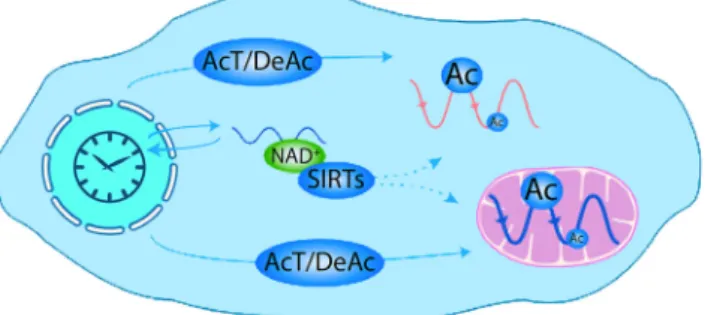
by-product of aerobic metabolism. Red blood cells express peroxiredox-in rhythms that are entraperoxiredox-inable by temperature cycles, and are tem-perature compensated. Circadian rhythms occur in the availability of nicotinamide adenine dinucleotide, a coenzyme for energy conver-sion in the cell, controlling the timing of oxidative metabolism in mammalian mitochondria (Peek et al., 2013). These data suggest that an underlying rhythmic capacity exists in the cytoplasm, not directly reliant on nascent gene expression. The implication is that, in nucleated cells, at a post-translational level, metabolic rhythms interact reciprocally with transcriptional and translational feedback loop elements known to regulate circadian timekeeping (Rey & Red-dy, 2013) (Fig. 4). Circadian cross-talk between metabolic and tran-scriptional clock components probably co-ordinates genome-wide temporal, cell type-specific programs of gene expression. In this way, circadian clocks exert regulatory control over almost every aspect of physiology, with disruptions leading to disease states, and their understanding lending opportunities for the analysis of novel mechanisms of diagnosis and treatment.
Sex differences in circadian rhythms
An aspect of the circadian regulation of genetic, metabolic and cyto-solic clocks that has received relatively little attention is how these processes might be affected by sex differences. Also, sex differences may confound the results of studies in which the sex of the animal or cell is not taken into account. Within the brain, widespread sex differences in gene expression and splicing have been detected in all major brain regions and involve 2.5% of all expressed genes (Trabz-uni et al., 2013). Furthermore, a diffusion tensor imaging study
indicates widespread sex differences in regional and global network characteristics of the brains of youths (Ingalhalikar et al., 2014). The sparsity of circadian studies may be attributed in part to the expense and work load associated with undertaking studies of both males and females, especially when ovulatory cycle-associated changes must also be taken into account (Morin et al., 1977). That said, there are tremendous sex differences in the circadian timing system (reviewed in Bailey & Silver, 2013). A salient example is seen in sleep regulation. Women go to sleep later and later until the age of around 19.5 years, whereas men continue to delay their sleep until around the age of 21 years (Roenneberg et al., 2007). Further-more, throughout adulthood, men tend to go to sleep later than women. This sex difference disappears at around the age of 50, at around the time of menopause.
A key symptom of major depressive disorder is the disruption of circadian patterns. In a study applying time-of-death analysis to gene expression data from postmortem brains, cyclic patterns of gene expression were much weaker in the brains of patients with major depressive disorders due to shifted peak timing and potentially dis-rupted phase relationships between individual circadian genes (Li et al., 2013). As noted above, sleep disturbance is associated with major depressive disorders. Turning to the question of sex differ-ences, Plante et al. (2012) found that women, but not men, with major depressive disorders demonstrate significant increases in slow wave activity in multiple cortical areas relative to control subjects. In conclusion, sex differences become important when they can pro-vide clues to the mechanisms conferring protection to one sex or susceptibility to the other, and in those research areas where sex dif-ferences are salient, attention to the underlying mechanisms is espe-cially warranted.
Circadian chronotherapeutics
It is now generally accepted that disruptions to the circadian timing system hasten the progression, or contribute to the etiology, of a number of disease states including cancer, cardiovascular disease, neural cell death, peptic ulcers, obesity, rheumatoid arthritis, bipolar disorder, depression and schizophrenia (reviewed in Golombek et al., 2013). In addition to the impact of circadian disturbances on disease, numerous studies in animal models and human clinical trials indicate that there is pronounced impact on the efficacy of a variety of treatments based on the timing of their delivery. Early work in
rats and mice, for example, provided evidence that cancer chemo-therapy was more efficacious if delivered at times of greatest drug tolerance (Halberg et al., 1980; Levi, 1987; Reinberg et al., 1987). Later, it was recognized that cancer cells exhibit daily rhythms in mitotic activity, and cytotoxic chemotherapeutic agents could be most effectively applied during peak mitotic activity, ideally when cell division is at a nadir in marrow and mucosal cells to avoid damage to healthy tissues (Ortiz-Tudela et al., 2013). Despite repeated clinical trials for a number of cancers revealing enormous increases in response rate and survivorship and decreased negative side-effects, it has been challenging to incorporate a chronotherapeu-tic strategy into oncological pracchronotherapeu-tice. Part of the challenge arises from the fact that sex, lifestyle, and genetic background influence the most appropriate time of delivery across individuals (Ortiz-Tu-dela et al., 2013). Thefinding of high-throughput, reliable circadian biomarkers for host and cancerous tissues, along with the implemen-tation of timed drug-delivery systems, is currently being explored to bring chronotherapeutic approaches to the clinic.
More recently, it has become clear that vast improvements in the efficacy of pharmacological, in addition to chemotherapeutic, agents can be gained by considering the timing of delivery. One strategy that has met with success is to administer medication at a time of greatest risk (e.g. myocardial infarction risk is greatest in the morn-ing) or at the daily peak in the manifestation of the ailment (e.g. asthma symptoms exhibit marked daily changes) (Bairy, 2013). A more effective strategy is to consider daily changes in drug pharma-codynamics and to deliver medications at a time when the drug is best tolerated and metabolism and elimination are lowest. For over 300 drugs, prominent daily changes in absorption, distribution, metabolism, and elimination have been noted (reviewed in Levi & Schibler, 2007). By considering these daily changes in pharmacoki-netics, striking increases in plasma concentrations of a drug can be achieved simply by altering the timing of administration (e.g. Ollag-nier et al., 1987; Smolensky et al., 1987; Bruguerolle, 1998) (Fig. 5). In addition to maximizing the concentration of drugs and minimizing their toxicity, drug targets exhibit daily changes that alter the response, including erythrocyte permeability (Levi et al.,
Fig. 4. Circadian acetylation in the cell. Scheme representing plausible mecha-nisms that the circadian clock might use to generate cytosolic and mitochondrial acetylation rhythms. The activity of AcT, including CLOCK, together with DeAc, might be controlled through transcriptional and post-transcriptional mech-anisms. In addition, the circadian clock may also use the nicotinamide adenine dinucleotide (NAD+)-dependent deacetylases SIRTs to modulate protein Ac, because NAD+ levels are rhythmic in the cytosol. Reproduced from Rey & Reddy (2013).© National Academy of Sciences of the United States of America. Ac, acetylation; AcT, acetyltransferases; DeAc, deacetylases; SIRTs, sirtuins.
Fig. 5. Chronopharmacokinetics of indomethacin (75 mg/h) after oral administration of a sustained release preparation; plasma concentrations of indomethacin according to the time of administration in patients. Reproduced from Bruguerolle (1998), with permission (Adis International Limited).
1987; Bruguerolle & Prat, 1989) and receptor numbers/binding affinity (Redfern, 2003). Such strategies have been effectively applied to the treatment of a variety of ailments that show marked dailyfluctuations, including hypertension (Nayak et al., 2009; Her-mida et al., 2014), asthma (Smolensky et al., 1987; Nainwal, 2012) and rheumatoid arthritis (Cutolo, 2012). Given that differences in the timing of symptoms for many conditions are similar across indi-viduals, implementing chronotherapeutic strategies for the treatment of some diseases is quite feasible and researchers and pharmaceuti-cal companies are developing strategies to effectively deliver medi-cations in a time-dependent fashion thorough time-release oral administration, implants, and pumps (reviewed in Maroni et al., 2010). Given the rapid advances in this emerging knowledge and technology, it will be important to educate the medical community in the magnitude of such effects and practical implementation of chronotherapeutic approaches.
Concluding remarks
The cells of our brains and bodies have evolved in a 24 h solar sys-tem in ways that enable optimal coordination of our internal and external circadian cycles. Transcription–translation feedback loops are modified by post-transcriptional regulatory processes, enabling a central master clock to signal peripheral clocks that then exert local control of cellular function specific to each organ and gland. Making optimal use of circadian timing mechanisms within specific brain regions and tissues will enable the understanding of interindividual differences and development of pharmacological modulators of cir-cadian timing identified from high-throughput screens. The hope is that the robustness and resilience of circadian oscillation can be enhanced, dysfunctional clocks can be repaired, and personalized treatment regimens developed for age-related declines and treatment of disease. Further information on mechanisms whereby the SCN signals rhythmic gene expression in the rest of the brain and body requires new genetic, mathematical and statistical tools to under-stand the spatial and temporal changes in the circadian timing system that underlie its normal and disrupted neural function.
Acknowledgements
We thank Dr Matthew Butler and unidentified reviewers for their comments on earlier drafts of this article. Support during the writing of this review and research from our laboratories reported herein was provided by NSF 1256105 and NIH NS37919 (R.S.), and NIH HD050470 and NSF IOS-1257638 (L.J.K.).
Abbreviations
Cry, cryptochrome; DMH, dorsomedial hypothalamus; FAA, food anticipa-tory activity; LD, light:dark; Per, Period; ROR, retinoid-related orphan recep-tor; SCN, suprachiasmatic nucleus; VLPO, ventrolateral preoptic nucleus.
References
Abe, M., Herzog, E.D., Yamazaki, S., Straume, M., Tei, H., Sakaki, Y., Me-naker, M. & Block, G.D. (2002) Circadian rhythms in isolated brain regions. J. Neurosci.,22, 350–356.
Acosta-Galvan, G., Yi, C.X., van der Vliet, J., Jhamandas, J.H., Panula, P., Angeles-Castellanos, M., Del Carmen Basualdo, M., Escobar, C. & Buijs, R.M. (2011) Interaction between hypothalamic dorsomedial nucleus and the suprachiasmatic nucleus determines intensity of food anticipatory behavior. Proc. Natl. Acad. Sci. USA,108, 5813–5818.
Aguilar-Roblero, R., Morin, L.P. & Moore, R.Y. (1994) Morphological cor-relates of circadian rhythm restoration induced by transplantation of the suprachiasmatic nucleus in hamsters. Exp. Neurol.,130, 250–260.
Akhtar, R.A., Reddy, A.B., Maywood, E.S., Clayton, J.D., King, V.M., Smith, A.G., Gant, T.W., Hastings, M.H. & Kyriacou, C.P. (2002) Circa-dian cycling of the mouse liver transcriptome, as revealed by cDNA microarray, is driven by the suprachiasmatic nucleus. Curr. Biol.,12, 540– 550.
Akiyama, M., Yuasa, T., Hayasaka, N., Horikawa, K., Sakurai, T. & Shibata, S. (2004) Reduced food anticipatory activity in genetically orexin (hypo-cretin) neuron-ablated mice. Eur. J. Neurosci.,20, 3054–3062.
Archer, S.N., Laing, E.E., Moller-Levet, C.S., van der Veen, D.R., Bucca, G., Lazar, A.S., Santhi, N., Slak, A., Kabiljo, R., von Schantz, M., Smith, C.P. & Dijk, D.J. (2014) From the Cover: mistimed sleep disrupts circa-dian regulation of the human transcriptome. Proc. Natl. Acad. Sci. USA, 111, E682–E691.
Bailey, M. & Silver, R. (2013) Sex differences in circadian timing systems: implications for disease. Front. Neuroendocrinol.,35, 111–139.
Bairy, L.K. (2013) Chronotherapeutics: a hype or future of chronopharmacol-ogy? Indian J. Pharmacol.,45, 545–546.
Balsalobre, A., Damiola, F. & Schibler, U. (1998) A serum shock induces circadian gene expression in mammalian tissue culture cells. Cell, 93, 929–937.
Bamshad, M., Aoki, V.T., Adkison, M.G., Warren, W.S. & Bartness, T.J. (1998) Central nervous system origins of the sympathetic nervous system outflow to white adipose tissue. Am. J. Physiol., 275, R291–R299. Bartness, T.J. & Bamshad, M. (1998) Innervation of mammalian white
adi-pose tissue: implications for the regulation of total body fat. Am. J. Phys-iol.,275, R1399–R1411.
Bartness, T.J., Song, C.K. & Demas, G.E. (2001) SCN efferents to periph-eral tissues: implications for biological rhythms. J. Biol. Rhythm.,16, 196– 204.
van der Beek, E.M., Wiegant, V.M., van der Donk, H.A., van den Hurk, R. & Buijs, R.M. (1993) Lesions of the suprachiasmatic nucleus indicate the presence of a direct vasoactive intestinal polypeptide-containing projection to gonadotrophin-releasing hormone neurons in the female rat. J. Neuroen-docrinol.,5, 137–144.
Berson, D.M., Dunn, F.A. & Takao, M. (2002) Phototransduction by retinal ganglion cells that set the circadian clock. Science,295, 1070–1073. Blum, I.D., Patterson, Z., Khazall, R., Lamont, E.W., Sleeman, M.W.,
Horv-ath, T.L. & Abizaid, A. (2009) Reduced anticipatory locomotor responses to scheduled meals in ghrelin receptor deficient mice. Neuroscience, 164, 351–359.
Borbely, A.A. (1982) A two process model of sleep regulation. Human Neu-robiol.,1, 195–204.
Bruguerolle, B. (1998) Chronopharmacokinetics. Current status. Clin. Phar-macokinet.,35, 83–94.
Bruguerolle, B. & Prat, M. (1989) Temporal variations in the erythrocyte permeability to bupivacaine, etidocaine and mepivacaine in mice. Life Sci., 45, 2587–2590.
Buijs, R.M., Hermes, M.H. & Kalsbeek, A. (1998) The suprachiasmatic nucleus-paraventricular nucleus interactions: a bridge to the neuroendo-crine and autonomic nervous system. Prog. Brain Res.,119, 365–382. Buijs, R.M., Wortel, J., Van Heerikhuize, J.J., Feenstra, M.G., Ter Horst,
G.J., Romijn, H.J. & Kalsbeek, A. (1999) Anatomical and functional dem-onstration of a multisynaptic suprachiasmatic nucleus adrenal (cortex) pathway. Eur. J. Neurosci.,11, 1535–1544.
Bunney, B.G. & Bunney, W.E. (2013) Mechanisms of rapid antidepressant effects of sleep deprivation therapy: clock genes and circadian rhythms. Biol. Psychiat.,73, 1164–1171.
Butler, M.P. & Silver, R. (2011) Divergent photic thresholds in the non-image-forming visual system: entrainment, masking and pupillary light reflex. P. Roy. Soc. B-Biol. Sci., 278, 745–750.
Butler, M.P., Rainbow, M.N., Rodriguez, E., Lyon, S.M. & Silver, R. (2012) Twelve-hour days in the brain and behavior of split hamsters. Eur. J. Neu-rosci.,36, 2556–2566.
Cheng, M.Y., Bullock, C.M., Li, C., Lee, A.G., Bermak, J.C., Belluzzi, J., Weaver, D.R., Leslie, F.M. & Zhou, Q.Y. (2002) Prokineticin 2 transmits the behavioural circadian rhythm of the suprachiasmatic nucleus. Nature, 417, 405–410.
Chou, T.C., Bjorkum, A.A., Gaus, S.E., Lu, J., Scammell, T.E. & Saper, C.B. (2002) Afferents to the ventrolateral preoptic nucleus. J. Neurosci., 22, 977–990.
Christian, C.A. & Moenter, S.M. (2010) The neurobiology of preovulatory and estradiol-induced gonadotropin-releasing hormone surges. Endocr. Rev.,31, 544–577.
Cutolo, M. (2012) Chronobiology and the treatment of rheumatoid arthritis. Curr. Opin. Rheumatol.,24, 312–318.