UNIVERSIDADE DO ALGARVE
Faculdade de Ciências do Mar e do Ambiente
Characterization of sea bream (Sparus aurata L.)
prolactin and control of release from pituitary gland
Lília Figueiredo Brinca Faro, 2004
UNIVERSIDADE DO ALGARVE
Faculdade de Ciências do Mar e do Ambiente
Characterization of sea bream (Sparus aurata L.)
prolactin and control of release from pituitary gland
Lília Figueiredo Brinca Faro, 2004
x?) 01 ZoôT (cri 50G T^ -T. {
This dissertation was submitted to the Universidade do Algarve for obtention of a PhD degree in Biological Sciences,
Declaration
I hereby declare that my dissertation contains material that has not been submitted for a degree or diploma or any other qualification at any other university. The contents of this dissertation is of the exclusive responsibility of the author
Acknowledgments
I want to acknowledge PRODEP, Programa de Desenvolvimento Educativo para Portugal (medida 5/concurso 4/5.3) forfunding this research.
I would like to thank my supervisor and friend, Dr. Deborah Power, for the constant support throughout these years, never a closed door and always available, for her commitment and endless encouragement, for her contagious enthusiasm, for ali the scientific discussions that made me think, for being so open-minded and understanding that the not-so-young need to be challenged. Bem haja!
I also would like to thank Dr. Adelino Canario for his help and support throughout these years.
I want to thank the "F6 team" namely Nádia Silva, Rita Jacinto and my special friend Dulce Estêvão for their friendship, support, and the unforgettable morning coffee with poetry.
I also thank MARESA (Ayamonte/Spain) for allowing me to use ali the facilities in the lab and a special thank to Rafael Duran Gomez and Manuel Jesus Garcia for their help and support.
I also thank João Reis, Adriana Silva and Miguel Ovídio who contributed significantly to the work carried out at the Estação Experimental do Ramalhete.
I would like to thank Dr. Juan Fuentes for generously helping to accomplish a significant part of this thesis.
I would also like to thank Elsa Couto, Dr. Josep Rotllant, Dr. Peter Flubbard, Liliana Anjos and Dr. Pedro Guerreiro for their contributions to this thesis.
I thank my friend Dr. Ana Freitas for cheering me up when my emotions were down.
A very special thanks to my family for their support and encouragement throughout these years.
Abstract
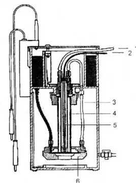
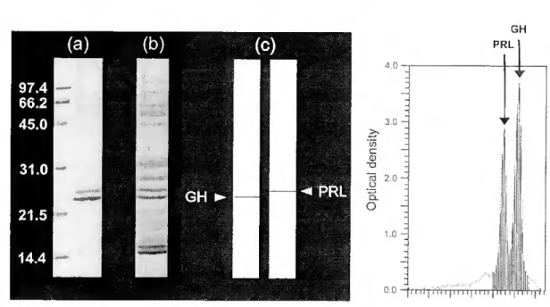
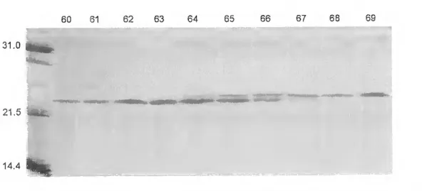
The sea bream {Sparus aurata L.) is an important aquaculture species along the Mediterranean coast. Prolactin (PRL) released by the pituitary gland play a central role in controlling several aspects of sea bream physiology. In this study, we examined the effect of several internai and externai factors on the activity of PRL cells in cultured pituitary glands. Sea bream pituitaries were cultured during 18hrs at 210C and the hormones present in the culture médium separated by SDS-PAGE. PRL was identified by Western blotting using antiserum directed against chum salmon PRL and quantified by optical densitometry.
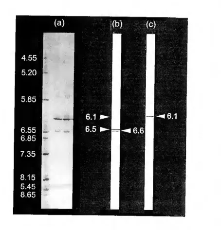
A highly purified PRL was isolated from the médium of cultured pituitaries by continuous elution electrophoresis performed using a Model 491 Prep Celi. The yield of purified hormone was 3mg/g wet weight of pituitary. Sea bream PRL had an estimated molecular weight of 25kDa after SDS- PAGE. PRL did not show size heterogeneity, but multiple charge variants were detected with isoelectric points varying between 6.1 and 6.7. The partial amino acid sequence established did not reveal genetic differences between PRL molecules. Post-translational modifications were not clearly demonstrated but a potential phosphorilation at S^s is proposed on the basis of the present data.
Seasonal and diurnal changes of PRL release to the culture media and pituitary content were investigated in immature seabream. A marked annual cycle in the pituitary gland activity was observed. This is highlighted by the seasonal variation in the basal pituitary PRL release rates but also by a variation in the relative concentration of PRL charge variants released from the pituitary gland. Circadian changes were also observed. Temperature (but not photoperiod) influences sea bream PRL cells activity, with higher temperature increasing in vitro PRL secretion and lower temperature having the opposite effect.
The effect of estradiol-17/S' (E2) implants on the in vitro secretion of PRL and its modulation by galanin (Gal) was determined. Experiments were conducted during winter and spring and it was observed that PRL cells responsiveness to E2 varied with season. In control fish Gal caused a dose-dependent stimulation of PRL secretion in vitro, but on E2 primed fish Gal had no detectable effect on the secretion of PRL.
The effect of E2 implants on the in vitro secretion of PRL and its modulation by vasoactive intestinal peptide (VIP) was determined. Experiments were conducted during winter and spring and PRL cells responsiveness to E2 varied with season. In E2 primed fish VIP caused a dose- dependent inhibition of PRL secretion in vitro. VIP had no detectable effect on the secretion of
PRL from control pituitaries. Anatomical evidence of abundant VIP immunoreactive nen/e fibres in neurohypophysial (NH) tissue penetrating the rostral pars distalis provide further evidence supporting an action for VIP in the regulation of PRL cells.
PRL is believed to be involved in the adaptation of fish to changes in environmental salinity. The effect of freshwater challenge on in vitro PRL release was studied in fish sampled 7 days after the onset of seawater dilution. Experiments were conducted during winter, spring, and autumn. Results indicate that cultured pituitaries of fish challenged with extremely low values of salinity (2ppt) released significantly more PRL that pituitary glands from seawater-adapted fish. This suggests a potential role for PRL in FW adaptation of sea bream. Moreover, the study indicates that the success of adaptation may depend on the time of year at which transfer to freshwater occurs.
Resumo
A dourada {Sparus aurata L.) é uma espécie importante para a aquacultura ao longo de toda a costa mediterrânea. A prolactina (PRL) é uma hormona libertada pela glândula pituitária e regula de um modo fundamental alguns aspectos da fisiologia deste teleósteo. No presente trabalho foi estudado o efeito que diferentes factores, externos e internos, exercem na actividade in vitro das células de PRL. Para este estudo foram feitas culturas de pituitárias de dourada com uma duração de 18 horas a uma temperatura média de 210C. A PRL, após separação por SDS- PAGE, foi identificada por "Western blotting" utilizando anticorpos da PRL do salmão e, posteriormente, quantificada por densidade óptica.
Por electroforese de eluiçáo contínua e utilizando uma "Prep Celi, modelo 491", foi purificada a PRL secretada In vitro, tendo-se obtido um rendimento aproximado de 3mg/grama de peso fresco de pituitária. A hormona purificada tinha um peso molecular aparente de 25kDa em SDS- PAGE e formas com pontos isoeléctricos variando entre 6.1 e 6.7. A identificação da proteína purificada foi feita por "Western blotting" e pela sequência parcial de amino ácidos. Não foi possível demonstrar a existência de variantes genéticas nas moléculas de PRL isoladas, tendo sido identificado um local potencial de fosforilação no resíduo Siss-
A secreção in vitro de PRL apresenta um evidente ciclo anual, não só na quantidade de hormona secretada mas também nas formas com diferentes pontos isoeléctricos que são produzidas ciclicamente.. Também foi observado um nitido ciclo diário na secreção in vitro de PRL. A temperatura influencia directamene a actividade das células de PRL, mas não foi demonstrada nenhuma influência do fotoperíodo sobre a actividade daquelas células.
Foi estudado o efeito do tratamento in vivo com estradiol-17b (E2) modulado in vitro pela galanina (Gal). As experiências decorreram durante o inverno e primavera, tendo-se verificado uma diferença estacionai na resposta das células de PRL aos implantes do estrogénio. O efeito da Gal depende do nível do estrogénio no corpo dos animais, uma vez que estimula a secreção in vitro de PRL de pituitárias colhidas de peixes sem implantes de E2 mas não tem qualquer efeito nas pituitárias colhidas de animais tratados com o estrogénio.
Também foi estudado o efeito do tratamento in vivo com E2 modulado in vitro pelo peptídeo vaso- intestinal (VIP). O efeito do VIP depende do nível de E2 no corpo dos animais, uma vez que inibe in vitro a secreção de PRL de pituitárias collhidas de animais tratados com o E2 e não tem qualquer efeito nas pituitárias colhidas de animais sem implantes do estrogénio.
A PRL está relacionada com a adaptação dos teleósteos às mudanças da salinidade do meio. No presente trabalho, foi estudado o efeito sobre a secreção in vitro da PRL da transferência de douradas adaptadas a água salgada para água doce .(2ppt). Foram realizadas experiências no inverno, primavera e outono em que os animais foram sacrificados 7 dias após o início da mudança gradual para a água doce. Os resultados indicam que as células de PRL libertam in vitro quantidades significativamente maiores da hormona em animais transferidos para a água doce em comparação com as dos animais mantidos em água salgada. A PRL parece assim ter um papel importante na adaptação da dourada à diminuição da salinidade do meio, estando o sucesso da adaptação dependente da época do ano em que é feita a transferência para a água doce.
List of figures Chapter 1. Figure 1.1 4 Figure 1.2 9 Figure 1.3 14 Figure 1.4 28 Figure 1.5 30 Figure 1.6 36 Figure 1.7 39 Chapter 2 Chapter 3 Figure 3.1 62 Figure 3.2 66 Figure 3.3 67 Figure 3.4 68 Figure 3.5 69 Figure 3.6 69 Chapter 4 Figure 4.1 88 Figure 4.2 93 Figure 4.3 94 Figure 4.4 95 Figure 4.5 96 Figure 4.6 97 Figure 4.7 98 Chapter 5 Figure 5.1 112 Figure 5.2 113 Chapter 6 Figure 6.1 125 Figure 6.2 125 Figure 6.3 127 Chapter 7 Figure 7.1 144 Chapter 8 Figure 8.1 155
List of tables Chapter 1. Table 1.1 19 Table 1.2 26 Table 1.3 31 Table 1.4 32 Table 1.5 33 Table 1.6 34 Table 1.7 37 Chapter 2. Table 2.1 52 Chapter 3 Table 3.1 63 Table 3.2 70 Table 3.3 71 Table 3.4 73 Table 3.5 74 Chapter 4 Table 4.1 89 Table 4.2 91 Table 4.3 92 Table 4.4 97 Chapter 5 Table 5.1 111 Chapter 6 Table 6.1 124 Chapter 7 Table 7.1 141 Table 7.2 142 Table 7.3 143
List of Abbreviations
ACTH Adrenocorticotropic hormone
aa Amino acid
a-MSH (x- melanophore-stimulating hormone
ATP Adenosine triphosphate
BSA Bovine serum albumin
CAs Catecholamines
cDNA Complementary DNA
DA Dopamine
E2 Estradiol 17/?
EGF Epidermal growth factor
ETs Endothelins
FW Freshwater
GABA Gamma-aminobutyric acid
Gal Galanin
GaIR Galanin receptor
GH Growth hormone
GH3 Celi line (rat pituitary tumor)
GHRH Growth hormone releasing hormone
GIP Glucose-dependent insulin
GnRH Gonadotropin releasing hormone
GtH Gonadotropin
IgG-PAP Immunoglobulin G-peroxidase-antiperoxidase
IT Isotocin
LHRH Luteinizing hormone releasing hormone
mRNA Messenger RNA
MSH Melanocyte-stimulating hormone
NGF Nerve growth factor
NH Neurohypophysial
NPY Neuropeptide Y
NT Neurotensin
OT Oxytocin
PBS Phosphate buffered saline
PI Pars intermédia
PIF Prolactin inhibiting factor
PL Mammalian placenta! lactogen
POMC Proopiomelanocortin
PPD Proximal pars distalis
PRL Prolactin
PRLR Prolactin receptor
RPD Rostral pars distalis
SDS Sodium duodecyl sulfate
SDS-PAGE SDS-poIyacrylamide gel electrophoresis
SL Somatolactin
SRIF Somatostatin release inhibiting factor
SS Somatostatin
SW Seawater
TEMED N' N' N' N' - tetramethyethylemediamine TGFs Transforming growth factors
TRH Thyrotropin releasing hormone TRIS T ris(hidroxymethyl)aminomethane TSH Thyroid stimulating hormone VIP Vaso-intestinal peptide
Table of contents
Chapter 1. General Introduction 1 1.1 Structure and evolution of PRL 3
1.2lsoforms 4
1.3 Sites of synthesis and secretion of PRL 9 1.3.1 Pituitary PRL 9 1.3.2 Extrapituitary PRL 11 1.4 PRL receptors (PRLRs) 11 1.5 PRL actions on target tissues 12 1.6 Regulation of PRL synthesis and secretion 13 1.6.1 Biogenic amines 15 1.6.2 Acetylcholine 17 1.6.3 Neuropeptides 18 1.6.4 Hormones 37 1.6.5 Other pituitary and hypothalamic factors 40 1.6.6 Externai factors 41 1.7 Pattems of pituitary PRL release 43 1.8 Biological actions of PRL 44 1.9 Objectives of project 49 Chapter 2. General Materials and Methods 50 2.1 Incubation of pituitary glands 51 2.2 Electrophoretic separation of PRL 52 2.2.1 SDS-PAGE 52 2.2.2 Staining procedures for SDS-PAGE gels 53 2.2.3 Isoelectric focusing 53 2.3 Quantification of PRL 54 2.4 Western blotting 55 2.5 Estimation of PRL molecular weight and isoelectric point 56 Chapter 3. Isolation and Characterization of PRL and GH 57 3.1 Introduction 58 3.2 Additional methods 80 3.2.1 Identification and characterization of PRL and GH 60 3.2.2 PRL isolation 62 3.2.3 Identification of PRL and GH by amino acid sequencing 63 3.2.4 Molecular weight estimation of purified PRL and GH 65 3.3 Results 85 3.4 Discussion 74 Chapter 4 Circannual and circadian rhythms of PRL in vitro release 79 4.1 Introduction 80 4.2 Additional methods 85 4.3 Results 92 4.4 Discussion 98
Chapter 7 PRL release in response to freshwater challenge 7.1 Introduction
7.3 Results 7.4 Discussion
Chapter 8 General Discussion Future work
105 106 Chapter 5 Effect of Gal on PRL in vitro release
5.1 Introduction 5.2 Additional methods 5.3 Results 5.4 Discussion 113 117 118 Chapter 6 Effect of E2 and VIP on PRL in vitro release
6.1 Introduction 6.2 Additional methods ^ 6.3 Results 6.4 Discussion 126 134 135 ' -1 "37 7.2 Additional methods ^ 145 149 158 References 161
Chapter 1 General Introduction
CHAPTER 1: General Introduction
The gilthead seabream {Sparus auratus) is an important aquaculture species in the Mediterranean area (FAO, 2002). Its commercial production started in the early 1980s and since then production areas have spread and in southern Europe its cultivation has become predominant over other finfish species. This expansion is related to its relatively straight forward husbandry and the initial high market price of this species, although more recently with the increase in its availability prices have declined.
The gilthead sea bream is a marine teleost and belongs to the Perciformes, a group which includes a number of other commercially interesting species. such as couch's sea bream (Pagrus pagrus), sea bass {Dicentrarchus labrax), Nile tilapia {Oreochromis nilotica), and Mozambique tilapia {Oreochromis mossambicus). Due to the commercial value of gilthead seabream the ecology and biology of this species has been extensively studied. The sea bream is found in both marine and brackishwater environments such as coastal lagoons and estuarine areas, in particular during the initial stages of its life cycle (Moretti et ai, 1999). Born in the sea during wintertime, the fingerlings typically migrate in early spring towards protected coastal waters in search for abundant food and milder temperatures, and return to the open sea in late autumn, where the adult fish breed (Moretti et ai, 1999).The sea bream is a protandrous hermaphrodite, i.e., individuais spawn as males during the first breeding season but may undergo sex reversal in one of the subsequent seasons. The spawning season in southern Portugal extends from October until February (Condeça, 2001).
Chapter 1 General Introduction
Homeostasis of a range of physiological processes in fish, in common with other vertebrates, is regulated by the endocrine and neuroendocrine systems. The latter system regulates development and physiology in fish. Reproduction, development, immune function and environmental adaptation are regulated through the orderly release of hormones by the neuroendocrine system, which integrates information from genes and the environment. One of the most versatile hormone is prolactin (PRL) which has numerous biological actions (reviewd by Sinha, 1995 and Bole-Feysot et a/., 1998;). Since the discovery, by Stricker and Grueter in 1928, of a pituitary factor capable of stimulating milk secretion in rabbits, a wealth of knowledge has accumulated about prolactin (PRL).
1.1 Struture and evolution of PRL
PRL belongs to a family of polypeptide hormones, which includes growth hormone (GH), mammalian placenta! lactogen (PL), and teleostean somatolactin (SL). Analysis of their amino acid sequences demonstrates that these hormones are highly conserved, and it has been proposed that they evolved from a common ancestral gene by duplication and subsequent divergence about 4x10⁸ years ago (Millerand Eberhardt, 1983; Nicoll et ai, 1986).
Mammalian PRL is a single polypeptide chain of approximately 190-200 amino acids (aa) and is synthesized as a prohormone containing a signal peptide of approximately 28 aa (Bole-Feysot et a/., 1998). The full-length amino acid sequence of PRL has been determined in mammals, birds, reptiles and amphibian. Ali PRLs identified so far in tetrapods are 197-199 aa and contain six
Chapter 1 General Introduction
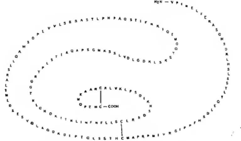
cysteines forming three intramolecular disulfide bonds, one at the N-terminus, one in the middle, and one at the C-terminal (Bole-Feysot et a/., 1998 and Manzon, 2002 for reviews). The amino acid sequence of PRL has also been characterised in a variety of teleostean and nonteleostean fish (reviewed by Manzon, 2002). Fiscine PRLs are also synthesized as prohormones with a signal peptide of 23-24 aa. Ali teleostean PRLs lack the N-terminal disulfide bond due to the absence of 12-14 aa at the N-terminus (see Manzon, 2002 for review) (Fig. 1.1). PRL from sea bream is a protein of 212 amino acids with a putative signal peptide of 24 residues and a mature protein of 188 amino acids (Santos et ai, 1999). MíN - v f , " t c QAPSCyn, r <• v D 1 C o „ t s « 1 ' s j I F M c - COOH 1 ' ' o L S S T M C
Figure 1.1 - Teleost PRLs (i.e. Nile tilapia PRLiss) lack the N-terminal disulfide bridge present in mammals (Manzon, 2002).
1.2 Isoforms
PRL is characterized by structural as well as functional polymorphism. The existence of both size and charge variants of PRL has been shown in mammals
Chapter 1 Gener^ntroduc^
1989; Martinat et ai, 1990; Sinha et ai, 1991; Briski et ai, 1996;), and two molecular forms of PRL have been isolated from pituitary glands of two reptiles, alligator and crocodile (Noso et ai, 1992), and from Xenopus laevis (Yamashita et ai, 1993). In turkeys, PRL is present in the pituitary gland as three different isoforms, a nonglycosylated form of approximately 22.5 KDa and two glycosylated forms of 24.5 KDa, which comigrate on SDS-PAGE (Corcoran and Proudman, 1991). In some teleosts, chum salmon {Oncorfiynchus keta), common carp (Cyprínus carpio), Japanese eel (Anguilla japonica), Mozambique tilapia {Oreochromis mossambicus) and Nile tilapia {Oreochromis niloticus), two different forms of PRL have been identified (Manzon, 2002 for review). The two salmon, carp, and eel PRLs are highly homologous, whereas the two forms of PRL secreted by tilapia pituitary share only 69% sequence identity and are designated tPRL177 and tPRL188 to indicate the number of amino acid residues in each isoform (Specker et ai, 1993). The biological activities of the two variants in the maintenance of the hydromineral balance are different as is their effect on growth. In addition to their differing biological activities, tPRL177 and tPRL188 are also differentially regulated during development and in response to alterations in environmental salinity (see Manzon, 2000 for review). To date only a single form of pituitary PRL has been isolated in sea bream (Santos et ai, 1999).
The source of prolactin variants may be by mechanisms such as alternative splicing of the primary transcripts, proteolytic cleavage of the protein,
Chapter 1 General Introduction
and other posttranslational modifications, such as glycosylation and phosphorylation of the amino acid chain.
ALTERNATIVE SPLICING - Alternative splicing of PRL mRNA has been proposed as one source of the variants (Sinha, 1995). Indeed, evidence suggestive of the existence of an alternatively spliced prolactin variant has been described in the rat anterior pituitary (Emanuele et a/., 1992; Wilson et a/., 1992). However, in general alternative splicing is not considered a major source of prolactin variants.
PROTEOLYTIC CLEAVAGE - Some PRL size variants may be the products of kallikrein enzymatic activity. Studies in vitro have shown that kallikrein is an estrogen-induced, trypsin-like serine protease, that is found in the Golgi cisternae and secretory granules of rat lactotrophs, which cleaves the 25KDa form to a 22KDa form in a thiol-dependent manner (Powers, 1993). Thiol alters the conformation of PRL such that kallikrein recognizes it as a substrate (Anthony and Powers, 1993). These proteolytic fragments were found to be released in substantial amounts during short incubations of rat pituitaries (Anthony ef a/., 1993).
OTHER POSTTRANSLATIONAL MODIFICATIONS - The majority of prolactin variants are probably the result of other posttranslational processing of the mature molecule in the pituitary gland or the plasma. These modifications
Chapter 1 General Introduction
include dimerization and polymerization, phosphorylation, glycosylation, sulfation, and deamidation.
Dimerization and polymerization: macroprolactins.
High molecular weight forms of PRL are encountered in significant amounts both in the pituitary gland and plasma. They represent dimers, polymers and aggregales of PRL, and PRL associated with binding proteins. They arise by both covalent (disulfide linkages and linkages between the sugar moieties of the glycosylated monomer) and noncovalent bonding. In general, these forms have reduced biological activity in comparison to the monomer (Sinha, 1995).
Phosphorylation.
PRL phosphorylation occurs within the secretory vesicle of lactotrophs just before exocytosis and involves esterification of hydroxyl groups of serine and threonine residues (Freeman et a/., 2000 for review). Phosphorylation of serine or threonin may occur when glutamic or aspartic acid is situated two residues away C-terminally (Ser/Thr - X - acidic). Serine residues that occur C-terminally to groups of basic amino acid residues may also be phosphorylated (basic - basic - X - Ser/Thr) (Dimaline, 1988). Ocurrence of phosphorylated isoforms has been demonstrated in the case of rat, bovine, murine, and avian PRLs (Sinha, 1995 for review). Phosphorylation generally lowers the biological activity of PRL, but interaction of heterogenous hormonal forms at the levei of target cell receptors may influence the magnitude of the biological effect. For example,
Chapter 1 GeneralIMct^
Wang and Walker (1993) reported that the biopotency of nonphosphorylated PRL variants is diminished as a consequence of coincubation with phosphorylated PRL, findings which suggest that posttranslational phosphorylation of the native molecule may generate a natural "antagonist" to the biological activity of non- modified variants. In rats, in vivo secretion of phosphorylated and nonphosphorylated PRL or their ratio, varies at different stages of the estrous cycle (Sinha, 1995 for review).
Glycosylation.
Glycosylated PRL has been found in the pituitary glands of mammalian, amphibian, and avian species. The linkage of the carbohydrate moiety may be either through nitrogen (N-glycosylation) or oxygen (O-glycosylation). The consensos sequence for N-glycosylation of aspargine is Asn - X - Thr/Ser (Dimaline, 1988). In several mammals and reptiles, in which glycosylated PRL occurs is N-glycosylation. In rat and turkey, the carbohydrate chains are attached through O-linkage (Sinha, 1995 for review). The carbohydrate residues of the oligosaccharide chain may contain varying ratios of sialic acid, fructose, mannose, and galactose that differ considerably between species and also vary with physiological and pathological states. As observed for other PRL variants, glycosylation also lowers biological activity and receptor binding, and alters the metabolic clearance rate of PRL (Freeman et a/., 2000 for review).
There is no published evidence in sea bream for the presence of glycosylated or phosphorylated forms of PRL. However, analysis of the primary
Chapter 1 General Introduction
ammo acid sequence reveals a consensus sequence for N-linked glycosylation at Asn 148 (N-l-S) and for phosphorylation at Ser 166 (R-R-D-S) (Santos et ai,
1999).
1.3 Sites of synthesis and secretion of PRL 1.3.1 Pituitary PRL
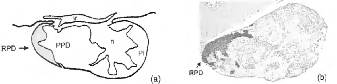
The principal site of production of PRL is the pituitary gland (Fig 1.2). In ali vertebrates, the pituitary gland consists of two parts, separable on the bases of embryology, structure, and function. These are the neurohypophysis, a downgrowth from the floor of the diencephalon, and the adenohypophysis originating as an ectodermal upgrowth (Rathke's pouch) from the roof of the embryonic buccal cavity (Ball and Baker, 1969). The adenohypophysis is divided into the pars distalis, site of secretion of most adenohypophysial hormones, and the pars intermédia (PI).
RPD PPD
rim
■ \ . , ' ^ ^
(b)
Figure 1.2 - (a) Diagram showing a sagital section through the pituitary gland of the adult sea bream with the distribution of PRL-cells mdicated by a ^aded area ( ) Immunohistochemistry of a sagital section of sea bream pituitary 9la"^ounter®'^ with haematoxylin with PRL cells revealed by anti-chum salmon Pra^tm (magnification xlOO). (n) neurohypophyseal tissue; (RPD) rostral pars distalis. (PPD) próxima!pars distalis-, (PI) pars intermédia-, (ir) mfudibular recess.
General Introduction Chapter 1 —
The adenohypophysis pare distalis is subdivided into rostral pare distahs (RPD) and proximal pare distalis (PPD). on the basis of cell types. The fish neurohypophysis consists essentially of a hypophysial stalk, suspending the gland from the ventral region of the diencephalon (hypothalamus) and contaming an extension of the third ventricle {infudibular recess). and at the distai end of the stalk an enlargement, the neurohypophysial lobe or core (Ball and Baker, 1969). The adenohypophysis is a complex and specialized endocrine tissue that contains a functionally heterogeneous population of hormone secretmg cells. At the dorsal surface of the piturtary lies the hypothalamus which is responsible for integration of a whole range of inputs from other brain centers and for the contrai of visceral functions, in many cases via its effects on the secretions of the pituitary gland (Chester-Jones et ai., 1987). In teleosts, the hypophysial stalk, which suspends the piturtary gland from the hypothalamus, do not have the median eminence-portal system of other vertrebrates. The blood to the pituitary is supplied by one (or two) hypophyseal artery (ies), and although this runs along the floor of the hypothalamus for a variable distance, no neurosecretory terminations on the vessels in this region have ever been observed (Chester- Jones et a/., 1987).
The hypothalamus regulates the synthesis and secretion of six major hormones: prolactin (PRL), growth hormone (GH). thyroid stimulatmg hormone (TSH), adrenocorticotropin (ACTH), melanocyte-stimulating hormone (MSH), and gonadotropin (GtH). In the adult sea bream, PRL cells are confmed to the RPD and occupy a relatively small proportion of the pituitary (Fig. 1.2). They are oval
^ , . General Introduction Chapter 1
in shape and do not appear to be arranged in follicles (Power and Canano, 1992).
1.3.2 Extrapituitary PRL
In higher vertebrates the principal site of production of PRL is the pituitary gland, but, as reviewed by Ben-Jonathan et ai (1996), the PRL gene is also expressed in several PRL target tissues and PRL occurs in the mammary gland of rat, human, goat, sheep, and rabbit (Nolin and Witorsch, 1976; Fields et ai. 1993; Kurtz et ai, 1993; Le Provost et ai, 1994; Gabou et ai, 1996) and in mammalian brain and spinal cord (Emanuele et ai. 1992; Wilson et ai, 1992).
Recently, PRL transcripts were also detected at some extrapituitary sites (liver, intestina, and gonads) of the sea bream (Santos et ai, 1999) and in the liver, kidney, spleen, gill, muscle, gonads, and brain of goldfish (Imaoka et ai, 2000).
1.4 PRL receptors (PRLRs)
The effects of PRL on target tissues are mediated by the PRL receptor (PRLR), which belongs to the cytokine receptor family (Bole-Feysot. 1998). In mammals, there are long, intermediate, and short PRLR isoforms (Freeman et at 2000). Ali fish PRLR cDNA encode for a mature protein of approximately 600 aa in length and are most similar in appearance to the long form of mammalian PRLRs (Manzon, 2001). The isolation and characterization of fish PRLRs has revealed that several important functional domains, receptor activation
Chapter 1 genera/ Introduction
mechanisms, and signal transduction pathways have been conserved between fish and mammals. Generally. the highest PRLR expression leveis were observed in the primary osmoregulatory organs (gills, kidney, and intestine). However. PRLR transcripts were also detected in other tissues such as the brain, gonad, liver, muscle, skin, spleen, head kidney, lymphocytes, and bone of some fish (Manzon, 2001 for review). In the sea bream, PRLR expression was also detected in early post-hatching stages of larvae (Santos et a/., 2003).
!.5 PRL actions on target tissues
Pituitary PRL acts via a classic endocrine pathway, i. e., it Is secreted by the pituitary gland, transported by the circulatory system, and acts on target cells at some peripheral sites via specific receptors located on the plasma membrane. The interaction of PRL with its receptor in various target cells leads to the activation of intracellular events that ultimately promote PRL-responsive genes responsible for a biological activity in the animal.
In mammals, PRL binding to its receptor leads to dimerization and activation of an intracellular cascade mediated by JAK/STAT signal transduction pathway (Ihle, 1996). The Janus kinase JAK2 is constitutively associated with the PRLR and is activated upon PRL binding. Following activation of JAK2, tyrosine residues on the PRLR and the transcription factor STAT5 are phosphorylated (Han et a/., 1997). Activated Stat proteins translocate into the nucleus where they bind DNA consensus motifs to mediate activation of target genes (Bole-Feysot et ai, 1998).
Chapter 1 General Introduction
PRL is derived primarily from the anterior pituitary and acts on tissues widely distributed through the body (endocrine action). But locally produced PRL can act on adjacent cells (paracrine action) or on the PRL-secreting cell itself (autocrine action). Intriguingly a paracrine and autocrine mechanisms could activate many of the actions associated with PRL, without affecting the circulating concentration of the hormone (Bole-Feysot et a/., 1998) and may explain the apparent discrepancies of some experiments where despite unchanging leveis of circulating hormone specific biological activities are observed.
1.6 Regulation of PRL synthesis and secretíon
The secretory cells of the anterior pituitary are influenced by a wide array of factors. The general and well-accepted view is that PRL secreting cells have a spontaneously high secretory activity. Therefore, pituitary PRL secretion is under a tonic and predominantly inhibitory control exerted by a hypothalamic PRL- inhibiting factor (PIF) that restrains in vivo PRL secretion (reviewed by Freeman et a/., 2000) (Fig. 1.3). But PRL release is also influenced by other secretagogues and gene regulators. The old notion that secretagogues act rapidly while gene regulators act slowly has been questioned when results on the induction of genes such as c-fos are considered. The two types of regulators may be better classified by their preferred utilization of cellular compartments, i.e. secretagogues act on the cell membrane and activate calcium-dependent exocytosis while gene regulators, directly or indirectly, utilize the nuclear compartment (Ben-Jonathan et ai, 1996 for review). These secretagogues and
Chapter 1 General Introduction Circadian input Environmental stimuli; Light Odor Sound Stress Internai millieu: Estradiol/progesterone Glucocorticoids Plasma osmolarity z- Regulatory Circuit Hypothalamic + Neuroendocrme neurons PRF Median eminence Lactotroph' Pituitary (PRL)M" Celi PRL Anterior lobe Intermediate lobe Peripheral blood
Fiqure 1.3 An overview of the regulation of PRL secretion in mammals. PRL secretion is paced by a light-entrained circadian rhythm. which is modified by environmental input. with the internai milieu affecting the inhibitory or stimulatory elements of the hypothalamic regulatory circuit. The final common pathways of the central stimulatory and inhibitory control of PRL secretion are the neuroendocrme neurons producmg PRL inhibiting factons (PIF), or PRL releasing factors (PRF). PIF and PRF from he neuroendocrine neurons can be released either at the median em.nence or at the neurointermediate lobe and reach the PRL-cells in the anterior lobe of the pituitary gland. Thus PRL-cells are regulated by blood-bome agents of central nervous
or by factors released from neighboring cells (paracrine regulation), or from the PRL- cells themselves (autocrine regulation) (adapted from Freeman et a/., 2000).
General Introduction Chapter 1
gene regulators are multiple hypothalamic factors, feedback signals from the target organs of the pituitary hormones and an increasing number of factors produced and secreted within the pituitary (other hormones, particularly steroids), and environmental factors.
1.6.1 Biogenic amines
DOPAMINE (DA) - In mammals, DA is considered the major PIF. Several well-defmed dopaminergic systems have been described in the mammalian brain.DA receptors have been detected in preparations of pituitary membranes by immunocytochemical methods. which include labelling the receptors with the DA antagonist haloperidol, and detection of it with an antibody agamst haloperidol and the peroxidase anti-peroxidase technique. Ample experimental evidence shows that DA inhibits PRL release from pituitary lactotrophs both in vivo and in vitro (reviews by Ben-Jonathan and Hnasko, 2001; Freeman et ai, 2000).
In avian species, DA-ir cells were detected in the developing chicken adrenal-gland (Sánchez-Montesinos et ai, 1996), In teleosts, the existence of dopaminergic nerve fibers innervating PRL cells has not been clearly demonstrated. However, the presence of DA in pituitary bioassays reduces PRL cell activity in some species suggesting it may also act as a PIF in some fish
Chapter 1 General Introduction
NOREPINEPHRINE AND EPINEPHRINE - In rats, adrenergic modulation, mediated by either norepinephrine or epinephrine, plays an important role in stress-induced PRL secretion (see Freeman et ai, 2000 for revision).
In the teleost fish Poecilia latipinna, the respective a adrenergic and (3 adrenergic agonists phenylephrine and isoproterenol inhibited PRL secretion In vitro, but the adrenergic blockers phentolamine and propranolol had no direct effect on their own, although they did oppose the inhibitory action of DA (Wigham, 1992). Interestingly in the saddleback wrasse {Thalassoma duperrey), norepinephrine stimulates both initiation and completion of sex reversal (Larson et a/., 2003).
SEROTONIN - In mammals, serotonin facilitates suckling-induced PRL release and regulates the estrogen-induced PRL secretion (Freeman et a/., 2000). Although receptors for serotonin are present in the anterior lobe of the pituitary gland, serotonin does not stimulate PRL release in vitro, suggesting that it functions as a neurotransmitter rather than a neurohormone (Freeman et a/., 2000).
In avian species, serotonin-ir was localized in the endocrine cells of the digestive and respiratory systems (Yamada et a/., 1985; Yamaguchi et a/., 1987, Adriaensen et ai, 1994; Lucini et ai, 1996). In the chicken, serotonin-ir is expressed in the sympathoadrenal system (Garcia-Arrarás and Martinez, 1990).
In some teleosts, namely clingfish {Lepadogaster ca n do Hei), fire eel {Mastacembelus erythrotaenia), goldfish (Carassius auratus), and turbot
. General Introduction Chapter 1
(Scophthalmus maximus), serotonin-ir endocrine cells and fibers were observed throughout the gastro-intestinal tract, in the skin and in some nerve projections to the retina (Fasulo et ai, 1993; Reinecke et ai, 1997; Lima and Urbina , 1998). Moreover, the brain, the pituitary, and the ventral spinal cord contam a dense innervation of serotonergic fibers in sailfin molly (Poecilia latipinna). garfish (Lepisosteus prvductus), and European eel (Anguilla anguitla) (Wigham, 1992, Batten et ai, 1993; Chiba and Oka, 1999). Relatively few studies have been carried out with serotonin in vitro, in the trout serotonin produces an increase of PRL cell activity and stimulates PRL secretion but te action on the PRL cells in other species remains to be studied (Wigham, 1992). In the saddleback wrasse {Thalassoma duperrey), serotonin inhibits both initiation and completion of sex reversal (Larson et a/., 2003).
1.6.2 Acetylcholine
|n GH3 cells, the activation of the (muscarinic) acetylcholine receptor decreases PRL secretion (Freeman et ai, 2000). Moreover. cholinergic stimulation by administration of cholinergic agonists in rats causes a decrease in serum PRL concentrations (Freeman et a/., 2000).
Experiments with the teleost fish rainbow trout (Oncorfiyncus mykiss), in vitro, showed that the acetylcholine agonist. carbachol, inhibits PRL synthesis and release. Moreover, there is evidence which suggests that the cholinergic inhibition of the PRL cells in teleosts operates via muscarinic receptors (Wigham,
Chapter 1 General Introduction
1.6.3 Neuropeptides
A variety of peptides are known to be involved in the regulation of PRL secretion. The list of this type of PRL secretagogues is rather long, but in the present study more attention is paid to the following peptides: (1) galanin (Gal), which in terms of its potential physiological activity is one of the better characterized paracrine factors in the pituitary, (2) vasoactive intestinal peptide (VIP), which has long been characterized as a likely local factor influencing function of lactotrophs, and recently has been shown to interact with galanin, (3) somatostatin (SS) and (4) thyrotropin-releasing hormone (TRH), which have received much attention as they are respectively, a potent inhibitor and stimulator of PRL secretion in mammals.
In addition to the aforementioned neuropeptides the other putative PRL regulatory factors which will be reviewed in the introduction are those which have been studied in teleost fish and include oxytocin and isotocin, neuropeptide Y, neurotensin, substance P, and urotensin.
GALANIN (Gal) - Gal is a 29-amino acid peptide originally isolated from porcine small intestine (Tatemoto et ai, 1983). It shares little homology with other known peptides. It has been demonstrated that the N-terminal sequence of Gal, comprising amino acids 1-15 are highly conserved in ali species from which it has been isolated as this region is probably responsible for receptor binding (Crawley, 1995). In contrast, the C-terminal region exhibits substantial
interspecies variability, suggesting that it might be responsible for the species- specific activity of Gal (Crawley, 1995) (Table 1.1).
Table 1.1 - Endogenously occurring Gal sequences (Bartfai, 2000; Chartrel eia/.. 1995; Wang and Conlon, 1994).
1 -15 16-25 26-30 Man GWTLNSAGYLLGPHA VGNHRSF SD K N G L T S ** Pig GWT L NSAGYLLGPHA I DNHRSFHDK Y G L A * Rat GWTLNSAGYLLGPHA l DNHRSFSDK H G L T * Chicken GWTLNSAGYLLGPHA VDNHRSF ND K H G F T * Frog GWTLNSAGYLLGPHA l DNHRSFNDK H G L A * Dogfish GWTLNSAGYLLGPHA VDNHRSF ND K H G L A * *C-terminalamide
"C-terminal free acid
Boldface denotes aa which were not conserved between species
Immunohistochemical studies have demonstrated that Gal-like immunoreactivity is widoly distributed in the central nervous system and the gastroenteric, respiratory, urinary and reproductive system of different mammals (Crawley, 1995). Gal and its mRNA have been shown so far to be expressed prevalently in neurons, but a number of studies have colocalized Gal with PRL in at least a fraction of the lactotrophs (see Schwartz, 2000 for review). These and other studies have reported colocalization in other cells in addition to or instead of lactotrophs. One study, employing cellular immunoblotting technology with rat pituitary cells, localized Gal with PRL or ACTH and in wild type and transgenic (human GHRF-expressing) mice, immunocytochemical techniques were used to demonstrate the colocalization of Gal with GH, PRL, or TSH (see Schwartz, 2000 for review).
In humans, Gal secretion has been measured in cultures of ACTH- secreting tumours (Invitti ef a/., 1999). Recently, using immunocytochemical
techniques, Gal has also been reported to be present in nerve fibers in monkey and canine pituitários in close proximity to ali types of secretory cells (Liu and Gao, 1998).
In reptiles and amphibians, Gal immunoreactivity was observed in brain, hypothalamus, heart, bladder, small intestino and oviducts (Crawley, 1995; Lamanna et a/., 1999). In avian species, Gal-ir cells and neurons were detected in the adrenal-gland of the chick embryo and the adult, in the quail brain, in the chicken medulla oblongata, and in the pancreatic islets of the bustard (Azumaya and Tsutsui, 1996; Sánchez-Montesinos et ai, 1996; Wang et al., 1997; Ohmori, 1998; Mensah-Brown et ai, 2000). Avian galanin mRNA was expressed in the quail brain, ovary, and intestine (Kohchi and Tsutsui, 2000).
Some teleost fish revealed similar extensivo systems of Gal immunoreactive neurons in brain and pituitary (Moons et ai, 1989; Batten et ai, 1990; Cornbrooks and Parsons, 1991; Moons et ai, 1991; Olivereau and Olivereau, 1991a; Power et ai, 1995; Batten et ai, 1999). In european seabream, Gal immunoreactive fibers were observed infiltrated between growth hormone, prolactin, and adrenocorticotropin cells (Power et ai, 1995). Studies on seabass pituitary revealed that Gal immunoreactive fibers abutted with ACTH, PRL, TSH, GtH, and GH cells (Moons et ai, 1989; Batten et ai, 1990; Moons et ai, 1991), suggesting it may directly influence the activity of such cells.
In mammals, Gal receptors have been identified in localized sites in the central nervous system, pituitary, pâncreas, stomach and intestine (Crawley, 1995). Effector systems linked to the Gal receptor in different tissues include
inhibition of adenylate cyclase, biockage of voltage-dependent calcium channels and activation of ATP-sensitive potassium channels (Crawley, 1995). Recently, two Gal receptors - Gal receptor-1 (GalR1) and Gal receptor-2 (GalR2) - have been characterized and shown to have different amino acid sequences, pharmacology, and second messenger signalling systems. In rats, Gal-R1 receptors have a broad role in normal synaptic transmission, while Gal-R2 receptors, in addition to a similar role in particular pathways, seem to be involved in processes prominent during the establishment and maturation of synaptic connections in developing brain and during neural damage and repair in the mature nervous system (Burazin and Gundlach, 1998; Burazin et ai, 2000). In three species of teleosts sailfin mollies sea bass {Poecilia latipinna), (Dicentrarchus labrax), and North African catfish {Ciarias gahepinus), the distribution of binding sites for Gal were described in the pituitary (Batten et ai, 1999). Moreover, in Atlantic salmon {Salmo salar), a specific Gal receptor was identified in the brain (Holmqvist and Carlberg, 1992) strengthening the idea that it has a direct action on the nervous system.
The regulation of Gal synthesis and secretion in pituitary cells is an area of ongoing investigation. Estrogens exert the most important influence on Gal activity in the pituitary, where estradiol positively regulates Gal-expressing cells, either increasing the amount of Gal protein and mRNA or the number of Gal- secreting cells. Pituitary Gal content is also controlled by other hormones such as thyroid hormones, vasoactive intestinal peptide (VIP) and PRL (see Schwartz, 2000 for review).
Chapter 1 General Introduction
In mammalian species, the intensity of Gal innervation has been observed to be dependent on the physiological status of the animal. For example, in rats, gonadal steroids have a dramatic activational effect on the numbers of visibly stained Gal cells in the hypothalamus and pituitary gland (Bloch et a/., 1993; Leibowitz et ai, 1998; Rugarn et ai, 1999). Moreover, the capacity of cells located in the hypothalamus to express Gal after testosterone or estradiol exposure in ferrets shows sexual divergence (Park et ai, 1997). In rats, hypothalamic and pituitary leveis of Gal mRNA, increased significantly after treatment with estrogen (Gabriel et ai, 1992; Brann et ai, 1993, Hyde et ai 1993; Crawley, 1995; Tseng et ai, 1997; Shen et ai, 1999; Degerman et ai, 2002). Gonadotropin-primed rat ovarian tissue cultured in vitro with galanin secreted significant amounts of estradiol, progesterone, and androstenedione into the médium (Fox et ai, 1994). Moreover, Gal mRNA leveis in the rat ovary are increased by treatment with human chorionic gonadotropin (Crawley, 1995).
In rats, combined autoradiographic and immunohistochemical studies have shown colocalization of receptors for estrogens and the neuropeptide Gal (Hõsli and Flõsli, 1999). Studies with immortalized LHRH neurons suggest that the estrogenic control of Gal gene expression in these neurons is transduced by estrogen receptors (Shen et ai, 1998). Studies in the estrogen receptor alpha- knock-out mouse revealed that the estrogen receptor subtype alpha is essential to estrogen-evoked Gal gene expression in the anterior pituitary of these animais (Shen et ai, 1999).
Chapter 1 General Introduction
Studies in vivo of the involvement of Gal in reproduction corroborate observations made in vitro. Exogenously administered Gal regulates the reproductive axis by acting as a growth regulator of the lactotrophs in rats (Wynick et al., 1993) or by stimulating the lutenizing hormone in monkeys (Finn et al., 2000). In female rats, Gal has both direct and indirect effeots on gonadal hormone release and this response is impaired in starved animais (Baranowska et a/., 2001).
In reptiles, the evidence which exists does not indicate if the action of Gal on the reproductive axis is direct or indirect. Administration of ly^-estradiol to non-reproductive female lizards induced a significant increase in neurons containing Gal immunoreactivity in the oviduct (Lamanna et al., 1999). Gal administration to pre-ovulatory lizard females induced premature oviposition, suggesting that Gal could be involved in the egg laying process (Lamanna et al., 1999). In avian species, estradiol induces an increase in Gal binding sites in mature quail oviducts, while 17/5'-estradiol and progesterone induced a marked increase in Gal binding sites (Tsutsui et al., 1998).
In teleost fishes Gal innervation is also dependent on the physiological status of the animal. For example, in eels treated with estradiol or methyl testosterone, increased Gal immunoreactive material was observed in some perikarya and brain fibers (Olivereau and Olivereau, 1991b). Moreover, a Gal-like peptide has been shown to have a sexually dimorphic distribution in the brain of the sailfin molly (Cornbrooks and Parsons, 1991).
Chapter 1 General Introduction
In addition to its action on reproduction a myriad of other physiological functions have been attributed to Gal. One important role is related to the control of appetite. Administration of Gal has been shown to induce feeding in satiated rats and ground squirrels; the effect is dose-related, with threshold doses at approximately 0.3 mmol (Crawley, 1995). In female rats, animais showing a preference for a fat-rich diet or animais with greater body fat, independent of the diet, exhibit higher leveis of hypothalamic Gal. This evidence suggests that in the female rat, Gal may contribute to the overeating and increased weight gain that is associated with a fat-rich diet (Leibowitz et ai, 1998). Interestingly in teleosts Gal has also been observed to influence feeding and in goldfish, where stimulation of food intake is mediated by the a2-adrenergic system, Gal affects the central
regulation of feeding, (de Pedro et ai, 1995).
Gal also has an important role in the mechanism of growth hormone release. Gal caused an in vitro increase in release of rat growth hormone (Gabriel et ai, 1988; Crawley, 1995) and LHRH (Lopez and Negro-Vilar, 1990), an effect proposed to be mediated via an effect on GHRH neurons (Meister et ai, 1987; Murakami et ai, 1989; Hulting et ai, 1991). Support for this proposal comes from in situ hybridisation studies which demonstrate that Gal mRNA is present in GnRH neurons (Marks et al., 1994; Selvais et ai, 1995). Intraventricular injections of Gal in the rat produce rapid increases of PRL (Crawley, 1995). Moreover, Gal and PRL are colocalized within secretory granules of the pituitary after estrogen treatment (Hyde et ai, 1991). Results from studies in mice carrying a loss-of-function mutation of the endogenous Gal
Chapter 1 General Introduction
gene, support the hypothesis that Gal acts as a paracrine regulator of PRL expression (Wynick et ai, 1998).
Gal is a potent regulator of a number of neurotransmitters and hormones. Its actions in some systems have been shown to involve a decrease in the cytosolic Ca2+ concentration. This may be the consequence of the hyperpolarization brought about by opening of Gal-receptor-coupled K+-channels or it may be a result of the Gal-receptor-mediated closure of some Ca2+ channels - or of a combination of both effects (Bartfai, 2000). A Gal suppressing calcium current and activating inwardly rectifying potassium current was also demonstrated in the enteric neurons of the guinea-pig small intestine. Suppression and activation of these channels was dependent on Gal concentration (Ren et ai, 2001). An alternative mechanism is observed for Gal inhibition of insulin secretion in a number of species (Crawley, 1995), this action is brought about by interference of Gal with adenylate cyclase activation and the activity of protein kinase C and cyclic AMR (Lindskog and Ahrén, 1991).
Several other biological activities have been attributed to Gal. In mammals, this neuropeptide produces direct effects on smooth muscle activity at several sites in the gastrointestinal tract (Botella et ai, 1992; Crawley, 1995). The multiple coexistence of Gal with most of the pituitary hormones during the fetal development of the rat (Cimini et ai, 2000), could be an indication that Gal may also have a role in cytodifferentiation. In elasmobranches, Gal causes differential vasoconstriction in vascular beds (Preston et ai, 1995).
Chapter 1 General Introduction
VASOACTIVE INTESTINAL PEPTIDE (VIP) - VIP is a vasoactive peptide which belongs to the secretin/glucagon family, other members include glucose- dependent insulin-releasing peptide (GIP), growth hormone releasing-factor (GRF), PHI (peptide having N-terminal histidine and C-terminal isoleucine) and the reptilian peptides helodermin and helospectins. It was first isolated from porcine intestinal extracts (Said and Mutt, 1972) but has subsequently been shown to have a widespread distribution in the central and peripheral nervous systems in a range of vertebrates. The sequence of VIP appears to have been highly conserved during evolution and it is composed of 28-amino acids (Table 1.2).
VIP has a wide spectrum of biological activities in mammals (Dockray, 1987). It acts on cardiovascular, reproductive. pulmonary, immune, and gastrointestinal systems. The general physiological effects include vasodilation, bronchodilation, immunosuppression, increases in gastric motility and hormonal secretion (Brenneman et a/., 2000 for review). VIP is a potent stimulator of PRL release from mammalian pituitaries both in vivo and in vitro (see Freeman et a/., 2000 and Schwartz, 2000 for reviews).
Table 1.2 - Mammalian amino acid sequence of VIP (Brenneman et a/., 2000) Pig H S D A V F T DNYTRLRKQ MAVKKYLNS ILN
Dog HSDAVFTDNYSRI RKQMAVKKY INSLLA Chicken |HSDAVFTDNYSRFRKQMAVKKYLNSVLT
In reptiles, VIP-ir peptidergic nerves were detected in the pâncreas (Buchan et a/., 1982) but PRL actions have not been reported. In amphibians,
Chapter 1 General Introductíon
avian species, VIP-ir fibers are localized in the brain, hypothalamus, gut, and pancreatic islets (Mikami and Yamada, 1984; Epstein and Poulsen, 1991; Erichsen et ai, 1991; Mensah-Brown et ai, 2000). VIP is a potent stimulator of PRL release in avian species in vivo and in vitro (Hall and Chadwick, 1985a: MacNamee et ai, 1986). Recently VIP was also shown to be associated with a significant rise in PRL during the breeding season of birds (Youngreen et ai, 1994; Bédécarrats et ai, 1999; Maney et ai, 1999). In amphibians, the results from in vitro studies suggest VIP may be a PRL-releasing factor (Koiwai et ai,
1986),
In teleosts relatively few studies of the action of VIP exist. VIP-ir nerves have been detected within both the mucosa and muscularis mucosa of the swimbladder of cod {Gadus morhua) (Lundin and Holmgren, 1986) and it has been shown to cause a slight relaxation in strips of coeliac and swimbladder artery). In addition, endocrine cells and fibers were observed throughout the gastro-intestinal tract of the turbot (Reinecke et ai, 1997) and VIP influences ion and water transport in the intestine of freshwater-adapted Mozambique tilapia {Oreochromis mossambicus) (Mainoya and Bem, 1984). Moreover, in cod VIP induces potent and persistent inhibition of gastric acid secretion (Holstein and Humphrey, 1980). In Mozambique tilapia, VIP appears to inhibit PRL secretion (Wigham, 1992). In the sea bream, VIP modulates PRL secretion from E2 primed pituitary glands (Brinca et ai, 2002 and present thesis).
Chapter 1 General Introduction
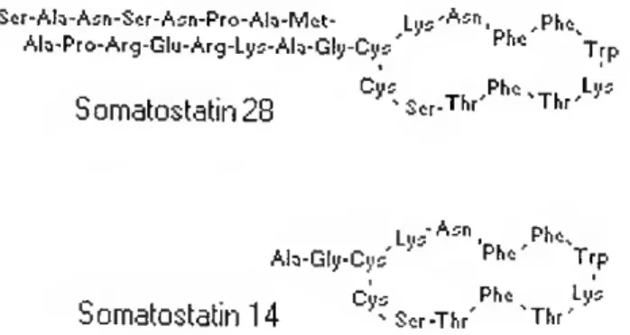
SOMATOSTATIN RELEASE-INHIBITING FACTOR (SRIF) - SRIF is a multifunctional hormone which inhibits the secretion of growth hormone. SRIF is a tetradecapeptide (SRIF-14) but multiple N-terminally extended forms such as SRIF-28, SRIF-22, SRIF-25, SRIF-34, and SRIF-37 have been isolated from hypothalamus, pancreas and intestinal tissue of mammals and non-mammals (King and Miller, 1979; Conlon et ai, 1985; Plisetskaya et ai, 1986; Cutfield et ai, 1987) (Fig.1.4).
Scr-Alí-Asn-Scr-Asn-Pro-Ala-Met- ,Phcs Ah-Pro-Arg-Glu-Arg-Lys-Ah-Gly-Cys' Trp
Cys Pho %T Lys Somatostal:in28 %Scr-Thr Thr
Lu5'A5n. ,Ph^ Alo-Gly-Cyc ' Ph,::' Trp _ „ < Cus Pta Ly- Somatostatm 14 ^ Scr-iV vThr'
Figure 1.4-Amino acid sequence of SS-14 and SS-18 (Rubinow et ai, 2000).
In mammals, in vitro and in vivo studies have shown that SRIF-14 and SRIF-28 not only inhibits GH secretion, but also secretion of PRL, TSH, and ACTH (Freeman et ai, 2000 for review). In rat and chick, it has also been shown to inhibit the enzyme activities of the small intestine (Taboada et ai, 1985).
In reptiles, SRIF immunoreactive endocrine cells have been detected in the pancreas (Buchan et ai, 1982). In avian species, SRIF-ir fibers and endocrine cells are localized in the brain, hypothalamus, proventriculus, gut, pancreas, and sympathoadrenal system of embryos and adults (Mikami and Yamada, 1984; Yamada et ai, 1985; Yamaguchi et ai, 1987; Garcia-Arrarás and
Chapter 1 General Introduction
Martinez, 1990; Erichsen et ai, 1991; Epstein and Poulsen, 1991; Erichsen et a/., 1994; Lucini eia/., 1996; Sánchez-Montesinos et ai, 1996; Takayanagi et ai,
1996).
In teleosts, immunocytochemical investigations have revealed the presence of SRIF-like material in the hypothalamus, pituitary, and gastro- intestinal tract of several species (Wigham, 1992; Zupanc et ai, 1994; Becerra et ai, 1995; Groff and Youson, 1997; Reinecke et ai, 1997; Batten et ai, 1999), including the sea bream (Power et ai, 1996). In rainbow trout (Oncorhynchus mykiss), preprosomatostatin (a SRIF precursor) has been isolated and characterized (Moore et ai, 1995) and has been shown to be expressed in the pancreas, stomach, intestine, and brain (Kittilson et ai, 1999). The fruit-eating fish, the pacu (Piaractus mesopotamicus), expresses two SRIF genes (de Lima, 1999). SRIF inhibits PRL synthesis and release in the trout (Wigham, 1992), and also appears to regulate the development of new neurons produced in response to injuries in the cerebellum (Zupanc, 1999).
THYROTROPIN-RELEASING HORMONE (TRH) - A hypophysiotrophic factor that stimulates thyroid-stimulating hormone (TSF!) secretion from pituitary cells was first isolated in 1966 (Schally et ai, 1966). In 1969, a group led by Guillemin (Burgus et ai, 1970) and another by Schally (Boler et ai, 1969) announced that the hypothalamic substance that causes the anterior pituitary gland to release TSH is L-pyroglutamyL-L-histidyl-L-prolineamide (L-pGlu-L-His-
Chapter 1 General Introduction
L-ProNH2) (Fig.1.5). This tripeptide is now called thyrotropin-releasing hormone (TRH) (Mason et ai, 2000).
In mammals, TRH-like immunoreactivity is widely distributed in the CNS, and TRH receptors have been identified on lactotrophs. TRH stimulates PRL release in a dose-dependent manner both in vitro and in vivo. Pharmacological blockade of VIP receptors attenuates the PRL response to TRH, consistent with TRH acting, at least partially, via local production of VIP (see Benker et ai, 1990,
Freeman et ai, 2000 and Schwartz, 2000 for reviews).
In avian species, TRH is distributed in the brain and hypothalarnus (Józsa et ai, 1988; Geris et ai, 1999). In the chicken, a TRH receptor has been cloned and characterized (Sun et ai, 1998). TRH stimulates PRL release from pituitary glands of fowl (Hall et ai, 1985b). and this effect is greatly increased in turkeys treated with estradiol benzoate (Saeed and el Halawani, 1986). In amphibians, TRH also stimulates PRL release (Preece and Licht, 1987).
In some teleosts, TRH immunoreactivity is detected in the brain, pituitary, and retina (Wigham, 1992; Anadón et ai, 2001; Diaz et ai, 2002). Recently, in the brain of embryos, alevin, and juveniles of brown trout {Salmo trutta fario),
Chapter 1 General Introduction
vitro studies have demonstrated that TRH plays an important role in both PRL synthesis and release in teleosts (Kagabu eia/., 1998; Wigham, 1992). Studies in chum salmon (Oncorhynchus keta) suggest TRH may be related to changes in olfactoryfunction during migration (Hamano eia/., 1996).
OXYTOCIN (OT) AND ISOTOCIN (IT) - The neurohypophyseal hormone oxytocin (OT) was originally identified as a nonapeptide with an amidated C- terminus (Acher et a/., 1970) (Table 1.3). In mammals it is primarily associated with the contraction of uterine and mammary smooth muscle during birth and lactation. Recently, plasma hyperosmolality has been identified as a stimulus for rat pituitary OT secretion (Rinaman et a/., 2000). OT is also involved in the regulation of PRL secretion, and an OT receptor has been localized on lactotrophs (Freeman et ai, 2000; Schwartz, J., 2000). In teleosts, oxytocin-ir fibers and binding sites are observed in the pituitary (Batten et ai, 1999) and in rainbow trout {Oncorhynchus mykiss), oxytocin increases PRL release from pituitaries in vitro (Wigham, 1992).
Table 1.3 - Amino acid sequence and similarities between the peptides isotocin and oxytocin
Isotocin CYISNC PIG* Oxytocin CYIQNCPLG* G*, amidated glycine residue
The functional role of isotocin the non-mammalian counterpart of OT (Table 1.3), is less well defined. The distribution of isotocin-ir fibers and binding
Chapter 1 General Introduction
sites have been described in the pituitary of three species of teleost sailfin mollies (Poecilia latipinna), sea bass (Dicentrarchus labrax), and North African catfish {Ciarias gariepinus) (Batten et a/., 1999), but no physiological studies of the activity of this peptide exist in fish.
NEUROPEPTIDE Y (NPY) - NPY is a member of the pancreatic polypeptide family isolated by Tatemoto in 1982 (Table 1.4). In mammals, NPY is distributed in the CNS, particularly in the hypothalamus, and it is a substance that alters the function of several different pituitary cell types (reviewed by Freeman et al., 2000). In rats, NPY inhibits PRL secretion and attenuates both the PRL- secretory and intracellular calcium flux responses to TRH in pituitary cells. The activity of NPY varies as a function of the estrous cycle, and expression leveis also change according to the steroid environment (reviewed by Schwartz, J., 2000).
Table 1.4- NPY amino acid sequence
YPSKPDNPGEDAPAEDLARYYSALRHYINLITRQRY-NH2
In avian species, NPY-ir fibers were seen in the brain and in the endocrine cells of the pancreas (Erichsen et al., 1991; Erichsen et al., 1994; Lucini et al., 2000; Mensah-Brown et al., 2000). In the chick embryo, NPY-ir cells were detected in the developing adrenal-gland (Sánchez-Montesinos et al., 1996). In an immunochemical study of the pituitary gland of some teleosts sailfin mollies, {Poecilia latipinna), sea bass (Dicentrarchus labrax), and North African catfish
were described in the pituitary (Batten et a/., 1999). In turbot, NPY-ir endocrine cells and fibers were observed throughout the gastro-.intestinal tract (Reinecke et a/., 1997), although no study of the brain was carried out.
NEUROTENSIN (NT) - Neurotensin (NT) is a tridecapeptide that was originally isolated from bovine hypothalamus (Carraway and Leeman, 1973) (Table 1.5) and was originally thought to be a vasoactive peptide. Subsequent studies have shown in mammals that NT is involved in a range of physiological processes, including blood flow, digestion, temperature regulation and nociception (Leeman and Carraway, 1982).
Table 1.5 - NT amino acid sequences of some mammals, avian, and fish species (Carraway and Bhatnagar, 1980; Carraway and Leeman, 1973; Hammer et al., 1980; Rodriguez-Bello et al., 1993; Warner et al., 1998)
Bovine, canine, human elyenkprrpyil-oh Chicken, alligator ELHVNKARRPYIL - OH
Toad E A 1 VSKARRPYIL - OH
Boldface denotes aa which are different from bovine sequence
Immunocytochemical studies have shown that NT is present in the mammalian brain and anterior pituitary (Uhl et al., 1977; Emson et al., 1982). One of the most potent central effects of NT in mammals is its analgesic action which can be blocked by thyroxine releasing hormone (Hernandez et al., 1984). Intravenous injection of NT leads to an increase in circulating PRL leveis while intracerebroventricular injections cause an inhibition of PRL release (Vijayan and McCann, 1979). In contrast NT increases PRL secretion in a dose-dependent manner in vitro. The opposite effects of NT in vivo and in vitro have been taken to
Chapter 1 General Introduction
indicate that NT can affect PRL secretion at multiple leveis (Freeman et a/., 2000).
In avian species, NT-ir endocrine cells were localized in the brain, proventriculus, and gizzard (Yamada et a/., 1985; Yamaguchi et a/., 1987; Esposito et a/., 1997). In vivo and in vitro studies in chickens showed that NT has an effect on the motility of the lower gut (Rawson et a/., 1990).
In the turbot (Scophthalmus maximus) and in the rosy barb (Barbus conchonius), NT-ir endocrine cells and fibers were observed throughout the gastro-intestinal tract (Rombout and Reinecke, 1984; Reinecke et ai, 1997). In the goldfish (Carassius auratus), extensive NT-ir was observed in the brain and pituitary (Bello et ai, 1994). Excitatory effects of NT on fish gut smooth muscle has been described (Holmgren, 1985).
SUBSTANCE P - Substance P was first described in 1931 by von Euler and Gaddum who demonstrated brain extracts contained substances that caused contractions of the intestinal preparations and lowered blood pressure. It was only in 1971 that the amino acid sequence of this undecapeptide was determined in extracts of bovine hypothalamus (Chang et ai, 1971) (Table 1.6). Substance P belongs to a family of neuropeptides known as tachykinins that share in common C-terminal sequence; Phe-X-Gly-Leu-Met-Nhb.
Table 1.6 - Amino acid sequence of substance P
Chapter 1 General Introduction
Substance P has a wide distribution in the nervous system of both vertebrates and invertebrates. In primates and rodents numerous immunoreactive cell bodies and fibers are present in the hypothalamus. Moreover, a high levei of expression of substance P receptors has been detected in the hypothalamus and pituitary. Substance P has been shown to regulate both in vitro and in vivo PRL secretion in primates (rhesus monkey) and rats, but paradoxical effects have been obtained - it appears that the actual effect of this peptide on PRL secretion depends on the dose and route of administration (reviewed by Freeman et a/., 2000).
In avian species, substance P-ir fibers are localized in the hypothalamus and brain (Mikami and Yamada, 1984; Erichsen ef a/., 1991; Erichsen et ai, 1994). In the teleosts, substance P-ir fibers have been described in the pituitary of sailfin mollies {Poecilia latipinna), sea bass {Dicentrarchus labrax), and North African catfish {Ciarias gariepinus) (Batten et ai, 1999). Substance P-ir endocrine cells and fibers have been observed in the gastro-intestinal tract of a range of teleosts in the elasmobranches and cyclostomes (Jensen, 1989). However, an authentic substance P has yet to be found in non-mammalian species (Severini et ai, 2002).
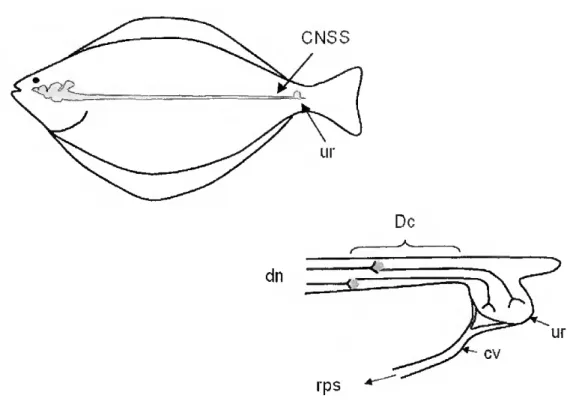
UROTENSIN - The caudal neurosecretory system of teleost fish (Fig. 1.6) which terminates in the urophysis, secretes two major regulatory peptides: urotensin I and urotensin II (Onstottk and Elde, 1986; Chester-Jones et al, 1987; Larsonefa/., 1987), a cyclic 12-amino acid residue peptide that has some
f
Dc
rps
Figure 1.6 - Diagrammatic representation of the caudal neurosecretory system of fish (CNSS). Descendings neurons in the caudal spinal cord (dn) project axons to the urophysis (ur).where the neurosecretory nerve terminais link to the caudal vein (cv) of the renal portal system (rps). Dahlgren cells secrete two osmoregulatory peptides (urotensin I and urotensin II) (adapted from an original drawing by Dr. Peter Hubbard).
sequence similarity, but is not homologous, to somatostatin-14 (Pearson ef a/., 1980). Urotensin II is a 12-amino acid peptide, and the structural characterization of this peptide from several species of fish has shown that the cyclic region of the peptide has been strongly conserved (Le Mevel ef a/., 1996) (Table 1.7).
Urotensin II is not confined to the caudal neurosecretory system of fish. and has been identified in anterior spinal cord and brain of several species (Yulis and Lederis, 1988; Wigham, 1992; Chartrel etal., 1998).
Chapter 1 General Introduction
Table 1.7 - Amino acid sequence of Urotensin II from two teleosts (trout and carp) and an elasmobranch (Waugh and Conlon, 1993)
Trõut G G N SE CFW KYCV-OH Carp GGN TECFWKYCV-OH Skate NN FSDCFWKYCV-OH Boldface denotes aa which are different from trout sequence
Although the precise physiological role of urotensin II remains unclear, the diverse actions of urotensin II suggest the possibility of cardiovascular and renal effects on human, rat, frog, eel and trout (Chan, 1975; Gibson et a/., 1986; Itoh et ai, 1987; Yano et ai, 1995; Le Mevel et ai, 1996; Bõhm and Pernow, 2002; Zhang et ai, 2003 ), as well as a role in lipid and carbohydrate metabolism in coho salmon (Oncoriiynchus kisutch) and dogfish {Torpedo marmorata) (Sheridan et ai, 1987; Conlon et ai, 1994). Moreover, urotensin II inhibited PRL release, in a dose-related manner from Mozambique tilapia {Oreochromis mossambicus) pituitaries cultured in vitro (Rivas et ai, 1986).
1.6.4 Hormones
OVARIAM STEROIDS - In mammals the modulation of PRL by ovarian steroids is well documented (Labrie et ai, 1978). In rats, estradiol-17í3 (Ez) increases the mitotic potency of PRL cells in the pituitary gland and has a stimulatory effect on PRL gene expression in the hypothalamo- neurohypophyseal system and a concomitant inhibitory action on PRL proteolysis at this site (Takahashi and Kawashima, 1986; Torner et ai, 1999). In mink, Mustela vison, a high systemic ratio of progesterone to Ez has been shown to be
Chapter 1 General Introduction
a prerequisite for increasing the expression of uterine PRL receptors (Rose et a/., 1996).
Ovarian steroids also affect the pituitary in birds, modulating in vitro PRL release (Knapp et a/., 1988). In teleost fish the regulation of PRL cells by ovarian steroids has been less extensively studied although there is evidence indicating their involvement, for example, pituitary PRL content and in vitro secretion is elevated by treatment with E2 in Mozambique tilapia (Borski et ai., 1991; Wigham, 1992; Poh et a/., 1997). Moreover, preincubation of tilapia pituitary glands with E2 in vitro appears to increase the sensitivity of PRL cells to stimulation by TRH and GnRH in vitro (Barry and Grau, 1986; Weber et ai, 1997). In contrast, E2 treatment of rainbow trout pituitary cultures stimulated in vitro PRL synthesis but did not affect release (Wigham, 1992; Williams and Wigham, 1994).
CORTISOL - In fish, cortisol production is located in the interrenal cells. These cells do not form a compact gland comparable to the mammalian adrenal córtex, but are located in layers, strands, and cords around the walls of the posterior cardinal veins and its branches run through the head kidneys (Fig. 1.7) (Wendelaar-Bonga, 1997). Studies by Young (1993) on hypophysectomized coho salmon (Oncorhynchus kisutch) have indicated that the pituitary gland dominates the endocrine control of cortisol secretion. a-Melanophore-stimulating hormone (a MSH) and ACTH are the main candidates for cortisol regulation with perhaps (3 endorphin as a potentiating factor (Wendelaar-Bonga, 1997).