1. Department of Physical Medicine and Rehabilitation, Faculty of Medicine, Mustafa Kemal University, Hatay, Turkey
2. Iskenderun Marine Regiment Medical Center, Hatay, Turkey 3. Division of Rheumatology, Department of Internal Medicine, Faculty of Medicine, Hacettepe University, Ankara, Turkey 4. Department of Biochemistry, Faculty of Medicine, Mustafa Kemal University, Hatay, Turkey
Sclerostin and Dkk-1 in patients
with ankylosing spondylitis
ACTA REUMATOL PORT. 2014:39;146-151
AbstrAct
Objective: To determine the serum Dickkopf-related
protein 1 (Dkk-1) and sclerostin levels, and their rela-tionship to structural damage and disease activity in pa-tients with ankylosing spondylitis (AS), as well as to compare the serum Dkk-1 and sclerostin levels in pa-tients receiving and not receiving anti-TNF-a treatment.
Materials and Methods: This cross-sectional study
in-cluded 44 AS patients and 41 healthy age- and -matched controls. Demographic data, disease activity parameters, and Bath Ankylosing Spondylitis Radiolo -gic Index (BASRI) scores were recorded. Serum Dkk-1 and sclerostin levels were measured using commercial-ly available ELISA.
Results: Serum Dkk-1 levels were lower (P > 0.05) and
sclerostin levels were significantly lower (P < 0.05) in the AS patients than in the controls. Dkk1 and sclerostin le -vels were similar in the patients that did and didn’t re-ceive anti-TNF-a treatment, and in the patients with ac-tive and inacac-tive disease (P > 0.05). There wasn’t a correlation between serum Dkk1 or sclerostin le vels, and di -sease activity indices (P > 0.05). BASRI scores did not correlate with serum Dkk-1 or sclerostin levels (P > 0.05).
Discussion: Sclerostin expression is impaired in AS,
but this is not the case for Dkk-1. The lack of an asso-ciation between Dkk-1 or sclerostin levels, and anti--TNF-a treatment, disease activity indices, and radio-logical damage might indicate that neither the Dkk-1 nor sclerostin level induce inflammation and radiolo gical damage in AS patients. Pathologic bone formation in AS might be due to molecular dysfunction of scle-rostin and Dkk-1 at the cellular level.
Ustun N1, Tok F2, Kalyoncu U3, Motor S4, Yuksel R4, Yagiz AE1, Guler H1, Turhanoglu AD1
Keywords: Ankylosing spondylitis; Structural damage;
Anti-TNF-a; Dkk-1; Sclerostin.
IntroductIon
Ankylosing spondylitis (AS) – a chronic inflammatory disease that predominantly affects axial joints and in-tervertebral spaces - is characterized by new bone for-mation and is thereby associated with syndesmophytes and ankylosis1. Several biomarkers, including compo-nents of the Wnt pathway signaling cascade that regu-late bone formation, have been evaluated in order to determine their role in the clinical prognosis of AS and the response to treatment2-9. The most commonly stu -died secreted Wnt inhibitors are sclerostin and Dick-kopf-related protein 1 (Dkk-1). Dkk-1 appears to be the most important biologically, as it was recently re-ported that Dkk-1 is a regulator of joint remodeling in animal models of arthritis, and that elevated Dkk1 le vels were linked to bone resorption and low Dkk1 le -vels were associated with new bone formation10. Scle-rostin, a soluble inhibitor of the Wnt pathway that is closely related to Dkk-1, is a bone-specific molecule produced by osteocytes that prevents binding of Wnt proteins, leading to inhibition of the Wnt pathway8. Loss-of-function mutations of the gene encoding scle-rostin are linked to diseases characterized by increased bone mass.
Numerous studies have sought to identify and clari -fy the mechanisms of bone turnover in AS, focusing on exploration of the potential role of Dkk-1 and sclerostin levels, and anti-TNF-a treatment in new bone forma-tion in AS patients2-9; however, the findings have been inconsistent. Some studies reported elevated serum sclerostin and Dkk-1 levels in AS patients, as compared to controls, whereas others reported lower levels and others reported similar levels in AS patients and healthy controls2-9. Findings regarding the effects of anti-TNF-a
treatment on serum sclerostin and Dkk-1 levels, and the relationship between these Wnt inhibitors, and di -sease activity status and structural damage, are also in-consistent2-9. As such, the present study aimed to add to the limited available data via examination of the re-lationship between sclerostin and Dkk-1 levels, and structural damage in AS patients.
mAterIAls And methods
This cross-sectional study included 44 AS patients re-cruited from the physical medicine and rehabilitation clinic of our hospital, and 41 age- and gender-matched healthy controls. All the AS patients included fulfilled the modified New York criteria11. The study protocol was approved by the Local Ethics Committee.
Demographic data and disease-specific data were recorded. Disease activity was assessed using the Bath Ankylosing Spondylitis Disease Activity Index (BAS-DAI)12. Patients with a BASDAI score ≥4 were consi dered to have active disease. Markers of inflammation (the ery-throcyte sedimentation rate [ESR] and C-reactive pro-tein [CRP] level) were measured in all patients and con-trols. Radiographic assessment was performed using the Bath Ankylosing Spondylitis Radiologic Index (BASRI)13. Serum samples were obtained from all participants and were stored in aliquots of 200 µL at –20 °C.
serum dkk-1 And sclerostIn meAsurement
Serum Dkk-1 levels were measured using a commer-cially available ELISA kit (Human Dkk-1 ELISA Kit), according to the manufacturer’s instructions (Adipo Bioscience, Santa Clara, USA). Serum sclerostin levels were measured using a commercially available ELISA kit (Human Soluble Sclerostin [SOST] ELISA Kit), ac-cording to the manufacturer’s instructions (Adipo Bio-science, Santa Clara, USA). All measurements were performed in triplicate for each sample, and a mean value was calculated.
stAtIstIcAl AnAlysIs
Statistical analysis was performed using SPSS v.13.0 for Windows (SPSS, Inc., Chicago, IL). Variables were tes ted for normality via the Shapiro-Wilk test.
Corre-lations between Dkk-1, sclerostin, and other variables were analyzed via Pearson’s or Spearman’s test, as ap-propriate. The Mann-Whitney U test was used for group comparisons. The level of statistical significance was set at P < 0.05.
results
The study included 34 (77%) male and 10 (23%) fe-male patients. In all, 19 (43%) patients were using an anti-TNFa agent for a mean 2.74 ± 1.52 years and 25 (57%) were receiving sulfasalazine - a disease-modify-ing anti-rheumatic drug. Among the patients, 11 (25%) had a BASDAI score ≥4, 14 (32%) had an ESR >20 mm h–1, and 20 (44%) had a CRP >0.8 mg dL–1. BASDAI scores were significantly lower in the patients that were using an anti-TNF-a agent than in those that were not (1.97 ± 2.38 vs. 3.16 ± 1.57, P = 0.012). Patient de-mographic and clinical data are presented in Table I.
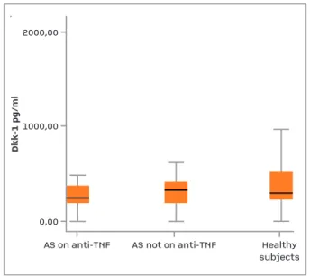
Serum sclerostin levels were significantly lower in the patients with AS than in the healthy controls (P = 0.037) (Table II). Although numerically lower, the difference between the Dkk-1 level in the AS patients and controls was not significant (314.96 pg mL–1 vs. 613.34 pg mL–1, P = 0.062). Serum sclerostin and Dkk-1 levels were similar in the patients that did (n = 19) and did not (n = 25) use an anti-TNF-a agent (P > 0.05) (Figures 1 and 2, and Table II). Serum sclerostin and Dkk-1 levels were similar in the patients with active disease and inactive disease (P > 0.05) (Figures 1 and
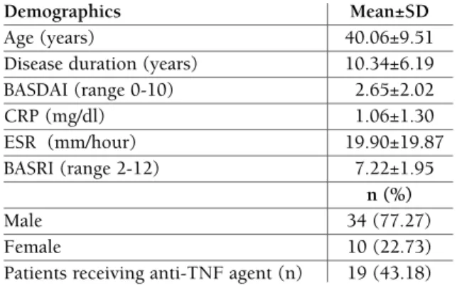
tAble I. demogrAphIc And clInIcAl dAtA of the pAtIents
Demographics Mean±SD
Age (years) 40.06±9.51
Disease duration (years) 10.34±6.19
BASDAI (range 0-10) 2.65±2.02 CRP (mg/dl) 1.06±1.30 ESR (mm/hour) 19.90±19.87 BASRI (range 2-12) 7.22±1.95 n (%) Male 34 (77.27) Female 10 (22.73)
Patients receiving anti-TNF agent (n) 19 (43.18)
BASDAI, Bath Ankylosing Spondylitis Disease Activity Index; CRP, C-reactive Protein; ESR, erythrocyte sedimentation rate; BASRI, Bath Ankylosing Spondylitis Radiologic Index
2, Table II). There wasn’t an association between the serum sclerostin or Dkk-1 level, and disease activity in-dices (BASDAI, ESR, and CRP) (P > 0.05). Structural damage assessed via BASRI did not correlate with serum sclerostin or Dkk-1 levels (P > 0.05).
dIscussIon
In the present cross-sectional study it was observed
that the serum sclerostin level was significantly hi gher in the AS patients than in the healthy controls. Al-though numerically lower, the difference between the Dkk-1 level in the AS patients and controls was not significant. Additionally, the sclerostin and Dkk1 le -vels were similar in the patients with active disease and inactive disease. Moreover, sclerostin and Dkk-1 le vels were similar in the patients that were using an anti--TNF-a treatment and those that were not, and struc-tural damage, as assessed via BASRI, did not correlate with serum Dkk-1 or sclerostin levels.
*p<0.05.
AS, ankylosing spondylitis; anti-TNF-a, anti–tumor necrosis factor-a.
tAble II. serum sclerostIn And dkk-1 levels of the pAtIents And heAlthy subjects
n Sclerostin ± SD (pg/ml) P Dkk-1 ± SD (pg/ml) P
Patients with AS 44 427.69 ±368.10
0.037* 314.96±196.73 0.062
Healthy subjects 41 656.32±643.51 613.34±861.86
Receiving anti-TNF-a agent 19 393.21±305.80
0.507 275.07±120.42 0.586
Not eceiving anti-TNF-a agent 25 453.90±413.50 345.28±237.17
Patients with active disease 11 449.15±327.94
0.574 331.85±267.65 0.504
Patients with inactive disease 33 420.54±385.05 309.33±385.05
AS on anti-TNFAS not on anti-TNFHealthysubjects
2000,001000,000,00
Sclerostin pg/ml
3000,00
AS on anti-TNF AS not on anti-TNF Healthy subjects 2000,00 1000,00 0,00 S cl e ro st in p g /m l 3000,00
fIgure 1.Serum sclerostin levels of the subjects Patients with AS had significantly lower sclerostin levels compared with controls (p<0.05). Patients receiving
anti-TNF-a agents and who were not receiving anti-TNF-a agents had similar sclerostin levels (p>0.05).
Values are presented as mean and SD.
AS, ankylosing spondylitis; anti-TNFa, anti–tumor necrosis factor a AS on anti-TNFAS not on anti-TNFHealthysubjects
2000,001000,000,00
Dkk-1 pg/ml
AS on anti-TNF AS not on anti-TNF Healthy subjects 2000,00 1000,00 0,00 D kk -1 p g/ m l
fIgure 2.Serum Dkk-1 levels of the subjects
Although numerically lower, the difference between level of Dkk-1 in AS patients and healthy subjects did not reach statistical significance (314.96 vs. 613.34 pg/ml, P=0.062). Patients receiving anti-TNF-a agents and who were not receiving anti-TNF-a agents had similar Dkk-1 levels (p>0.05).
Values are presented as mean and SD.
The present findings regarding sclerostin levels are in accordance with those of Appel et al.6and Saad et al.3. Appel et al. were among the first researchers to evalu-ate serum sclerostin levels in AS patients. They com-pared 46 AS patients and 50 healthy controls, and re-ported that sclerostin levels were lower in the AS pa-tients. Moreover, they reported correlation with for-mation of new syndesmophytes6; however, in the present study an association between structural dama -ge and the sclerostin level was not observed. This dif-ference in findings might be due to difdif-ferences in the indices used to evaluate the radiological status of AS patients; whereas Appel et al. used the modified Stoke Ankylosing Spondylitis Spine Score (mSASSS), the pre-sent study used BASRI for structural damage assessment. These 2 indices use completely different parame -ters for scoring. Saad et al. also reported lower scle-rostin levels in AS patients than in controls; however, they reported a significant increase in the sclerostin le -vel after 12 months of anti-TNF-a treatment in their cohort3, whereas in the present study an association between the sclerostin level and anti-TNF-a treatment was not noted. This difference in findings might be due to the differences in the 2 study’s methodologies.
Although in the present study 19 patients received anti-TNF-a treatment for a mean 2.74 years, their pre-treatment sclerostin levels were not known. Moreover, the sclerostin levels in the patients in Saad et al. study after 2 years are not known. Resistance or insensitivity always occurs with anti-TNF-a treatment, which can decrease the sclerostin level to the pretreatment level, which makes direct comparison between studies diffi-cult. Nonetheless, Taylan et al. reported that sclerostin levels were similar in AS patients and healthy controls4, whereas, sclerostin and Dkk-1 levels did not vary ac-cording to disease activity. In contrast, Korkosz2 re-ported that sclerostin levels were significantly higher and Dkk-1 levels lower in patients with high disease activity; this contradiction might indicate that the in-teraction between disease activity and inhibitors of bone formation in AS is complex.
Daoussis et al.9reported higher Dkk-1 levels in AS patients, Kwon et al.7reported lower Dkk-1 levels in AS patients, and Taylan et al.4reported similar Dkk1 le -vels in AS patients, as compared to healthy controls. The present findings are in accordance those of Taylan et al. Daoussis et al. reported that patients receiving anti-TNF-a treatment had higher Dkk-1 levels than those that were not9; whereas, both Kwan et al. and Taylan et al. reported that Dkk-1 levels did not vary
ac-cording to anti-TNF-a treatment4,7, which is in agree-ment with the present findings. The inconsistency of findings might indirectly indicate that neither TNF-a per se, nor inflammation in general, are the primary inducers of Dkk-1.
In an animal model of chronic inflammatory arthri-tis TNF-a was shown to induce skeletal expression of Dkk-1, which in turn triggered sclerostin production14 - both molecules being potent inhibitors of new bone formation15-17; however, the present findings show that serum levels of sclerostin and Dkk-1 did not differ ac-cording to disease activity or use of anti-TNF-a treat-ment, which might indicate that there are molecular mechanisms other than those related to acute-phase response and anti-TNF-a treatment that are responsible for modulation of serum Dkk1 and sclerostin le -vels in AS patients10. The link between TNF-a, and scle-rostin and Dkk-1 warrants additional research, as it could have pathogenic and clinical implications in AS.
TNF-a blockers have been successfully used to sup-press inflammation in AS18-20. In the present study the patients that were using an antiTNFa agent had signi -ficantly lower disease activity. The hypothesis that ankylosis is invariably preceded by inflammation in AS21is yet to be definitively proven by prospective stu -dies, but might be due to either the presence of un-derlying inflammation that is not detectable via MRI22,23 or the role of a noninflammatory pathway. The deve -lopment of new bone in the spine in the form of syn-desmophytes and ankylosis is still evaluated via plain radiography24. A minimum of 2 years is required before radiographic changes can be reliably detected25. The hypothesis that anti-TNF-a agents might not prevent the development of spinal ankylosis in AS26,27 also mains to be definitively proven via prospective re-search, which indicates that even in AS patients in-flammation and effective suppression of inin-flammation could be more relevant than a minor increase in osteo-proliferation. The AS patients in the present study that were using anti-TNF-a agents used them for about 3 years (on average), which is not considered to be long--term use, and the present study employed a cross-sec-tional - not prospective - design.
Lastly, in the present study an association between structural damage and the serum Dkk-1 level was not observed, which is in agreement with Taylan et al.4and Korkozs et al.2. Moreover, Dkk-1 and sclerostin levels in the present study did not correlate with radiological damage. Although Dkk-1 and sclerostin have been shown to be potent inhibitors of bone remodeling, the
lack of association between structural damage, and sclerostin and Dkk-1 in the present study and earlier studies might indicate that serum sclerostin and Dkk-1 levels are not the primary predictors of structural da mage in AS patients, but might be indicative of a glo -bal bone metabolism. More comprehensive cellular mechanisms and receptorial dysfunction of sclerostin and Dkk-1 might be factors that play a more important role in structural damage in AS patients.
The present study has some limitations; primarily, the moderate sample size and the lack data on pre-treatment sclerostin and Dkk-1 levels in the patients that were receiving anti-TNF-a agents, as well use of a cross-sectional rather than prospective design, and lack of examination of additional Wnt pathway in-hibitors, bone mineral density values, and bone turnover markers. The remaining issue is whether or not serum sclerostin and Dkk-1 levels are stable enough to warrant the conclusion that structural dama ge is not associated with these molecules. Final-ly, the lack of data on patient HLA-B27 positivity or negativity is another limitation. Nevertheless, we think the present findings are clinically important.
conclusIon
Sclerostin expression is impaired in AS, but this is not the case for Dkk-1. Lack of an association between Dkk-1 or sclerostin levels, and anti-TNF-a treatment, disease activity indices, and radiological damage might indicate that neither Dkk-1 nor sclerostin level are fac-tors associated with inflammation and radiological damage in AS. Lastly, pathologic bone formation in AS might be due to molecular dysfunction of sclerostin and Dkk-1 at the cellular level.
correspondence to
Fatih Tok
Iskenderun Deniz Alayi
Saglik Merkezi, Iskenderun, Hatay E-mail: drfatihtok@gmail.com
references
1. Schett G. Bone formation versus bone resorption in ankylosing spondylitis. Adv Exp Med Biol 2009; 649:114–121. 2. Korkosz M, Gąsowski J, Leszczyński P, Pawlak-Buś K, Jeka S,
Kucharska E, Grodzicki T. High disease activity in ankylosing spondylitis is associated with increased serum sclerostin level and decreased wingless protein-3a signaling but is not linked with greater structural damage. BMC Musculoskelet Disord 2013:19; 14:99.
3. Saad CG, Ribeiro AC, Moraes JC, Takayama L, Goncalves CR,
Rodrigues MB, et al. Low sclerostin levels: a predictive marker of persistent inflammation in ankylosing spondylitis during anti-tumor necrosis factor therapy? Arthritis Res Ther 2012; 14:216.
4. Taylan A, Sari I, Akinci B, Bilge S, Kozaci D, Akar S, et al. Bio-markers and cytokines of bone turnover: extensive evaluation in a cohort of patients with ankylosing spondylitis. BMC Mus-culoskelet Disord 2012; 13:191.
5. Heiland GR, Appel H, Poddubnyy D, Zwerina J, Hueber A, Hai-bel H, et al. High level of functional dickkopf-1 predicts pro-tection from syndesmophyte formation in patients with anky-losing spondylitis. Ann Rheum Dis 2012; 71:572-574. 6. Appel H, Ruiz-Heiland G, Listing J, Zwerina J, Herrmann M,
Mueller R, et al. Altered skeletal expression of sclerostin and its link to radiographic progression in ankylosing spondylitis. Arthritis Rheum 2009; 60:3257-3262.
7. Kwon SR, Lim MJ, Suh CH, Park SG, Hong YS, Yoon BY, et al. Dickkopf-1 level is lower in patients with ankylosing spondy-litis than in healthy people and is not influenced by anti-tumor necrosis factor therapy. Rheumatol Int 2012; 32:2523-2527. 8. Daoussis D, Andonopoulos AP. The emerging role of
Dickkopf-1 in bone biology: is it the main switch controlling bone and joint remodeling? Semin Arthritis Rheum 2011; 41:170-177. 9. Daoussis D, Liossis SN, Solomou EE, Tsanaktsi A, Bounia K, Ka-rampetsou M, et al. Evidence that Dkk-1 is dysfunctional in ankylosing spondylitis. Arthritis Rheum 2010; 62:150-158. 10. Diarra D, Stolina M, Polzer K, Zwerina J, Ominsky MS, Dwyer
D, et al. Dickkopf-1 is a master regulator of joint remodeling. Nat Med 2007; 13:156-163.
11. Van der Linden SM, Valkenburg HA, Cats A. Evaluation of diag-nostic criteria for ankylosing spondylitis: a proposal for modi-fication of the New York criteria. Arthritis Rheum 1984; 27:361–368.
12. Garrett S, Jenkinson T, Kennedy LG, Whitelock H, Gaisfrod P, Calin A. A new approach to defining disease status in ankylo-sing spondylitis: the Bath Ankyloankylo-sing Spondylitis Disease Ac-tivity Index. J Rheumatol 1994; 21:2286–2291.
13. MacKay K, Mack C, Brophy S, Calin A. The Bath Ankylosing Spondylitis Radiology Index (BASRI): a new, validated ap-proach to disease assessment. Arthritis Rheum 1998;41:2263--2270.
14. Heiland GR, Zwerina K, Baum W, Kireva T, Distler JH, Grisan-ti M et al. NeutralisaGrisan-tion of Dkk-1 protects from systemic bone loss during inflammation and reduces sclerostin expression. Ann Rheum Dis 2010; 69:2152–2159.
15. Rosen V. BMP and BMP inhibitors in bone. Ann N Y Acad Sci 2006; 1068:19–25.
16. Lories R, Luyten F. Bone morphogenic proteins in destructive and remodelling arthritis. Arthritis Res Ther 2007; 9:207–214. 17. Krishnan V, Bryant HU, Macdougald OA. Regulation of bone mass by Wnt signaling. J Clin Invest 2006; 116:1202-1209. 18. Braun J, Brandt J, Listing J, Zink A, Alten R, Golder W, et al.
Treatment of active ankylosing spondylitis with infliximab: a randomised controlled multicentre trial. Lancet 2002; 359:1187–1193.
19. Davis JC Jr, van der Heijde D, Braun J, Dougados M, Cush J, Clegg DO, et al, for the Enbrel Ankylosing Spondylitis Study Group. Recombinant human tumor necrosis factor receptor (etanercept) for treating ankylosing spondylitis: a randomized, controlled trial. Arthritis Rheum 2003; 48:3230–3236. 20. Van der Heijde D, Kivitz A, Schiff MH, Sieper J, Dijkmans BA,
Braun J, et al, for the ATLAS Study Group. Efficacy and safety of adalimumab in patients with ankylosing spondylitis: results of a multicenter, randomized, double-blind, placebo-control-led trial. Arthritis Rheum 2006; 54:2136–2146.
21. Maksymowych WP, Inman RD, Salonen D, Dhillon SS, Kris-hnananthan R, Stone M, et al. Spondyloarthritis Research Con-sortium of Canada magnetic resonance imaging index for as-sessment of spinal inflammation in ankylosing spondylitis. Arthritis Rheum 2005; 53:502–509.
22. Sieper J, Baraliakos X, Listing J, Brandt J, Haibel H, Rudwaleit M, et al. Persistent reduction of spinal inflammation as asses-sed by magnetic resonance imaging in patients with ankylo-sing spondylitis after 2 yrs of treatment with the anti-tumour necrosis factor agent infliximab. Rheumatology (Oxford) 2005; 44:1525–1530.
23. Appel H, Loddenkemper C, Grozdanovic Z, Ebhardt H, Drei-mann M, Hempfing A, et al. Correlation of histopathological findings and magnetic resonance imaging in the spine of pa-tients with ankylosing spondylitis. Arthritis Res Ther 2006; 8:143.
24. Braun J, Baraliakos X, Golder W, Hermann KG, Listing J, Brandt J, et al. Analysing chronic spinal changes in ankylosing spon-dylitis: a systematic comparison of conventional x rays with magnetic resonance imaging using established and new sco-ring systems. Ann Rheum Dis 2004; 63:1046–1055. 25. Wanders AJ, Landewe RB, Spoorenberg A, Dougados M, van
der Linden S, Mielants H, et al. What is the most appropriate radiologic scoring method for ankylosing spondylitis? A com-parison of the available methods based on the Outcome Mea-sures in Rheumatology Clinical Trials filter. Arthritis Rheum 2004; 50:2622–2632.
26. Van der Heijde D, Landewe R, Baraliakos X, Houben H, van Tu-bergen A, Williamson P, et al. Radiographic findings following two years of infliximab therapy in patients with ankylosing spondylitis. Arthritis Rheum 2008; 58:3063–3070. 27. Van der Heijde D, Landewe R, Einstein S, Ory P, Vosse D, Ni L,
et al. Radiographic progression of ankylosing spondylitis after up to two years of treatment with etanercept. Arthritis Rheum 2008; 58:1324–1331.