1. Department of Neurology, Chinese PLA General Hospital, Beijing *Both authors had the same contribution for this paper
gene sis of vascular diseases in AS patients. Further prospective study with more samples will be needed to confirm this hypothesis.
Keywords:Cerebrovascular disease; anklyosing spon -dylitis; inflammatory factors; risk factors.
IntroductIon
Ankylosing spondylitis (AS) is a systemic inflammato-ry rheumatic disease that affects the axial skeleton, causing characteristic inflammatory back pain, and can lead to spinal immobility1. AS predominantly affects
young adults, with a peak age of onset between 20 and 30yearsold, and AS is more prevalent in males1.
Atherosclerosis is a chronic progressive disease and one of the main causes of vascular diseases2. Against a
background of chronic inflammatory disease, such as in cases of AS, the development of atherosclerosis may be accelerated3-5, and an association between
prema-ture atherosclerosis and chronic inflammatory disease has been demonstrated6,7. With regard to AS
specifi-cally, previous studies reported significant differences in morbidity, mortality, and risk factors such as diabetes mellitus, hypertension, and altered lipid profiles be-tween cardiovascular disease patients with and with-out AS8-10. Moreover, additional studies have indicated
that patients with AS likely have an elevated risk of de-veloping coronary artery disease (CAD)11-13.
Cardiovascular diseases share the same etiology re-lated to atherosclerosis. However, the etiology of cerebrovascular disease is heterogeneous and may be rela -ted to atherosclerosis, vasculitis, and/or embolism. Some rheumatic diseases, such as polymyalgia rheuma -tica, were associated with higher risk of stroke14-16. It
re-mains to be determined whether this is also true in cere-brovascular disease patients with AS. A few studies in the field of clinical neurology have compared the fea-tures of cerebrovascular disease between patients with
Risk of premature cerebrovascular disease
in patients with ankylosing spondylitis
Zhang X1, Liu R1*, Wang J1, Zhang Y1, Liu Y1, Yu Z1, Yu S1
ACTA REUMATOL PORT. 2016;41:322-327
AbstrAct
Objectives: Patients with ankylosing spondylitis (AS) are at an elevated risk for the development of coronary artery disease, but the risk cerebrovascular disease among these patients remains incompletely under-stood. We investigated the cerebrovascular risk profiles of patients with a cerebrovascular disease and AS and compared these profiles to those of cerebrovascular di -sease patients without AS. Methods: We retrospective-ly anaretrospective-lysed 34 patients with ischemic cerebrovascular disease also diagnosed with AS and 597 controls with-out AS with respect to patient age, gender, cerebrovas-cular risk factors, and laboratory test results.
Results: AS patients were significantly younger than control patients in this study (56.2±13.5 years vs. 63.0±13.4 years, respectively; p=0.004). Logistic re-gression analysis did not indicate significant relation-ships between gender, cerebrovascular risk factors, and biochemical risk factors in AS patients, nor were any significant relationships found between erythrocyte sedimentation rate or Creactive protein and bioche -mical risk factors. A low frequency of large-artery atherosclerosis and high frequency of small-vessel oc-clusion according to Trial of ORG 10172 in Acute Stroke Treatment (TOAST) classification were found in AS patients with stroke.
-AS and those without -AS14-16. At present, there are no
published studies comparing the cerebrovascular risk profiles of cerebrovascular disease patients with and without AS receiving treatment in the same hospital, likely due to the rarity of such cases.
In the present study, we investigated the cerebro -vas cular risk profiles of patients with a cerebro-vascu- cerebrovascu-lar disease and AS and compared these profiles to those of cerebrovascular disease patients without AS.
Methods
This project was reviewed and approved by the Re-search Ethics Committee of Chinese PLA General Hos-pital. The need for informed consent was waived becau se the data used consisted of deidentified secon -dary data released for research purposes and were analy sed anonymously.
In this crosssectional study, we reviewed the re -cords of 34 consecutive patients with ischemic cere-brovascular disease who met the 1984 modified New York diagnostic criteria for AS and who were treated at our hospital between January 1, 2004 and January 31, 2014. The diagnoses of cerebrovascular diseases were confirmed by magnetic resonance imaging results. Control subjects were recruited at the same hospital, and the control group consisted of consecutive is-chemic cerebrovascular disease patients seen between January 1, 2013 and January 31, 2014 and diagnosed based on MRI. Patients were excluded based on the following criteria: hereditary dyslipidemia, other au-toimmune disease, active infection at the time of as-sessment, liver or renal disease, malignancy, pregnan-cy, or lactation.
The clinical records of patients in the AS group and the control group were analysed retrospectively, and the following data were recorded for each case: pa-tient’s age at cerebrovascular disease onset, gender, body mass index (BMI), personal history of cardio-vascular disease, family history of premature ischemic heart disease, and other cerebrovascular risk factors (including hypertension, diabetes mellitus, and smo king status), stroke subtype, laboratory findings inclu ding serologic tests (e.g., homocysteine [HCY], hae -mo globin A1c [HbA1c], total cholesterol [TC], low den sity lipoprotein [LDL], high density lipoprotein [HDL], triglycerides [TG], apolipoprotein AI[ApoAI], ApoAII, ApoB, ApoE, lipoprotein (a) [Lp(a)], erythro-cyte sedimentationrate [ESR], and C-reactive protein
[CRP]). The atherogenic index (TC/HDL) and ApoB/ /ApoAI ratio were also calculated.
The stroke subtypes were classified according to the original TOAST, including large-artery atherosclero-sis, cardioembolism, small-vessel occlusion, stroke of other determined cause, and stroke of undetermined cause17.
Comparisons were performed using two sample t-tests for parametric values, Wilcoxon Mann-Whi tney tests for non-parametric values, and Chi-square tests for categorical values. Based on the results of univaria -te analysis, binary logistic regression was used to iden-tify risk factors. Correlation analyses were performed with Pearson’s or Spearman’s rank order correlation coefficients, where appropriate, while a comparison for confounding factors was made using binary logis-tic regression analysis. For all tests, p-values less than 0.05 were considered significant. Data are presented as mean ± standard deviation unless otherwise noted. Sta-tistical analysis was performed using SPSS 16.0 (SPSS Inc, Chicago, IL, USA).
results
Thirty-four AS patients and 597 control patients were included in the analysis. The patients’ characteristics are shown in Table I. With an average age of 56.2±13.5 years (range, 33.4–83.4 years), AS patients were sig-nificantly younger than the control patients without AS (63.0±13.4 years; range, 18.25–90.05 years; p=0.004). However, the patient groups with and with-out AS did not differ in terms of gender. AS patients had a slightly, but not significantly (p= 0.347), lower BMI than control patients, and the mean BMI values for both groups were in the overweight range. The fre-quencies of current smoking, hypertension, and dia-betes mellitus were similar among patients with and without AS (Table I).
The prevalence of LAA was significantly lower in the AS group than in the control group, whereas the prevalence of SVO was significantly higher in the AS group than in the control group. Stroke subtype of AS group was significantly different than control group (p=0.047).
(Table I).
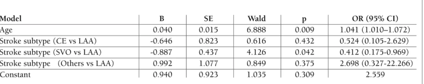
A subsequent binary logistic regression model, in which AS was the dependent variable and the variables with significant difference in univariate analysis (in-cluding age, stroke subtype, TC, HDL, and ApoAI) were entered as independent variables showed that age and stroke subtype (SVO vs. LAA) were independent-ly associated with AS (Table II). Onindependent-ly age (odds ratio [OR]=1.041) was found to be significant variables.
Among the AS patients, there were no significant re-lationships between ESR or CRP and biochemical risk factors (Table III).
dIscussIon
In this study, we evaluated the prevalences of the ma-jor cerebrovascular risk factors in patients with AS in comparison with those in a control group of patients without AS. Although the number of AS patients with a cerebrovascular disease included in our study was small due to the rarity of such cases, the mean age of these patients was significantly less than that of patients without AS. Thus, our results demonstrate the prema-ture onset of cerebrovascular disease in AS patients. In a study comparing AS patients with the general popu-tAble I. epIdeMIologIcAl, AnthropoMetrIc, And clInIcAl chArActerIstIcs, As well As
cerebrovAsculAr And bIocheMIcAl rIsk fActors, In the As And the control groups
Parameter AS group Control group p
N 34 597
-Male, % 76.5% 71.0% 0.494
Age, years 56.2±13.5 63.0±13.4 0.004
BMI, kg/m2 24.8±4.1 25.3±3.4 0.347
ESR, mm/h 24.63±25.35 – –
CRP, mg/L 2.62±3.44 – –
Current smokers, % 35.3% 40.4% 0.552
Hypertension, % 58.8% 72.5% 0.084
Diabetes mellitus, % 29.4% 34.7% 0.108
Personal history of cardiovascular disease, % 32.4% 28.6% 0.642
HCY, µmol/L 13.79±5.72 16.71±9.79 0.112
HbA1c, % 6.67±2.64 7.92±19.07 0.749
TC, mmol/L 4.58±1.27 4.16±1.10 0.037
TG, mmol/L 1.84±1.68 1.55±1.21 0.338
HDL, mmol/L 1.20±0.39 1.07±0.30 0.022
LDL, mmol/L 2.76±1.08 2.49±0.90 0.096
TC/HDL 4.17±1.69 4.06±1.22 0.739
ApoAI, mmol/L 1.26±0.27 1.15±0.26 0.034
ApoAII, mmol/L 26.31±5.10 24.23±6.95 0.223
ApoB, mmol/L 0.86±0.28 0.83±0.23 0.479
ApoB/ApoAI 0.72±0.29 0.75±0.25 0.540
ApoE, mmol/L 3.95±1.47 4.65±2.95 0.300
Lp(a), mmol/L 27.69±34.61 23.84±19.44 0.578
Stroke subtype-LAA 28.6% 49.0%
Stroke subtype-CE 5.7% 5.5%
Stroke subtype-SVO 57.1% 34.3% 0.047*
Stroke subtype-Others 8.6% 11.2%
Abbreviations: AS, ankylosing spondylitis; BMI, body mass index; ESR, erythrocyte sedimentation rate; CRP, C-reactive protein; LAA, large-artery atherosclerosis; CE, cardioembolism; SVO, small-vessel occlusion; HCY, homocysteine; HbA1c, hemoglobin A1c; TC, total cholesterol; TG, triglycerides; HDL, high-density lipoprotein; LDL, low-density lipoprotein; ApoAI, apolipoprotein AI; ApoAII, apolipoprotein AII; ApoB, apolipoproteinB; ApoE, apolipoproteinE; Lp(a), lipoprotein (a).
with AS had significantly higher levels of TC, HDL, and ApoAI. However, the logistic regression analysis did not indicate significant differences in these markers. Notably, the AS patients in our study were on average younger than the control patients, and if the mean ages of the groups had been more similar, the results regarding lipid levels may have been completely diffe -rent. Moreover, similar inconsistencies have been re-ported for AS patients with cardiovascular disease, specifically in relation to lower levels of TG8,21, TC8,23,24,
ApoB8, LDL9,24, ApoE8, Lp(a)8, and HDL8,9,21; a higher
TC/HDL ratio9,21; and no change in serum LDL and
HDL24,25. In addition, intensive lipid-lowering therapy
with a statin achieved comparable lipid-lowering ef-fects in patients with and without AS23.
The controversial results related to vascular risk fac-tors in AS patients may hint that these facfac-tors are not key indicators of premature cardiovascular or cere-brovascular disease. Traditional cardiovascular risk factors are involved in the pathogenesis of vascular disea -ses in AS patients, but such alterations cannot com pletely explain the enhanced vascular risk in AS patients26,27.
The stroke subtype according to the TOAST criteria was a determined etiologic classification17. We found
significant differences in the frequencies of TOAST clas-sifications (low frequency of large-artery atherosclero-sis and high frequency of small-vessel occlusion) in AS patients with stroke, but multivariate analysis showed that the small-vessel stroke subtype was independent-ly associated with AS. This may indicate that the fac-tors that resulted in small-vessel stroke are involved in the pathogenesis of vascular diseases in AS patients, such as small-vessel inflammation. Of cause the age ei-ther influence the consisting of stroke subtype.
The relationships between inflammatory mediators and lipid profiles have been explored, and no signifi-cant correlations were identified in our present study. tAble II. results of logIstIc regressIon AnAlysIs of pAtIents wIth And wIthout As
Model B SE Wald p OR (95% CI)
Age 0.040 0.015 6.888 0.009 1.041 (1.010–1.072)
Stroke subtype (CE vs LAA) -0.646 0.823 0.616 0.432 0.524 (0.105-2.629) Stroke subtype (SVO vs LAA) -0.887 0.437 4.126 0.042 0.412 (0.175-0.969) Stroke subtype (Others vs LAA) 0.992 1.077 0.849 0.375 2.698 (0.327-22.266)
Constant 0.940 0.923 1.035 0.309 2.559
Abbreviations: B, regression coefficient; SE, standard error; OR, odds ratio; 95% CI, 95% confidence interval
Abbreviations: CRP, C-reactive protein; ESR, erythrocyte sedimentation rate; HCY, homocysteine; TC, total cholesterol; TG, triglycerides; HDL, high-density lipoprotein; LDL, low-density lipoprotein; ApoAI, apolipoprotein AI; ApoB, apolipoproteinB; ApoE, apolipoproteinE; Lp(a), lipoprotein (a).
tAble II. AssocIAtIons between InflAMMAtory MArkers And bIocheMIcAl pArAMeters AMong
34 pAtIents wIth As
CRP ESR
Correlation Correlation coefficient p coefficient p
HCY 0.209 0.296 0.189 0.155
TC 0.115 0.553 -0.247 0.268
TG -0.051 0.793 -0.314 0.155
HDL -0.184 0.340 -0.317 0.150
LDL 0.098 0.613 -0.130 0.563
TC/HDL 0.117 0.547 -0.059 0.793 ApoAI -0.007 0.973 -0.353 0.138
ApoB 0.327 0.111 0.216 0.375
ApoE 0.022 0.929 0.252 0.365
Lp(a) 0.189 0.389 0.574 0.116
lation, AS patients were found to be at an increased risk for cerebrovascular diseases, and the excess risk is greatest in younger patients with AS14-16. Therefore, the
results of our present study are in agreement with those of studies in AS patients with cardiovascular disea -se6,10,11,13,18,19.
In the present study, there were no differences in smoking status, hypertension, or diabetes mellitus be-tween patients with and without AS. However, diffe rent results were reported by other studies of AS patients with cardiovascular disease, such as higher prevalences of smoking20, 21, hypertension10, 21, 22, and diabetes
mel-litus10, 22.
However, circulating inflammatory mediators such as interleukin-6, tumor necrosis factor-alpha, and CRP have been shown to negatively affect endothelial func-tion, ultimately leading to endothelial dysfunction9.
Previous studies of vascular structures in patients with AS have shown impaired endothelial function28,29.
Moreover, other studies have reported that treatment with tumor necrosis factor-alpha inhibitors may im-prove reduce the inflammatory response to imim-prove microvascular dysfunction30and promote endothelial
function31,32.
HCY levels did not differ between patients with and without AS. However, the disproportionate frequency of a methylenetetrahydrofolate reductase (C677T) gene polymorphism in patients with AS compared to those without AS may provide a potential prognostic factor for AS33.
Cyclooxygenase-2 selective inhibitor has been used to treat patients with inflammatory rheumatic diseases and has been associated with an increased incidence of cardiovascular events in recent clinical trials and ob-servational studies34. Therefore, such a drug may not be
a suitable for treating AS patients with a vascular disea -se. Also, nonselective nonsteroidal anti-inflammatory drugs, such as aspirin, may achieve a better response than cyclooxygenase-2 selective inhibitor in patients with AS.
Finally, a previous study reported that AS disease ac-tivity, functional and mobility limitations, and struc-tural damage may be the most influential risk factors for the premature onset of vascular diseases among AS pa-tients3.
With the combination of AS and stroke being a rare medical condition, only a few related reports have been published, and the sample sizes of these studies were small, just as in our study. Other limitations of our study include the retrospective nature and the fact that it was a single center study. A prospective, multi-cen-ter study with a larger sample size is needed to confirm our findings. Moreover, disease activity and treatment of AS should be considered in the study.
conclusIon
Cerebrovascular patients with AS were found to be signi ficantly younger than matched patients without AS. Our results indicate that AS patients may be more likely to experience premature onset of cerebrovascu-lar disease than patients without AS, and the lack of
signi ficant difference in traditional risk factors between the two groups suggests that premature atherosclero-sis occurs in patients with AS. Also, the high frequen-cy of the small-vessel stroke subtype in AS patients sug-gests that small-vessel inflammation may be involved in the pathogenesis of vascular diseases in AS patients. A further prospective study with more samples will be nee ded to confirm this hypothesis. The disease activi-ty and treatment of AS should be included in this study.
correspondence to
Shengyuan Yu
Department of Neurology Chinese PLA General Hospital Beijing 100853
China
E-mail: yusy1963@126.com
references
1. Braun J, Sieper J. Ankylosing spondylitis. Lancet. 2007;369 (9570): 1379-1390.
2. Galkina E, Ley K. Immune and inflammatory mechanisms of atherosclerosis. Ann Rev Immunol. 2009; 27: 165-197. 3. Hamdi W, Bouaziz MC, Zouch I, et al. Assessment of
Preclini-cal Atherosclerosis in Patients with Ankylosing Spondylitis. Journal of rheumatology. 2012; 39(2): 322-326.
4. Peters MJL, van Eijk IC, Smulders YM, et al. Signs of Accelera -ted Preclinical Atherosclerosis in Patients with Ankylosing Spondylitis. Journal of rheumatology. 2010. 37(1): 161-166. 5. Gonzalez-Juanatey C, Vazquez-Rodriguez TR, Miranda-Filloy
JA, et al. The High Prevalence of Subclinical Atherosclerosis in Patients With Ankylosing Spondylitis Without Clinically Evi-dent Cardiovascular Disease. Medicine (Baltimore). 2009;88(6): 358-365.
6. Hahn BH, Grossman J, Chen W, McMahon M. The pathogene-sis of atheroscleropathogene-sis in autoimmune rheumatic diseases: Roles of inflammation and dyslipidemia. Journal of autoimmunity. 2007. 28(2-3): 69-75.
7. Goodson NJ, Solomon DH. The cardiovascular manifestations of rheumatic diseases. Current opinion in rheumatology. 2006. 18(2): 135-140.
8. Mathieu S, Gossec l, Dougados M, Soubrier M. Cardiovascular profile in ankylosing spondylitis: a systematic review and meta--analysis. Arthritis care & research 2011;63(4): 557-563. 9. Heenernan S, Daemen MJAP. Cardiovascular risks in
spondy-loarthritides. Current opinion in rheumatology 2007;19(4): 358-362.
10. Han C, Robinson DW, Hackett MV, Paramore LC, Fraeman KH, Bala MV. Cardiovascular disease and risk factors in patients with rheumatoid arthritis, psoriatic arthritis, and ankylosing spondylitis. Journal of rheumatology 2006;33(11): 2167-2172. 11. Roifman I, Beck PL, Anderson TJ, Eisenberg MJ, Genest J. Chronic inflammatory diseases and cardiovascular risk: a sys-tematic review. Canadian journal of cardiology 2011; 27(2): 174-182.
24. Rollefstad S, Kvien TK, Holme I, Eirheim AS, Pedersen TR, Semb AG. Treatment to lipid targets in patients with inflamma-tory joint diseases in a preventive cardio-rheuma clinic. Annals of the rheumatic diseases. 2013;72(12): 1968-1974. 25. Caliskan M, Erdogan D, Gullu H, et al. Impaired coronary
mi-crovascular and left ventricular diastolic functions in patients with ankylosing spondylitis. Atherosclerosis 2008; 196(1): 306--312.
26. Scotece M, Conde J, Gomez R, et al. Role of adipokines in atherosclerosis: interferences with cardiovascular complications in rheumatic diseases. Mediators of inflammation. 2012 : 2012:125458. doi: 10.1155/2012/125458. Epub 2012 Jul 15 27. Onat A, Direskeneli H. Excess cardiovascular risk in
inflamma-tory rheumatic diseases: pathophysiology and targeted therapy. Current pharmaceutical design 2012;18(11): 1465-1477. 28. Bodnar N, Kerekes G, Seres I, et al. Assessment of subclinical
vascular disease associated with ankylosing spondylitis. Journal of rheumatology. 2011;38(4): 723-729.
29. Azevedo VF, Pecoits-filho R. Atherosclerosis and endothelial dysfunction in patients with ankylosing spondylitis. Rheuma-tology international 2010;30(11): 1411-1416.
30. van Eijk IC, Peters MJL, Serne EH, et al. Microvascular function is impaired in ankylosing spondylitis and improves after tumour necrosis factor a blockade. Annals of the rheumatic di -seases 2009;68(3): 362-366.
31. Syngle A, Vohra K, Sharma A, Kaur l. Endothelial dysfunction in ankylosing spondylitis improves after tumor necrosis factor--alpha blockade. Clinical rheumatology 2010;29(7): 763-770. 32. Soltesz P, Kerekes G, Der H, et al. Comparative assessment of vascular function in autoimmune rheumatic diseases: consi -derations of prevention and treatment. Autoimmunity reviews 2011;10(7): 416-425.
33. Gecene M, Tuncay F, Borman P, Yucel D, Senes M, Yilmaz BK. Atherosclerosis in male patients with ankylosing spondylitis: the relation with methylenetetrahydrofolate reductase (c677t) gene polymorphism and plasma homocysteine levels. Rheuma-tol Int 2013;33(6): 1519-1524.
34. White WB, West CR, Borer JS, et al. Risk of cardiovascular events in patients receiving celecoxib: a meta-analysis of ran-domized clinical trials. Am J Cardiol. 2007;99(1): 91-98. diseases 2010;69(3): 579-581.
14. Keller JJ, Hsu J, Lin S, et al. Increased risk of stroke among pa-tients with ankylosing spondylitis: a population-based matched-cohort study. Rheumatology international 2014; 34(2): 255--263.
15. Lin C, Huang Y, Chiu Y, Ho Y, Pan S. Increased risk of ischemic stroke in young patients with ankylosing spondylitis: a popu-lation-based longitudinal follow-up study. Plos one. 2014 Apr 8;9(4):e94027. doi: 10.1371/journal.pone.0094027. eCollec-tion 2014
16. Szabo SM, Levy AR, Rao SR, et al. Increased risk of cardiovas-cular and cerebrovascardiovas-cular diseases in individuals with ankylos-ing spondylitis a population-based study. Arthritis and rheuma-tism 2011;63(11): 3294-3304.
17. Adams HP, Bendixen BH, Kappelle LJ, et al. Classification of subtype of acute ischemic stroke. Definitions for use in a mul-ticenter clinical trial. Toast. Trial of org 10172 in acute stroke treatment. Stroke. 1993;24(1): 35-41.
18. Unverdi S, Goker B, Oktar S, et al. Assessment of the morpho-logic evidence of subclinical atherosclerosis by carotid b-mode ultrasonography in patients with ankylosing spondylitis. An-nals of the rheumatic diseases 2007;66: 411-411.
19. Papagoras C, Voulgari PV, Drosos AA. Atherosclerosis and car-diovascular disease in the spondyloarthritides, particularly ankylosing spondylitis and psoriatic arthritis. Clinical and ex-perimental rheumatology 2013;31(4): 612-620.
20. Zifman E, Amital H, Gilburd B, Shoenfeld Y. Antioxidants and smoking in autoimmune disease - opposing sides of the seesaw. Autoimmunity reviews 2008;8(2): 165-169.
21. Peters MJ, van der Horst-bruinsma IE, Dijkmans BA, Nurmo-hamed MT. Cardiovascular risk profile of patients with spondylarthropathies, particularly ankylosing spondylitis and psoria -tic arthritis. Seminars in arthritis and rheumatism 2004;34(3): 585-592.
22. Brophy S, Cooksey R, Atkinson M, et al. No increased rate of acute myocardial infarction or stroke among patients with anky-losing spondylitis-a retrospective cohort study using routine data. Seminars in arthritis and rheumatism 2012;42(2): 140--145.