Brazilian Journal of Physics, vol. 35, no. 3B, September, 2005 811
Measure of Nitrogen in Special Steel
J. F. Curado, N. Added, M. A. Rizzutto, and M. H. Tabacniks Pelletron Laboratory - DFN/IF - Universidade de S ˜ao Paulo, Caixa Postal 66318 - CEP 05315-970 - S ˜ao Paulo - SP - Brazil
Received on 11 August, 2005
Stainless steel is widely employed in different areas of modern industrial production, taking advantage of its corrosion resistance. In this work we use the elastic recoil detection analysis (ERDA) to determinate the profile of nitrogen in stainless steel samples. An incident beam of35Cl of 50 MeV was used for the analysis of sample components. The results have indicated the presence of thin films in the surface of some analyzed samples and have allowed the determination of the concentration and the thicknesses of these films.
I. INTRODUCTION
The development in the area of material science has been very significant in the last years, with special emphasis in the microelectronics and metallurgy industries generating a pa-rallel development in the area of analysis of materials due the necessity of better thickness definition of thin films and the depth profile of composites deposited on thick substrate.
The characterization of special steel samples, in special the profile of nitrogen, aims to improve the quality of the surface and to increase the durability of steel pieces. In this work, the samples have been characterized through ERDA [1, 2], getting the profile of the nuclei of the constituent elements in samples from the analysis of the energy spectra.
The method ERDA has high efficiency for the identifica-tion and quantificaidentifica-tion of materials with light elements in he-avy elements matrix (lighter than Ne) [3, 4]. Using hehe-avy elements projectiles and a∆E−E detection system [1, 2, 5– 8], this method has the advantage of high sensibility for the atomic number identification, thereby being applicable for the elementary analysis of thin films deposit on heavy element substrates [9, 10].
II. EXPERIMENTAL
The experimental setup was mounted in the scattering chamber at 30obeam line of the 8UD Tandem accelerator of the Pelletron Laboratory of the University of So Paulo. An ionization chamber was positioned at 40owith respect to the beam direction and the sample holder was set in a way that the angle between the normal direction related sample surface and the incident beam was 60o. The ionization chamber, with a polypropylene beam entrance window of 80 µg/cm2, uses P10 mixture (90% argon and 10% methane) with 17 Torr of pressure. For the measurements, an incident beam of 50 MeV Cl7+ions was used.
The data acquisition was a home mode one. That allows to check the experience through display of the spectrums. The digitalized information corresponding to E and∆E of the re-coil particles was stored in the event mode during the acqui-sition. The elementar composition relative to Fe was calcula-ted taking in account the difference in their scattering
cross-section. This composition was simulated and compared using the specific code SIMNRA[11].
The samples analyzed consist of small discs, with 15 mm of diameter and 6 mm of height. Each sample was treated in different conditions using plasma for addiction of nitrogen. The samples were given by Antonio Jorge Abdalla, researcher of Diviso de Fotoionica do IEAv (Instituto de Estudos Avana-dos).
III. RESULTS AND DISCUSSION
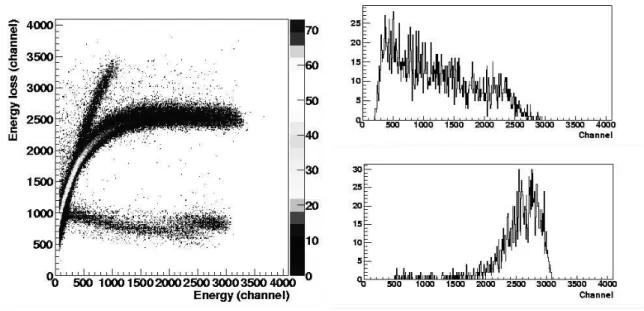
An E versus∆E spectrum for one of the samples is shown in figure 1. In this spectrum can be clearly identify the pre-sence of thin films in the surface of analyzed sample. The area for each element was selected and projected on the E axis to compare with SIMNRA simulations.
Analysis of all samples allows the identification of two groups: a) samples with thinner oxide film on the surface and a smaller carbon contribution in the bulk; b) samples with wi-der oxide film on the surface and a bigger carbon contribution in the bulk.
Figures 1 and 2 shows the energy spectrum for carbon, ni-trogen and oxygen particles for different samples. The com-position ratios were estimated using the integrated the area for each element.
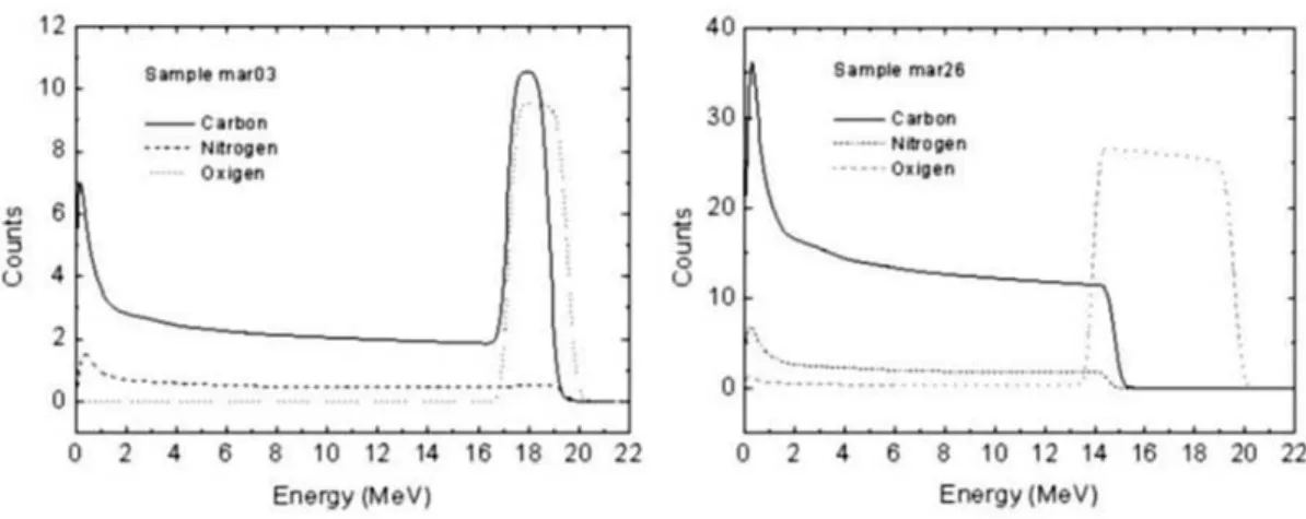
Figure 3 shows the results from simulation using SIMNRA for fitting energies spectra for each element.
IV. CONCLUSIONS
The experimental procedure using this ERDA system has enough resolution for the identification of light elements pre-sents in the surface of the samples.
However results show a limitation of few mm for the depth profile due to energy restriction related to the stopping power of the ions in steel.
812 J. F. Curado et al.
FIG. 1: Show biparametric spectra for sample with a small contribution of oxygen and carbon on surface and the projection on the Energy axis.
FIG. 2: Shows biparametric spectra with carbon and oxygen contribution on surface and the projection on the Energy axis.
Elem Concentration
Layer 1 600 x 1015 atoms/cm2
C N O Fe
0,0438±0,0034 0,00245±0,00076
0,0470±0,0070 0,9067±0,0095
Layer 2 Bulk
C N O Fe
0,00793±0,00098 0,00214±0,00064 0,00000±0,00002
0,990±0,011
Elem Concentration
Layer 1 1600 x 1015 atoms/cm2
C N O Fe
-0,2975±0,0096
0,702±0,017
Layer 2 Bulk
C N O Fe
0,00793±0,00098 0,1927±0,0076 0,00325±0,00064
0,793±0,020
Brazilian Journal of Physics, vol. 35, no. 3B, September, 2005 813
FIG. 3: Simulated energy spectrum using program SIMNRA using a35Cl beam with ELab= 50 MeV for the samples mentioned in table above: figure a) thin film of FeO with thickness of 1600 x 1015atoms/cm2; figure b) shows thin film (FeOCN) with thickness of 600 x 1015atoms/cm2.
[1] J. Lecuyer, C. Brassard, C. Cardinal, J. Chabbal, L. Deschenes, and J. P. Labrie, J. Appl. Phys. 47, 381 (1976).
[2] E. Arai, A. Zounek, M. Sekino, K. Takemoto, and O. Nittono, Nucl. Instr. and Meth. B 85, 226 (1994).
[3] R. J. Girnius, L. W. Anderson, Nucl. Instr. and Meth B 36, 283 (1996).
[4] W. Bohne, G. U. Reinsperger, J. R ¨uschert, B. Selle, and P. Staub, Nucl. Instr. and Meth B161-163, 467 (2000).
[5] N. Dytlewski, P. J. Evans, J. T. Noorman, L. S. Wielunski, and J. Bunder, Nucl. Instr. and Meth B118, 278 (1996).
[6] Y. Wang, B. Huang, D. Cao, D. Zhu, and K. Shen, Nucl. Instr. and Meth B84, 111 (1994).
[7] W. Assmann, J. A. Davies, G. Dollinger, J. S. Forster, H. Huber, T. Reichelt, and R. Siegele, Nucl. Instr. and Meth B 118, 424 (1996).
[8] R. Siegele, W. Assmann, J. A. Davies, and J. S. Forster, Nucl. Instr. and Meth B 118, 283 (1996).
[9] W. Hong, S. Hayakawa, K. Maeda, S. Fukuda, M. Yanokura, M. Aratani, K. Kimura, Y. Gohshi, and I. Tanihara, Nucl. Instr. and Meth B 124, 95 (1997).
[10] J. Tirira, P. Trocellier, and J. P. Frontier, P. Trouslard, Nucl. Instr. and Meth B 45, 203 (1990).