Short Report
Printed in Brazil - ©2012 Sociedade Brasileira de Química0103 - 5053 $6.00+0.00S
*e-mail: noronha@ufpr.br
Caavuranamide, a Novel Steroidal Alkaloid from the Ripe Fruits of
Solanum caavurana
Vell. (Solanaceae)
Nelissa Pacheco Vaz,a Emmanoel V. Costa,a,b Érica L. Santos,a Sandra Bos Mikich,c
Francisco A. Marques,a Raquel M. Braga,d Camila Delarmelina,e Marta C. T. Duarte,e
Ana Lúcia T. G. Ruiz,f Vanessa H. S. Souza,f João E. de Carvalhof and
Beatriz H. L. N. Sales Maia*,a
aDepartamento de Química, Universidade Federal do Paraná, Centro Politécnico,
CP 19081, 81531-990 Curitiba-PR, Brazil
bDepartamento de Química, Universidade Federal de Sergipe, Av. Marechal Rondon s/n,
49100-000 São Cristovão-SE, Brazil
cLaboratório de Ecologia, Embrapa Florestas, CP 319, 83411-000 Colombo-PR, Brazil
dInstituto de Química, Universidade Estadual de Campinas,
CP 6154, 13083-970 Campinas-SP, Brazil
eDivisão de Microbiologia and fDivisão de Farmacologia e Toxicologia,
Centro Pluridisciplinar de Pesquisas Químicas Biológicas e Agrícolas (CPQBA), Universidade Estadual de Campinas, CP 6171, 13083-970 Campinas-SP, Brazil
A investigação fitoquímica dos frutos maduros de Solanum caavurana Vell. (Solanaceae) forneceu um novo alcaloide esteroidal do tipo espirosolano, caavuranamida, juntamente com os alcaloides 4-tomatiden-3-ona e 5α-tomatidan-3-ona. Suas estruturas foram elucidadas através de métodos espectroscópicos. As atividades antiproliferativa e antimicrobiana para o extrato etanólico e subfrações provenientes de partição e de extração ácido-base foram investigadas. A caavuranamida apresentou atividade antibacteriana similar ao controle positivo cloranfenicol contra Rhodococcus equi.
Phytochemical investigation of the ripe fruits of Solanum caavurana Vell. (Solanaceae)
afforded a novel steroidal alkaloid with spirosolane-type skeleton, named as caavuranamide, together with the alkaloids 4-tomatiden-3-one and 5α-tomatidan-3-one. Their structures were elucidated on the basis of spectroscopic methods. The antiproliferative and antimicrobial activities for the ethanolic extract, sub-fractions obtained from partition and acid-base treatment were also evaluated. Caavuranamide showed antibacterial activity similar to the chloramphenicol positive control against Rhodococcus equi.
Keywords:Solanum caavurana, Solanaceae, caavuranamide, steroidal alkaloids, spirosolane
Introduction
The genus Solanum is the most representative of
Solanaceae family, comprising about 1400 species1 mainly
distributed in tropical and subtropical regions of Americas, Africa and Australia.2 In Brazil, about 350 Solanum species
have been identified, many of them endemic.3
Solanum caavurana Vell. grows as a bush or a small
tree with ca. 5 m high, and is widely distributed in
Brazil (in Ceará, Bahia, Mato Grosso do Sul, Paraná and Santa Catarina States), occurring in Paraguay and Argentina as well. It is popularly known as ‘laranjinha do mato’, ‘jurubebarana’2 or ‘jurubeba-branca’, being used
in folk medicine to treat anemia, liver disorders and as digestive as well.1 Although phytochemical studies on
Solanum species resulted in the isolation of a great variety
of steroidal alkaloids and glycoalkaloids,4,5 to the best
Here, we report the identification of a novel steroidal alkaloid, named as caavuranamide (1), along with the
known 4-tomatiden-3-one (2) and 5α-tomatidan-3-one (3),
isolated from the ethanolic extracts of the ripe fruits of
Solanum caavurana Vell.
Experimental
General experimental procedures
Melting point (mp) was determined on a Quimis Q-340S23 micromelting point apparatus. IR spectra were acquired on a BIORAD FTS-3500, Fourier transform infrared (FTIR) equipment. Optical rotation was measured by using a Rudolph Research (Flanders, New Jersey) Autopol III automatic spectropolarimeter. Nuclear magnetic resonance (NMR) data were recorded at 293 K in CDCl3
on a Varian Inova 500 operating at 11.7 T, observing 1H
at 500 MHz and 13C at 125 MHz. Chemical shifts (d) are
given in ppm relative to TMS (tetramethylsilane) (d 0.00) as the internal standard. The electron spray ionization mass spectra (HRESIMS and HRESIMS/MS) were acquired in positive ion mode and recorded on a quadrupole-time of flight instrument (UltrOTOF-Q, Bruker Daltonics, Billerica, MA). The conditions were: capillary voltage of 3400 V, dry gas temperature of 180 °C, dry gas flow of 4 L h-1,
nitrogen as nebulizer gas and 10 mmol L-1 NaTFA (sodium
trifluoroacetate) was used as the standard for internal and external calibration. For tandem mass spectrometry analysis (ESIMS/MS), nitrogen was used as the collision gas. Silica gel 60 (70-230 mesh) was used for column chromatography, aluminum precoated silica-gel plates (60 F254 Merck, 0.25 mm) were used for analytical thin layer chromatography (TLC), and glass precoated silica-gel plates (60 PF254 Merck, 1 mm) were used for preparative TLC. The spots were detected by spraying with Dragendorff’s or p-anisaldehyde
reagents, followed by heating.
Plant material
The ripe fruits of S. caavurana were collected in the Parque
Estadual de Vila Rica do Espírito SAnto (PEVR: 23°55' S, 51°57' W), in the municipal district of Fênix (Paraná State, Brazil), in January 2004. A voucher specimen (No. 28280) was deposited at the Herbarium of the Botany Department from Universidade Federal do Paraná (UPCB-UFPR).
Extraction and isolation
The ripe fruits of S. caavurana were dried at 40 °C,
crushed, pulverized (180.5 g) and successively extracted at
room temperature with petrol (30-60 °C) (5 days) and ethanol (5 days), with the solvent removed every 24 h. The ethanolic extract was submitted to liquid-liquid partition with CH2Cl2
followed by n-butanol (n-but). Removal of the solvents under
reduced pressure furnished 2.6 and 3.0 g of CH2Cl2 and
n-but fractions, respectively, both revealing positive TLC
Dragendorff’s test. CH2Cl2 fraction was dissolved in 3%
aqueous HCl and the acid liquid phase then extracted with CHCl3 (3 × 20 mL). The organic fractions were combined,
dried with anhydrous Na2SO4 and concentrated under reduced pressure to obtain the neutral CHCl3 fraction (947.0 mg).
The remaining aqueous acid solution was adjusted to pH 10-12 with 37% aqueous NH4OH followed by extraction
with CHCl3 (3 × 20 mL). The resulting organic fractions were treated as previously described to yield an alkaloid residue (122.5 mg), which was purified by preparative TLC eluted with EtOAc-MeOH 90:10 to yield 1 (6.6 mg).
The neutral CHCl3 fraction (947.0 mg) was subjected to silica-gel column chromatography, with gradient elution of CH2Cl2/EtOAc from 100:0 to 25:75, EtOAc/MeOH from 100:0 to 90:10 and MeOH, affording a total of 184 fractions (20 mL each), which were assembled in 18 subfractions according to their composition on TLC. Subfractions 10 (34.2 mg) and 9 (50.7 mg) were purified by preparative TLC eluted with CH2Cl2/MeOH 90:10 and CH2Cl2/MeOH 95:5,
respectively, to yield 2 (5.0 mg) and 3 (2.5 mg).
Caavuranamide (1)
Yellowish powder; 1H and 13C NMR data given in Table 1;
mp 115-117 ºC; [α]D25 + 6.3 (CHCl3; c 0.0027); IR (KBr)
νmax/cm-1 3429, 2930, 2852, 2362, 2341, 1656, 1543, 1456,
1383, 1264, 1037, 973, 872, 750 and 668; HRESIMS m/z
443.3705 [M + H]+ (calcd. for C
28H46N2O2 + H+, 443.6770).
4-Tomatiden-3-one (2)
Light-yellow amorphous powder; 1H and 13C NMR data
given in Table 1; HRESIMS m/z 412.3320 [M + H]+ (calcd.
for C27H41NO2 + H+, 412.6338).
5α-Tomatidan-3-one (3)
Yellow amorphous powder; 1H and 13C NMR data given
in Table 1; HRESIMS m/z 414.3443 [M + H]+ (calcd. for
C27H43NO2 + H+, 414.6496).
In vitro antimicrobial activity
(Bacillus subtilis ATCC 6051, Escherichia coli ATCC 11775,
Enterococcus faecium CCT 5079, Micrococcus luteus
ATCC 4698, Pseudomonas aeruginosa ATCC 13388, Rhodococcus equi ATCC 25729, Staphylococcus aureus
ATCC 6538, Salmonella choleraesuis ATCC 10708 and Staphylococcus epidermides ATCC 12228). Compound 1 was
tested against E. coli, R. equi, S. aureus, S. epidermides and S. choleraesuis.
The bacteria strains were subcultured overnight at 36 °C in Nutrient Agar (Merck). Inoculum for the assays were prepared by diluting a scraped cell mass in 0.85% NaCl solution, adjusted to McFarland scale 0.5 and confirmed by spectrophotometer reading at 580 nm. Cell suspensions were finally diluted to 104 cfu mL-1 for use in
the activity assays. Minimal inhibitory concentration (MIC) tests were carried out according to Eloff,7 using
Müller-Hinton broth on a tissue-culture test plate (96 wells). The stock solutions of crude extracts and fractions were diluted and transferred into the first well, and serial dilutions were made, so that concentrations in the range of 1.000-0.015 mg mL-1 were obtained. Chloramphenicol
(Merck) was used as the reference antibiotic control in the range of 0.250-0.002 mg mL-1. The inoculum was
added to all wells, and the plates were incubated at 36 °C for 48 h. Each concentration was screened in triplicate. Antimicrobial activity was detected by adding 20 µL of 0.5% TTC (triphenyltetrazolium chloride, Merck) aqueous solution. MIC was defined as the lowest concentration of the sample that inhibited visible growth, as indicated by TTC staining (dead cells are not stained by TTC).
In vitro antiproliferative activity assay
Human tumor cell lines U251 (glioma, CNS), UACC-62 (melanoma), MCF-7 (breast), NCI-H460 (lung, non-small cells), OVCAR-03 (ovarian), HT-29 (colon), 786-0 (renal), NCI-ADR/RES (ovarian expressing phenotype multiple drugs resistance) and K562 (leukemia) were kindly provided by National Cancer Institute (NCI, U. S. Department of Health and Human Services). Stock cultures were grown in medium containing 5 mL RPMI 1640 (Gibco-BRL) supplemented with 5% fetal bovine serum. Peniciline:streptomicine (1000 µg mL-1:1000 UI mL-1,
1 mL L-1) was added to experimental cultures. Cells
in 96 well plates (100 µL cells per well) were exposed
to sample concentrations in DMSO/RPMI (0.25, 2.5, 25 and 250 µg mL-1) at 37 °C, 5% of CO
2 in air for 48 h.
Final DMSO concentration did not affect cell viability. Afterwards, cells were fixed with 50% trichloroacetic acid and cell proliferation determined by spectrophotometric quantification (540 nm) of cellular protein content using
sulforhodamine B assay. Using the concentration-response curve for each cell line, TGI (concentration that produces total growth inhibition or cytostatic effect) was determined through non-linear regression analysis using software ORIGIN 7.5 (OriginLab Corporation).8
Results and Discussion
The ethanolic extract from the ripe fruits of Solanum caavurana afforded steroidal alkaloids 1-3 (Figure 1).
Caavuranamide (1) was isolated as a yellowish
powder and gave a positive Dragendorff reagent test. Its molecular formula C28H46N2O2 was calculated from
the HRESIMS quasi-molecular ion peak [H + H]+ at
m/z 443.3705. The HRESIMS/MS revealed two fragments
at m/z 255.2139 and 300.2349 corresponding to a difference
of 45.0210 Da, suggesting the loss of a formamide group (H2N-CHO, calcd. 45.041 Da).
The decoupled 13C NMR spectrum of 1 (Table 1)
displayed 28 carbon signals, mainly in the sp3 C field
characteristic of steroidal alkaloids,9 whereas DEPT-135
(distortionless enhancement by polarization transfer) analysis aided to attribute the presence of four methyl groups, one carbonyl (dC 160.3), eleven methylenes, nine methines
(including one oximethine) and three quaternary carbons. The carbinolic hydrogen H-16 (dH 4.14) was correlated
with C-16 (dC 78.5) in the HSQC (heteronuclear single
quantum correlation) spectrum, that also showed four methyl hydrogens at dH 0.82 (s), 0.83 (s), 0.86 (d, J 6.5 Hz) and
0.96 (d, J 6.5 Hz) correlated to carbons at dC 16.9 (C-18),
11.4 (C-19), 19.3 (C-27) and 15.8 (C-21), respectively, possessing characteristic chemical shifts for steroidal alkaloid methyl groups. The signals at dC 78.5 (C-16),
99.3 (C-22) and 50.2 (C-26) supported a spirosolane-type structure.9 Through 2D NMR spectral data [HSQC, HMBC
(heteronuclear multiple bond correlation) and NOESY (nuclear Overhauser effect spectroscopy)], the chemical
shifts of all hydrogens, as well as its correlations, were fully established (Table 1).
The 1H NMR spectrum revealed a signal at d H 8.12
(m, 1H) that was assigned to the formyl hydrogen of the formamide group, which existence was previously evidenced by HRESIMS analysis, in association with the presence of a downfield carbonyl group at dC 160.3. The
existence of a hydrogen at dH 3.86 and the remaining broad
low field signal at dH 5.36 (d, 1H, J 7.5 Hz) were assigned
to H-3 and the formamide N-hydrogen, respectively, by
analogy with solanopubamide A.10
The NOESY spectrum was used to confirm the proposed stereochemistry assignments made for caavuranamide 1
(Figure 2), as the trans junction of A/B ring and cis
junction of D/E ring, as well as (25S)-22βN-spirosolane
configuration (as observed for tomatidine)9 and the
orientation of 3β-N-formylamino group. A strong NOE
(nuclear Overhauser effect) was observed between the
signal of CH3-27 (dH 0.86) with the signal of H-26 (dH
2.74) and H-24ax (dH 1.37), indicating that CH3-27 is in
equatorial position. Strong correlations could also be observed among CH3-21 (dH 0.96) with H-17 (dH 1.60),
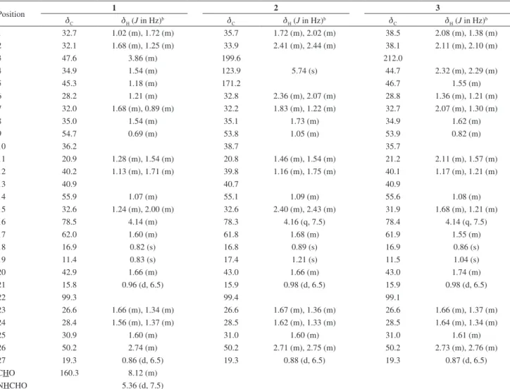
H-23 (dH 1.66 and 1.34) and H-16 (dH 4.14), suggesting Table 1. 1H and 13C NMR spectral dataa for compounds 1-3
Position 1 2 3
dC dH (J in Hz)b dC dH (J in Hz)b dC dH (J in Hz)b
1 32.7 1.02 (m), 1.72 (m) 35.7 1.72 (m), 2.02 (m) 38.5 2.08 (m), 1.38 (m)
2 32.1 1.68 (m), 1.25 (m) 33.9 2.41 (m), 2.44 (m) 38.1 2.11 (m), 2.10 (m)
3 47.6 3.86 (m) 199.6 212.0
4 34.9 1.54 (m) 123.9 5.74 (s) 44.7 2.32 (m), 2.29 (m)
5 45.3 1.18 (m) 171.2 46.7 1.55 (m)
6 28.2 1.21 (m) 32.8 2.36 (m), 2.07 (m) 28.8 1.36 (m), 1.21 (m)
7 32.0 1.68 (m), 0.89 (m) 32.2 1.83 (m), 1.22 (m) 32.7 2.07 (m), 1.30 (m)
8 35.0 1.54 (m) 35.1 1.73 (m) 34.9 1.62 (m)
9 54.7 0.69 (m) 53.8 1.05 (m) 53.9 0.82 (m)
10 36.2 38.7 35.7
11 20.9 1.28 (m), 1.54 (m) 20.8 1.46 (m), 1.54 (m) 21.2 2.11 (m), 1.57 (m)
12 40.2 1.13 (m), 1.71 (m) 39.8 1.16 (m), 1.75 (m) 40.1 1.17 (m), 1.21 (m)
13 40.9 40.7 40.9
14 55.9 1.07 (m) 55.1 1.09 (m) 55.6 1.08 (m)
15 32.6 1.24 (m), 2.00 (m) 32.6 2.40 (m), 2.43 (m) 31.9 1.68 (m), 1.21 (m)
16 78.5 4.14 (m) 78.3 4.16 (q, 7.5) 78.4 4.14 (q, 7.5)
17 62.0 1.60 (m) 61.8 1.68 (m) 61.9 1.55 (m)
18 16.9 0.82 (s) 16.8 0.89 (s) 16.9 0.86 (s)
19 11.4 0.83 (s) 17.4 1.21 (s) 11.5 1.04 (s)
20 42.9 1.66 (m) 43.0 1.66 (m) 43.0 1.74 (m)
21 15.8 0.96 (d, 6.5) 15.9 0.98 (d, 6.5) 15.9 0.98 (d, 6.5)
22 99.3 99.4 99.1
23 26.6 1.66 (m), 1.34 (m) 26.6 1.67 (m), 1.36 (m) 26.6 1.66 (m), 1.37 (m)
24 28.4 1.56 (m), 1.37 (m) 28.5 1.62 (m), 1.33 (m) 28.5 1.64 (m), 1.34 (m)
25 30.9 1.60 (m) 31.0 1.60 (m) 31.0 1.61 (m)
26 50.2 2.74 (m) 50.2 2.71 (m), 2.75 (m) 50.2 2.73 (m), 2.76 (m)
27 19.3 0.86 (d, 6.5) 19.3 0.88 (d, 6.5) 19.3 0.87 (d, 6.5)
CHO 160.3 8.12 (m)
NHCHO 5.36 (d, 7.5)
aExperiments were carried out at 500 MHz for 1H and 125 MHz for 13C in CDCl
3 and TMS as internal reference (d 0.00 ppm); bassignments confirmed by DEPT-135, HSQC, HMBC and NOESY experiments.
a S configuration for C-22 and the cis junction of the D/E
ring. The axial orientation of H-5 (dH 1.18) was sustained
by intense correlations with H-9ax (dH 0.69) and H-3ax
(dH 3.86). This later information denotes that the substituent
N-formylamino located in C-3 is found in 3β arrangement. The above findings supported the configuration (25S,22S
)-3β-N-formylamino-5α-spirostane which is in agreement with solanopubamide A10 and with data reported for
(22S,25S)-3β-amino-5α-spirosolane.11
It is known that steroidal alkaloids isolated from the Solanaceae possess almost exclusively a 3-hydroxyl substituent or its glycosylated derivatives. The only report of a N-formyl steroidal alkaloid in the Solanum10 genus
indicated a solanidane-type skeleton. Caavuranamide [(22S,25S)-3β-N-formylamino-5α-spirosolane] (1) is
the second report of a N-formyl steroidal alkaloid in this
genus, and the first with a spirosolane-type skeleton. Compounds 2 and 3 were identified on the basis of 1H and 13C NMR spectral data (Table 1) as
4-tomatiden-3-one and 5α-tomatidan-3-one, respectively. Although
substance 2 has been obtained before by microbial12
conversion of tomatidine, this is the first report of its isolation directly from plants. Compound 3 was previously
isolated13 from the roots of a hybrid plant between
Lycopersicon esculentum Mill. and L. hirsutum Humb. et
Bonpl. (Solanaceae). The 1H and 13C NMR data, as well
as the assignments of chemical shifts for both compounds, were incomplete in the literature, and are now supported by DEPT-135 and HSQC experiments.
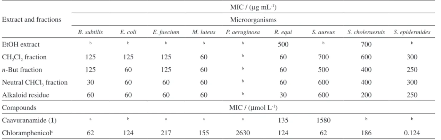
The crude extracts and fractions obtained from acid-base treatment were evaluated for antimicrobial (Table 2) and
in vitro antiproliferative activities (Table 3). According
to Table 2, the ethanolic extracts do not show significant activity for neither of tested microorganisms, whereas CH2Cl2 and n-but extracts, as well as those subfractions
obtained from acid-base treatment, showed potent activity for E. coli, M. luteus, R. equi, B. subtilis and E. faecium,
being similar and even in some cases, stronger than positive control chloramphenicol. Compound 1 (6.6 mg) exhibited
activity (MIC 135 µmol L-1) similar to chloramphenicol
Table 2. Antibacterial activity of extracts, fractions and compound 1 of ripe fruits of S. caavurana
Extract and fractions
MIC / (µg mL-1) Microorganisms
B. subtilis E. coli E. faecium M. luteus P. aeruginosa R. equi S. aureus S. choleraesuis S. epidermides
EtOH extract b b b b b 500 b 700 b
CH2Cl2 fraction 125 125 125 60 b 60 700 600 300
n-But fraction 125 60 125 60 b 60 500 400 250
Neutral CHCl3 fraction 30 60 60 60 b 60 600 400 300
Alkaloid residue 60 60 60 60 b 30 600 200 250
Compounds MIC / (µmol L-1)
Caavuranamide (1) a b a a a 135 1580 b b
Chloramphenicolc 62 124 217 155 2630 124 62 186 0.124
aNot tested; bconcentration > 1000 µg mL-1; cpositive control for bacteria.
Table 3. Antiproliferative activity of extracts and fractions of ripe fruits of S. caavurana against cancer cell lines
Extracts and fractions
TGI / (µg mL-1) Tumor cell lines
U251 UACC-62 MCF-7 NCI-H460 OVCAR-3 HT-29 786-0 K562 NCI-ADR/RES VERO
EtOH extract b 2.11 93.06 b 84.49 60.90 60.62 b 102.13 b
CH2Cl2 fraction 75.53 b 142.48 b 139.66 115.39 94.97 b b b
n-But fraction 82.45 b 100.40 b 117.84 62.13 46.04 b 243.35 b
Neutral CHCl3 fraction 90.47 8.09 57.17 100.27 100.27 73.67 80.63 b 82.37 b
Alkaloid residue 65.59 37.14 98.21 b 51.77 228.07 74.64 b 61.20 b
Doxorubicina 3.24 0.15 10.49 > 25 5.62 > 25 3.73 > 25 > 25 > 25
(MIC 124 µmol L-1) for R. equi, but it is less active than
the alkaloid residue that furnished 1.
For in vitro antiproliferative activity, the extracts and
respective subfractions showed to be active for a wide range of human tumor cell lines, with the ethanolic extract and neutral CHCl3 fraction presenting the strongest antiproliferative results for melanoma (UACC-62) with TGI values of 2.11 and 8.09 µg mL-1, respectively.
Conclusions
The present phytochemical investigation of the ripe fruits of S. caavurana Vell. afforded a novel steroidal
alkaloid with spirosolane-type skeleton, named as caavuranamide (1), together with the alkaloids
4-tomatiden-3-one (2) and 5-α-tomatidan-3-one (3). Compound (1)
showed significant antibacterial activity (MIC 135 mol L-1)
for R. equi, similar to chloramphenicol (MIC 124 mol L-1).
Supplementary Information
Supplementary information (Figure S1-S20) is available free of charge at http://jbcs.sbq.org.br as PDF file.
Acknowledgements
The authors are grateful to Prof. Dr. Norberto P. Lopes of the Faculdade de Ciências Farmacêuticas de Ribeirão Preto, USP (Ribeirão Preto-SP, Brazil) for the HRESIMS and HRESIMS/MS analysis, and to Coordenação de Aperfeiçoamento de Pessoal de Nível Superior (CAPES-PROCAD) and the Fundação Araucária (Paraná, Brazil), for financial support. F. A. M. is also grateful to Conselho Nacional de Desenvolvimento Científico e Tecnológico (CNPq-INCT). J. E. C. is grateful to CNPq for research
fellowship. N. P. V. is grateful to CAPES for scholarship. S. B. M. receives a Productivity Fellowship (process No. 308419/2008-1) from CNPq.
References
1. Nurit-Silva, K.; Agra, M. F.; Lat. Am. J. Pharm.2009, 28, 675. 2. Mentz, L. A.; Oliveira, P. L.; Pesquisas, Botânica2004, 54, 53.
3. Silva, T. M. S.; Camara, C. A.; Freire, K. R. L.; Silva, T. G.; Agra, M. F.; Bhattacharyya, J.; J. Braz. Chem. Soc. 2008, 19,
1048.
4. Lu, Y.; Luo, J.; Kong, L.; Phytochemistry2011, 72, 668.
5. Colmenares, A. P.; Rojas, L. B.; Usubillaga, A.; Bol. Latinoam. Caribe Plantas Med. Aromat.2010, 9, 80.
6. Barbosa-Filho, J. M.; Agra, M. F.; Oliveira, R. A. G.; Paulo, M. Q.; Trolin, G.; Cunha, E. V. L.; Ataíde, J. R.; Bhattacharrya, J.;
Mem. Inst. Oswaldo Cruz 1991, 86, 189. 7. Eloff, J. N.; Planta Med.1998, 64, 711.
8. Shoemaker, R. H.; Nat. Rev. Cancer 2006, 6, 813.
9. Radeglia, R.; Adam, G.; Ripperger, H.; Tetrahedron Lett. 1977, 11, 903.
10. Kumari, G. N. K.; Rao, L. J. M.; Rao, K. V. R.; Rao, N. S. P.; Kaneko, K.; Mitsuhashi, H.; Phytochemistry 1986, 25, 2003. 11. Maxwell, A.; Pingal, R.; Reynolds, W. F.; McLean, S.;
Phytochemistry 1996, 43, 913.
12. Belič, I.; Garberc-Porekar, V.; Sočič, H.; Žakelj, M.; Z. Allg. Mikrobiol. 1982, 22, 359.
13. Nagaoka, T.; Yoshihara, T.; Ohra, J.; Sakamura, S.;
Phytochemistry 1993, 34, 1153.
Submitted: May 16, 2011
Published online: December 13, 2011