Short Report
S
J. Braz. Chem. Soc., Vol. 25, No. 4, 788-794, 2014. Printed in Brazil - ©2014 Sociedade Brasileira de Química 0103 - 5053 $6.00+0.00http://dx.doi.org/10.5935/0103-5053.20140037
*e-mail: [email protected]
Rodriguesic Acids, Modified Diketopiperazines from the Gastropod Mollusc
Pleurobranchus areolatus
Fabio R. Pereira,a Mario F. C. Santos,a David E. Williams,b Raymond J. Andersen,b
Vinicius Padula,c Antonio G. Ferreirad and Roberto G. S. Berlinck*,a
aInstitutode Química de São Carlos, Universidade de São Paulo,
CP 780, 13560-970 São Carlos-SP, Brazil
bDepartmentsof Chemistry and Earth, Ocean & Atmospheric Sciences,
University of British Columbia, Vancouver, BC, V6T 1Z1, Canada
cSNSB-Zoologische Staatssammlung München, Münchhausenstrasse 21,
81247 München, Germany and Department Biology II and GeoBio-Center, Ludwig-Maximilians-Universität München, Germany
dDepartamentode Química, Universidade Federal de São Carlos,
Rodovia Washington Luiz, km 235, 13565-905 São Carlos-SP, Brazil
O presente estudo foi realizado com dois espécimes do molusco nudipleura Pleurobranchus areolatus que demonstraram acumular derivados oxidados da rodriguesina A. O ácido rodriguêsico apresenta um grupo ácido carboxílico no lugar do grupo metila de uma cadeia alquila terminal da rodriguesina A. Um grupo hidroxamato foi observado na porção dicetopiperazínica do ácido rodriguêsico. As estruturas do ácido rodriguêsico e do hidroxamato do ácido rodriguêsico foram estabelecidas por análise de seus dados espectroscópicos, inclusive sua configuração absoluta. Dois ésteres metílicos dos ácidos rodriguêsicos foram isolados como compostos majoritários, porém considerados como artefatos de isolamento.
In the present investigation, two specimens of the nudipleuran mollusc Pleurobranchus areolatus have shown to accumulate oxidized rodriguesin A derivatives. Rodriguesic acid presents a carboxylic acid replacing the terminal methyl group of the alkyl chain of rodriguesin A. A hydroxamate group was also present on the diketopiperazine moiety of a rodriguesic acid derivative. The structures of both rodriguesic acid and of rodriguesic acid hydroxamate have been established by analysis of spectroscopic data, including their absolute configuration. Two methyl esters of the rodriguesic acids have been isolated as major compounds, but were considered to be isolation artifacts.
Keywords: nudipleura, Pleurobranchus, diketopiperazine, hydroxamate, mollusc
Introduction
Shell-less heterobranch molluscs are usually predators of sessile invertebrates, or feed on marine algae or cyanobacteria, from which they frequently capture secondary metabolites, typically for defensive purposes.1-4
Among the secondary metabolites acquired through diet by sea slugs are modified peptides that exhibit potent biological activities. Kahalalides, obtained from molluscs belonging to the genus Elysia, are potently cytotoxic
modified peptides, for which the actual source is the alga
Bryopsis sp.5 Currently kahalalide F is undergoing Phase I
clinical trials for the treatment of a number of solid tumors.6
Cyanobacteria-derived peptides and lipopeptides are also frequently captured and stored by molluscs, such as sea-hares.7,8 Onchidins, modified cyclic peptides isolated from
the panpulmonate mollusc Onchidium sp., provide an example.9,10 The mixed polyketide-non ribosomal peptide
Pereira et al. 789 Vol. 25, No. 4, 2014
adenocarcinoma, displaying cytotoxic activities at sub-nanomolar concentrations.11,12
As part of our ongoing program for the search of bioactive secondary metabolites from opisthobranch molluscs occurring in Brazilian waters,13 we have
investigated the chemistry of two specimens of the nudipleuran mollusc Pleurobranchus areolatus. The
nudipleuran molluscs were found on the Didemnum
sp., from which we have previously isolated the related antibacterial modified diketopiperazines, rodriguesines A (1) and B (2).14 In the course of this study we aimed
to address whether P. areolatus not only accumulates the modified diketopiperazines 1 and 2 found in the ascidian but also whether these compounds are modified to generate related derivatives.
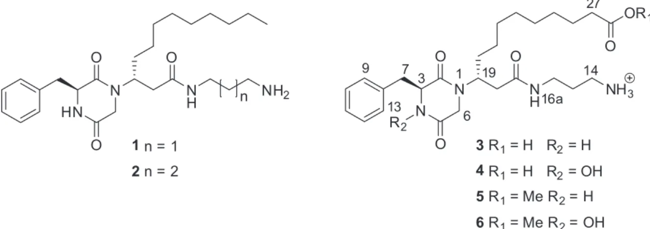
Herein we report the isolation and structures of two new modified diketopiperazines from P. areolatus, rodriguesic acids 3 and 4, and the respective esters 5 and 6 (Figure 1), that are closely related to the diketopiperazines 1 and 2
previously reported from Didemnum sp.
Experimental
General procedures
Optical rotations were measured using a Jasco P-1010 polarimeter with sodium light (589 nm). The 1H and 13C
nuclear magnetic resonance (NMR) spectra were recorded on a Bruker AV-600 spectrometer with a 5 mm CPTCI cryoprobe. 1H chemical shifts are referenced to the residual
DMSO-d6 signal (δ 2.49 ppm), and 13C chemical shifts are
referenced to the DMSO-d6 solvent peak (δ 39.5 ppm). Low- and high-resolution electrospray ionization quadrupole ion trap mass spectrometry (ESI-QIT-MS) spectra were recorded on a Bruker-Hewlett-Packard 1100 Esquire-LC system mass spectrometer. Solvents used for extraction
and flash chromatography were glass distilled prior to use. HPLC-grade solvents were utilized without further purification in high-performance liquid chromatography (HPLC) separations. HPLC semipreparative separations were performed either with a Waters quaternary pump 600, double beam UV detector 2487, and data module 746 or with a Waters 600E system controller liquid chromatography attached to a Waters 996 photodiode array detector, on which the UV spectra have been recorded as well. High-performance liquid chromatography/photo-diode array mass spectrometry (HPLC-PDA-MS) analyses were performed using a Waters Alliance 2695 coupled online with a Waters 2996 photodiode array detector, followed by a Micromass ZQ2000 mass spectrometry detector with an electrospray interface. The photodiode array scanned the sample between 205 and 254 nm. The mass spectrometer detector was optimized to the following conditions: capillary voltage, 3.00 kV; source block temperature, 100 °C; desolvation temperature, 350 °C, operating in electrospray positive mode; detection range: 200-800 Da with total ion count extracting acquisition. The cone and desolvation gas flow were 50 and 350 L h-1, respectively, and were obtained from a Nitrogen Peak
Scientific N110DR nitrogen source. Data acquisition and processing were performed using Empower 2.0.
Animal material
Pleurobranchus areolatus (identified by V. Padula, SNSB-Zoologische Staatssammlung München, Germany and Department Biology II and GeoBio-Center, Ludwig-Maximilians-Universität München, Germany) was collected at a depth of 8 m, at Ilha do Papagaio, Cabo Frio, Rio de Janeiro state, in December 2008. The specimens were immersed in 95% EtOH immediately after collection. A voucher specimen is kept at the Malacological collection,
Figure 1. Structures of rodriguesins A (1) and B (2) previously isolated from the ascidian Didemnum sp.14 and of rodriguesic acid derivatives 3-6 herein
Rodriguesic Acids, Modified Diketopiperazines from the Gastropod Mollusc Pleurobranchus areolatus J. Braz. Chem. Soc.
790
Museu Nacional, Universidade Federal do Rio de Janeiro (MNRJ 12312).
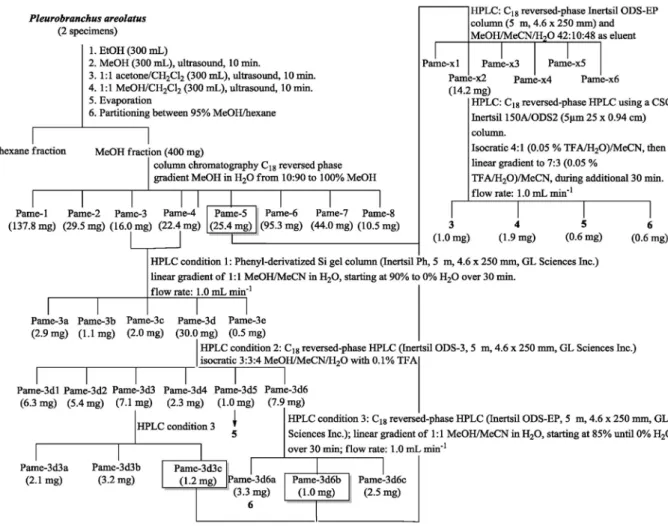
Extraction and isolation
Two specimens of P. areolatus, collected from the surface of Didemnum sp., were removed from the EtOH where they were preserved (300 mL) and sequentially extracted with MeOH (300 mL, 10 min in ultrasound bath), 1:1 acetone/CH2Cl2 (300 mL, 10 min in ultrasound bath) and
CH2Cl2/MeOH (300 mL, 10 min in ultrasound bath). The
extracts were pooled and evaporated. The resulting organic extract was suspended in MeOH 95% and defatted with hexane (3 × 200 mL). After evaporation, the MeOH extract (400 mg) was first analyzed by thin layer chromatography (TLC, 9:1 CH2Cl2/MeOH, ninhydrin) then fractionated
by chromatography on a reversed-phase C18-silica-gel
column (10 g) with a gradient of MeOH in H2O, from 9:1
H2O/MeOH to 100% MeOH. Eight fractions were obtained:
Pame-1 (137.8 mg), Pame-2 (29.5 mg), Pame-3 (16.0 mg), Pame-4 (22.4 mg), Pame-5 (25.4 mg), Pame-6 (95.3 mg), Pame-7 (44.0 mg) and Pame-8 (10.5 mg). All fractions were analyzed by HPLC-UV-MS, using an analytical C18
reversed-phase column (Waters X-Terra MS C18, 3.5 mm,
2.1 × 50 mm) with a linear gradient of 1:1 MeOH/MeCN in H2O (with 0.1% formic acid), starting at 85% to 10% H2O
over 30 min, at a flow rate of 0.5 mL min-1. Detection was
monitored by UV between 200 and 400 nm and by positive ion ESIMS with a cone voltage of 25 V monitoring ions between m/z 180 and 700. Fractions Pame-3 and Pame-4 were pooled (38.4 mg) and further fractionated using a phenyl-derivatized silica gel column (Inertsil Ph, 5 mm, 4.6 × 250 mm, GL Sciences Inc.) with a linear gradient of 1:1 MeOH/MeCN in H2O, starting at 90% to 0% H2O
over 30 min, at 1.0 mL min-1 flow rate. Five fractions were
obtained: Pame-3a (2.9 mg), Pame-3b (1.1 mg), Pame-3c (2.0 mg), Pame-3d (30.0 mg) and Pame-3e (0.5 mg), which were again analyzed by HPLC-UV-ESIMS. Fraction Pame-3d was separated by reversed-phase HPLC (Inertsil ODS-3, 5 mm, 4.6 × 250 mm, GL Sciences Inc.) using 3:3:4 MeOH/MeCN/H2O with 0.1% trifluoroacetic acid (TFA) as
eluent. Six additional fractions were obtained: Pame-3d1 (6.3 mg), Pame-3d2 (5.4 mg), Pame-3d3 (7.1 mg), Pame-3d4 (2.3 mg), Pame-3d5 (1.0 mg) and Pame-3d6 (7.9 mg) and these were analyzed by HPLC-UV-MS and dereplicated using SciFinder and MarinLit databases. Fraction Pame-3d5 was identified as a pure sample of rodriguesic acid methyl ester (5). Fraction Pame-3d6 was further purified by reversed-phase HPLC (Inertsil ODS-EP, 5 mm, 4.6 × 250 mm, GL Sciences Inc.) using a linear gradient of 1:1 MeOH/MeCN in H2O, starting at 85%
until 0% H2O over 30 min, at 1.0 mL min-1, to give 3.3 mg
of the methyl ester of rodriguesic acid hydroxamate (6). HPLC-UV-MS analysis of fractions Pame-3d6b (1.0 mg), Pame-3d3c (1.2 mg) and Pame-5 (25.4 mg) revealed the presence of both 5 and 6, as well as the minor compounds, the free acids 3 and 4. Pooling these fractions and separation by reversed-phase HPLC using a C18 Inertsil ODS-EP
column (5 mm, 4.6 × 250 mm) and MeOH/MeCN/H2O
42:10:48 as eluent, yielded fractions Pame-x1 to -x6. Fraction Pame-x2 (14.2 mg) was then further fractionated by C18 reversed-phase HPLC using a CSC-Inertsil 150A/
ODS2 (5 µm, 25 × 0.94 cm) column, initially under isocratic conditions for 30 min with 4:1 (0.05% TFA/H2O)/MeCN
as eluent, followed by a linear gradient to 7:3 (0.05% TFA/H2O)/MeCN over the course of an additional 30 min
(at 1.0 mL min-1), to give pure samples of compounds 3
(1.0 mg) and 4 (1.9 mg), 5 (0.6 mg) and 6 (0.6 mg).
Rodriguesic acid (3)
Colorless gum; [a]D
26 +24.5 (c 0.5, 3:1 MeOH/CH 2Cl2);
1H NMR (DMSO-d
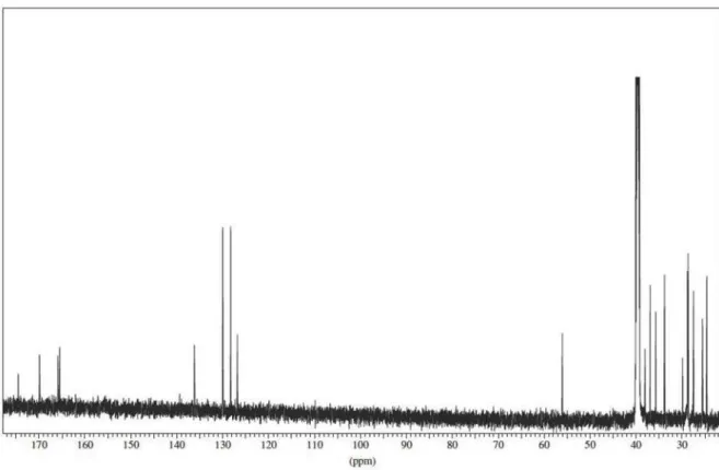
6, 600 MHz), see Table 1; 13C NMR
(DMSO-d6, 150 MHz) see Table 2; HRESIMS ([M+H]+)
calcd. for C26H41N4O5: 489.3077; found: 489.3080.
Hydroxamate of rodriguesic acid (4)
Colorless gum; [α]D
26 +27.4 (c 0.95, 3:1 MeOH/CH 2Cl2);
1H NMR (DMSO-d
6, 600 MHz), see Table 1; 13C NMR
(DMSO-d6, 150 MHz) see Table 2; HRESIMS ([M]+) calcd.
for C26H41N4O6: 505.3026; found: 505.3020.
Rodriguesic acid methyl ester (5)
Colorless gum; [α]D
26 +13.8 (c 0.05; MeOH); UV
(MeOH) λmax/nm (log ε) 206 (3.1); 1H NMR (DMSO-d6,
600 MHz), see Table 1; 13C NMR (DMSO-d
6, 150 MHz) see
Table 2; HRESIMS ([M]+) calcd. for C
27H43N4O5: 503.3233;
found: 503.3232.
Hydroxamate of rodriguesic acid methyl ester (6)
Colorless gum; [α]D26 + 9.09 (c 0.16; MeOH);
UV (MeOH) λmax/nm (log ε) 206 (3.1);
1H NMR
(DMSO-d6, 600 MHz), see Table 1; 13C NMR (DMSO-d6,
150 MHz) see Table 2; HRESIMS ([M]+) calcd. for
C27H43N4O6: 519.3183, found: 519.3179. HRFTMS/MS
([M]+) 519.31476; ([M−OH]+) 502.29129; 484.28073;
([M−C3H9N2]+) 445.23340; ([M–C5H12N2O]+) 403.22293;
([M–C11H13N2O3]
+) 298.20139; 282.20651.
Results and Discussion
Pereira et al. 791 Vol. 25, No. 4, 2014
acetone/CH2Cl2 and 1:1 CH2Cl2/MeOH. The pooled and
concentrated organic extracts were partitioned between hexane and 95% MeOH. The MeOH fraction was fractionated by reversed-phase C18 column chromatography.
HPLC-UV-MS analysis of the fractions obtained revealed compounds related to the rodriguesines A (1) and B (2) previously isolated from a Didemnum sp. ascidian.14
Subsequent HPLC purifications gave rodriguesic acid (3),
rodriguesic acid hydroxamate (4), and the methyl
esters 5 and 6.
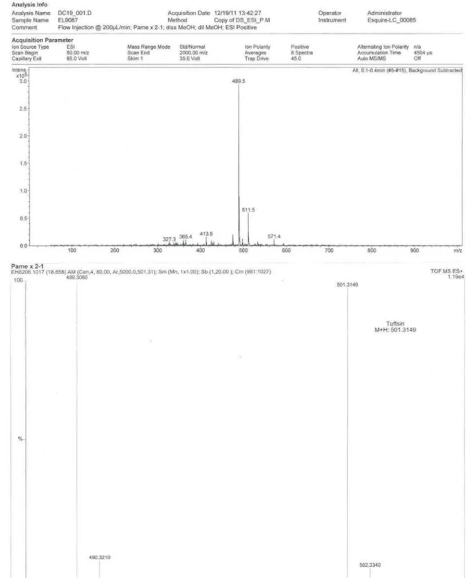
The HRESIMS analysis of rodriguesic acid (3)
displayed a [M]+ ion at m/z 489.3080, appropriate for
the molecular formula C26H41N4O5, that differs from the
ammonium salt of rodriguesine A (1) by the addition of two oxygens and the loss of two hydrogens, and requiring an additional site of unsaturation. The 1H and 13C NMR
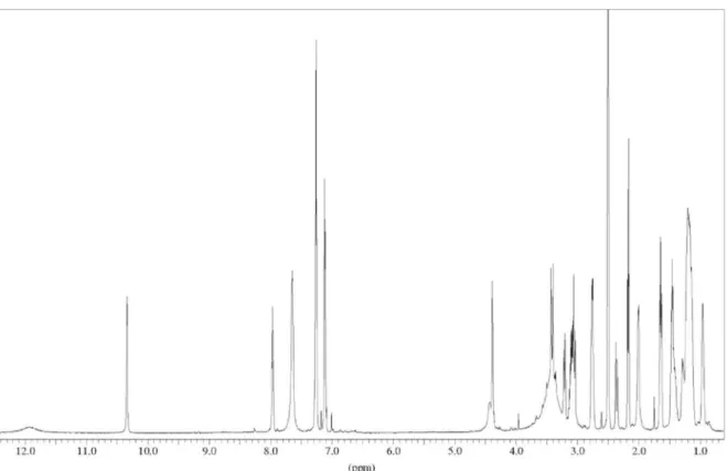
spectra (Tables 1 and 2) of 3 showed a marked similarity to the spectra obtained for rodriguesine A (1). The significant
difference was the absence of the terminal methyl triplet assigned to Me-28, resonating at δ0.93, in 1. Instead, an additional carbonyl resonance at δC 174.5, along with the
constraints imposed by the molecular formula, suggested the presence of a C-28 carboxylic acid functionality in 3. Correlations observed in the gHMBC spectrum between the alkyl chain methylenes assigned to H-26 (δH 1.46/δC 24.4)
and H-27 (δH 2.16/δC 33.6) and the C-28 carboxylate carbon
at δC 174.5 confirmed this assignment. The isolation of
rodriguesic acid (3) as its corresponding ammonium salt was indicated by the integration of the NH3
+ signal at δ 7.63,
and also by the fact that CH2-14 was observed as a sextet.
All other structural features of 3 were the same as 1. Rodriguesic acid hydroxamate (4) gave a [M]+ ion at m/z 505.3020 in the HRESIMS, that was appropriate for the molecular formula C26H41N4O6, which has one oxygen
more than that recorded for compound 3. Moreover, the doublet at δ 8.24 assigned to the H-4 resonance of
Table 1. 1H NMR data for compounds 3-6 in DMSO-d
6 [δ, multiplicity (J in Hz)] at 600 MHz
Position 3 4 5 6
3 4.07 (bd, 2.9) 4.37 (bs) 4.07 (bdd, 7.9, 4.9) 4.37 (tl, 4)
4 8.24 (bd, 2.4) – 8.25 (d, 3.1) –
6 2.95 (d, 17); 3.40 (d, 17) 2.35 (d, 17); 3.41 (d, 17) 2.95 (d, 17.2); 3.40 (d, 17.1) 2.35 (d, 17); 3.41 (d, 17)
7 2.88 (dd, 4.9, 13.6); 3.01 (dd, 5.6, 13.6)
3.04 (bd, 13.6); 3.20 (dd, 4.7, 13.6)
2.88 (dd, 5.0, 12.9); 3.00 (dd, 5.6, 13.5)
3.04 (dd, 2.9, 13.7); 3.21 (dd, 4.8, 13.8)
9/13 7.13 (d, 6.7) 7.11 (d, 6.0) 7.14 (m) 7.11 (m)
10/12 7.27 (m) 7.25 (m) 7.27 (m) 7.25 (m)
11 7.25 (m) 7.25 (m) 7.25 (m) 7.24 (m)
14 2.75 (bsex, 6.2) 2.74 (bsex, 6.5) 2.75 (sex, 6.2) 2.74 (sex, 7.4)
15 1.63 (qui, 7.4) 1.63 (qui, 7.4) 1.64 (qui, 6.6) 1.63 (qui, 7.3)
16 3.07 (m) 3.08 (m) 3.08 (m) 3.09 (m)
16a 7.99 (t, 5.6) 7.96 (t, 5.6) 7.99 (t, 6.0) 7.96 (t, 5.4)
18 2.10 (d, 6.7) 2.00 (d, 6.9) 2.11 (d, 6.5) 2.00 (bd, 7.7)
19 4.52 (bs) 4.42 (bs) 4.53 (bs) 4.42 (bs)
20 1.30 (m); 1.45 (m) 1.27 (m); 1.40 (m) 1.30 (m); 1.46 (m) 1.27 (m); 1.40 (m)
21 1.01 (m) 0.94 (m) 1.02 (m) 0.94 (m)
22 1.19 (m) 1.16-1.19 (m) 1.20 (m) 1.17 (m)
23 1.19 (m) 1.16-1.19 (m) 1.20 (m) 1.17 (m)
24 1.19 (m) 1.16-1.19 (m) 1.20 (m) 1.17 (m)
25 1.10 (m) 1.16-1.19 (m) 1.20 (m) 1.17 (m)
26 1.45 (bqui, 6.7) 1.45 (m) 1.46 (m) 1.47 (qui, 6.7)
27 2.16 (t, 7.4) 2.16 (t, 7.4) 2.26 (t, 7.4) 2.26 (t, 7.4)
29 – – 3.56 (s) 3.57 (s)
NH3+ 7.63 (bs) 7.64 (bs) 7.62 (bs) n.o.
CO2H 11.93 (vbs) 11.9 (vbs) – –
N-OH n.o. 10.33 n.o. 10.33
Rodriguesic Acids, Modified Diketopiperazines from the Gastropod Mollusc Pleurobranchus areolatus J. Braz. Chem. Soc.
792
the amide in 3 is missing in the 1H NMR spectrum of 4
(Table 1). Instead, a sharp singlet at δ10.33, which did not show gHSQC correlations to a carbon or a nitrogen atom, was observed. Additionally the significant chemical shift differences for the resonances assigned to the protons and carbons at positions CH-3 (δH 4.37/δC 64.3), C-5 (δC 159.9)
and CH2-7 (δH 3.04 and 3.20/δC 35.2) compared to those
observed for 3 (Tables 1 and 2) indicated a structural change at N-4. A weak three bond correlation observed in the gHMBC spectrum between the singlet at δ10.33 and the
C-5 carbonyl assigned to the resonance at δ 159.9, along with the additional oxygen required by the HRESIMS, allowed for the placement of a hydroxamate at position N-4.
A tROESY correlation between theN-OH hydroxamate
singlet (δ10.33) and the phenyl H-9/H-13 (δ 7.11) provided further support for this assignment, since an equivalent tROESY correlation was observed between the amide H-4 resonance (δ 8.24) and the phenyl H-9/H-13 resonances (δ 7.13) in the spectrum obtained for 3. Complete analysis of the 1D and 2D NMR data revealed that all other structural features of 4 were the same as 3 and the structure was defined as the rodriguesic acid hydroxamate (4). A closely related hydroxamate-modified diketopiperazine, etzionine, was previously isolated from an ascidian of the genus
Didemnum.15
The HRESIMS analysis of compound 5 displayed a [M]+ ion at m/z 503.3232, appropriate for the molecular
formula C27H43N4O5, that differs from rodriguesic acid (3)
by the addition of 14 mass units. Other than an additional oxygenated methyl singlet (δC 51.1/δH 3.56, s) and the loss
of the broad signal at δ11.93 assigned to the exchangeable carboxylate proton in 3, the 1H and 13C NMR spectra
obtained for 3 and 5 were essentially identical Therefore, the structure of compound 5 was assigned that of the methyl ester of rodriguesic acid. The structure of 6, for which the HRESIMS analysis showed a [M]+ ion at m/z
519.3179 corresponding to the formula C27H43N4O6, was
established based on analogous arguments as the methyl ester of rodriguesic acid hydroxamate (Tables 1 and 2). Furthermore, HRFTMS/MS analysis revealed fragments corresponding to the loss of hydroxyl at m/z 502.3, the loss of a diaminopropyl fragment at m/z 445.2, loss of CH2–(CO)–NH(CH2)3NH2 at m/z 403.2, as well as the loss
of the entire diketopiperazine moiety at m/z 298.2 (see Supplementary Information). The last three fragmentations provide additional support that the hydroxyl group in 6, and by inference in 4, is attached to the phenylalanine nitrogen rather than the nitrogens of the 1,3-diaminopropyl chain.
The absolute configuration of rodriguesines A (1) and B (2) was previously established as 3S,19R.14 The circular
dichroism (CD) spectrum of the inseparable mixture of 1
and 2 showed a well-defined, intense negative Cotton effect at λmax 215 nm (Figure S11, Supplementary Information).
The CD spectra of rodriguesic acid methyl ester (5) and of the hydroxamate 6 present almost identical negative Cotton effects (Figures S12 and S13, Supplementary Information). Therefore, both 5 and 6 and the free acids 3 and 4 are assumed to have the same absolute configuration as 1 and 2.
Previous investigations of the chemistry of
Pleurobranchus mollusc species have reported the membrenones, polypropionates from P. membranaceus,16
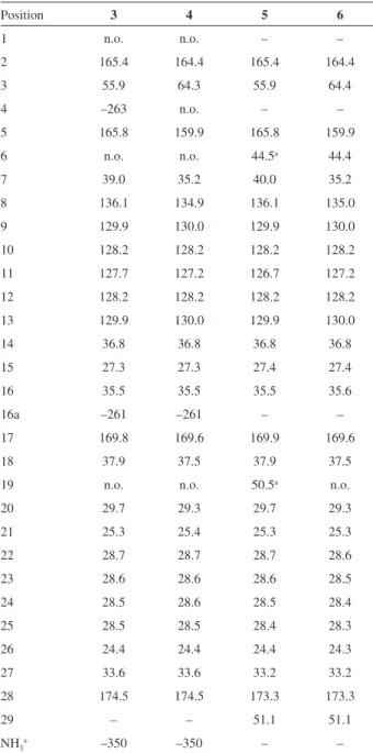
Table 2. 13C NMR and 15N data for diketopiperazines 3, 4, 5 and 6
(150 MHz, DMSO-d6)
Position 3 4 5 6
1 n.o. n.o. – –
2 165.4 164.4 165.4 164.4
3 55.9 64.3 55.9 64.4
4 –263 n.o. – –
5 165.8 159.9 165.8 159.9
6 n.o. n.o. 44.5a 44.4
7 39.0 35.2 40.0 35.2
8 136.1 134.9 136.1 135.0
9 129.9 130.0 129.9 130.0
10 128.2 128.2 128.2 128.2
11 127.7 127.2 126.7 127.2
12 128.2 128.2 128.2 128.2
13 129.9 130.0 129.9 130.0
14 36.8 36.8 36.8 36.8
15 27.3 27.3 27.4 27.4
16 35.5 35.5 35.5 35.6
16a –261 –261 – –
17 169.8 169.6 169.9 169.6
18 37.9 37.5 37.9 37.5
19 n.o. n.o. 50.5a n.o.
20 29.7 29.3 29.7 29.3
21 25.3 25.4 25.3 25.3
22 28.7 28.7 28.7 28.6
23 28.6 28.6 28.6 28.5
24 28.5 28.6 28.5 28.4
25 28.5 28.5 28.4 28.3
26 24.4 24.4 24.4 24.3
27 33.6 33.6 33.2 33.2
28 174.5 174.5 173.3 173.3
29 – – 51.1 51.1
NH3+ –350 –350 – –
Pereira et al. 793 Vol. 25, No. 4, 2014
keenamide A, a cytotoxic cyclic hexapeptide from
P. forskalii,17 testuninariols A and B, ichtyotoxic
triterpenes from P. testudinarius,18 as well as the
cytotoxic maleimide-bearing diterpenes haterumaimide L, M and 3β-hydroxychlorolissoclimide isolated from
P. albiguttatus and P. forskalii.19 More recent examples
of Pleurobranchus spp. metabolites include the cytotoxic macrocyclic peptide cycloforskamide,20 as well as the
highly modified ergot alkaloid ergosinine,21 both isolated
from P. forskalii. Considering that Pleurobranchus spp. molluscs are carnivores, the chemical diversity found in
P. forskalii may well be a result of the animals’ varied diet and possibly the presence of associated microorganisms that have the potential to transform sequestered metabolites into modified derivatives.
The methyl esters 5 and 6 may well be artifacts of isolation of the actual secondary metabolites, rodriguesic acid (3) and rodriguesic acid hydroxamate (4). A particular feature of compounds 3-6 is the presence of a carboxylic acid replacing the methyl group at the terminus of the aliphatic side chain. Only recently Hertweck’s group unveiled the biosynthesis of the potent microbial toxin bongkrekic acid. Bongkrekic acid also bears a carboxylic acid group in the place of a polyketide terminal methyl group. The results obtained by Hertweck’s team demonstrated that the bonL enzyme, in association with a cytochrome P450 monooxygenase (CYP), is responsible for the transformation of a terminal methyl group into a carboxylic acid through a six-electron oxidation. The enzyme bonL is assumed to be the first CYP reported to oxidize the terminal methyl group of a polyketide-derived putative precursor to a carboxylic acid.22 The isolation
of rodriguesic acids 3 and 4 perhaps represent additional examples of CYP oxidation, and suggests that a P. areolatus
symbiont may be responsible for this transformation. Recently diketopiperazines have been isolated from the culture media of a mollusc-derived actinobacterium.23 To
the best of our knowledge, rodriguesic acids 3 and 4 are the first diketopiperazine derivatives isolated from a mollusc. On the other hand, another possible biogenetic pathway that can be proposed for the β-amino-dicarboxylic acid moiety of both 3 and 4 has aspartic acid as a “starter” for the condensation with four malonate/acetate groups (Figure 2).
This pathway would require a transamination reaction with glycine in order to account for the subsequent condensation of the β-amino-dicarboxylic acid residue into the core diketopiperazine. In this scenario, compounds 3 and 4 would be made by the ascidian prior to sequestration by the mollusc rather than being formed by the mollusc (or an associated microorganism) transformation of 1 after ingestion.
Diketopiperazines exert an array of biological activities, such as antibiotic, antifungal, antiviral, biofilm formation inhibition, as well as acting as chemical signaling agents.24
The accumulation of rodriguesic acid (3) and of the hydroxamate of rodriguesic acid (4) by the shell-less mollusc P. areolatus may represent a strategy of chemical defense. Although we have been unable to test compounds
3 and 4 in bioassays, due to the limited amount of material, the esters 5 and 6 were evaluated in cytotoxicity assays against MCF-7 (breast), B16 (melanoma) and HCT8 (colon) cancer cell lines25 and antimicrobial assays
against different pathogenic strains of Staphylococcus aureus, oxacillin-resistant S. aureus, Escherichia coli,
P s e u d o m o n a s a e r u g i n o s a, C a n d i d a a l b i c a n s,
Enterococcus faecalis, Streptococcus sanguinis, and
Streptococcus mutans,14 but did not exhibit any significant
activity.
Conclusions
The isolation of rodriguesic acid (3) and of the rodriguesic acid hydroxamate (4) represents the first report of diketopiperazine derivatives from a mollusc. The presence of rodriguesic acids in P. areolatus presumably results from the sequestering of rodriguesin A (1) from the ascidian Didemnum sp., on which the mollusc was found, with a further modification of the terminal methyl group into a carboxylic acid group. However, secondary metabolites sequestration and subsequent transformation by marine opistobranchs are a rare metabolic capacity.4,26 The
presence of a carboxylic acid group substituting a terminal methyl group of an alkyl chain is also unusual among such compounds, and may constitute the second example of such a biosynthetic transformation in secondary metabolites. The results herein reported constitute an example of complex biological and biochemical interactions among marine
Rodriguesic Acids, Modified Diketopiperazines from the Gastropod Mollusc Pleurobranchus areolatus J. Braz. Chem. Soc.
794
organisms which deserve further investigation to uncover the metabolic origin of these metabolites.
Acknowledgements
Financial support was provided by a BIOTA/ BIOprospecTA FAPESP grant (2010/50190-2) to R. G. S. B. and by a NSERC grant to R. J. A.. Scholarships to F. R. P. by CAPES and to V. P. by CNPq-Brasil and DAAD (Germany) are also gratefully acknowledged.
Supplementary Information
Supplementary data (1H and 13C NMR for compounds
3-6, circular dichroism spectra of compounds 1, 2, 5 and 6, as well as HRFTMS/MS data for compound 6) are available free of charge at http://jbcs.sbq.org.br as a PDF file.
References
1. Karuso, P. In Bioorganic Marine Chemistry; Scheuer, P. J., ed.; Springer-Verlag: Berlin & Heidelberg, 1987, volume I, ch. 2, pp. 31-60.
2. Faulkner, D. J. In Ecological Roles of Marine Natural Products; Paul, V., ed.; Comstock Publishing Associates: Ithaca, NY, 1992, ch. 4, pp. 119-163.
3. Cimino, G.; Ghiselin, M. T. In Marine Chemical Ecology; McClintock, J. B.; Baker, B. J., eds.; CRC Press: Boca Raton, 2001, ch. 3, pp. 115-154.
4. Cimino, G.; Ghiselin, M. T.; Chemoecology1999, 9, 187. 5. Gao, J.; Hamann, M. T.; Chem. Rev. 2011, 111, 3208. 6. Salazar, R.; Cortés-Funes, H.; Casado, E.; Pardo, B.;
López-Martín, A.; Cuadra, C.; Tabernero, J.; Coronado, C.; G a r c í a , M . ; M a t o s - P i t a , A . S . ; M i g u e l - L i l l o , B . ; Cullell-Young, M.; Dios, J. L. I.; Paz-Ares, L.; Cancer Chemother. Pharmacol. 2013, 72, 75.
7. Luesch, H.; Harrigan, G. G.; Goetz, G.; Horgen, F. D.; Curr. Med. Chem. 2002, 9, 1791.
8. Leao, P. N.; Engene, N.; Antunes, A.; Gerwick, W. H.; Vasconcelos, V.; Nat. Prod. Rep. 2012, 29, 372.
9. Rodríguez, J.; Fernández, R.; Quiñoá, E.; Riguera, R.; Debitus, C.; Bouchet, P.; Tetrahedron Lett. 1994, 35, 9239. 10. Fernández, R.; Rodríguez, J.; Quiñoá, E.; Riguera, R.;
Muñoz, L.; Fernández-Suárez, M.; Debitus, C.; J. Am. Chem. Soc. 1996, 118, 11635.
11. Nakao, Y.; Yoshida, W. Y.; Takada, Y.; Kimura, J.; Yang, L.; Mooberry, S. L.; Scheuer, P. J.; J. Nat. Prod. 2004, 67, 1332. 12. Umehara, M.; Negishi, T.; Tashiro, T.; Nakao, Y.; Kimura, J.;
Bioorg. Med. Chem. Lett. 2012, 22, 7422.
13. Pereira, F. R.; Berlinck, R. G. S.; Rodrigues-Filho, E.; Veloso, K.; Ferreira, A. G.; Padula, V.; Quim. Nova2012, 35, 11, 2194.
14. Kossuga, M. H.; Lira, S. P.; McHugh, S.; Torres, Y. R.; Lima, B. A.; Gonçalves, R.; Veloso, K.; Ferreira, A. G.; Rocha, R. M.; Berlinck, R. G. S.; J. Braz. Chem. Soc. 2009, 20, 704. 15. Hirsch, S.; Miroz, A.; McCarthy, P.; Kashman, Y.; Tetrahedron
Lett. 1989, 30, 4291.
16. Ciavatta, M. L.; Trivellone, E.; Villani, G.; Cimino, G.;
Tehahedron Lett. 1993, 34, 6191.
17. Wesson, K. J.; Hamann, M. T.; J. Nat. Prod. 1996, 59, 629. 18. Spinella, A.; Mollo, E.; Trivellone, E.; Cimino, G.; Tetrahedron
1997, 53, 16891.
19. Fu, X.; Palomar, A. J.; Hong, E. P.; Schmitz, F. J.; Valeriote, F. A.; J. Nat. Prod. 2004, 67, 1415.
20. Tan, K. C.; Wakimoto, T.; Takada, K.; Ohtsuki, T.; Uchiyama, N.; Goda, Y.; Abe, I.; J. Nat. Prod. 2013, 76, 1388.
21. Wakimoto, T.; Tan, K. C.; Abe, I.; Toxicon2013, 72, 1. 22. Moebius, N.; Ross, C.; Scherlach, K.; Rohm, B.; Roth, M.;
Hertweck, C.; Chem. Biol. 2012, 19, 1164.
23 Kalinovskaya, N. I.; Kalinovsky, A. I.; Romanenko, L. A.; Dmitrenok, P. S.; Kuznetsova, T. A.; Nat. Prod. Commun. 2010,
5, 597.
24. Carvalho, M. P.; Abraham, W.-R.; Curr. Med. Chem. 2012, 19, 3564.
25. Seleghim, M. H. R.; Lira, S. P.; Kossuga, M. H.; Batista, T.; Berlinck, R. G. S.; Hajdu, E.; Muricy, G.; Rocha, R. M.; Nascimento, G. F.; Silva, M.; Pimenta, E. F.; Thiemann, O. H.; Oliva, G.; Cavalcanti, B. C.; Pessoa, C.; Moraes, M. O.; Hajdu, E.; Peixinho, S.; Rocha, R. M.; Rev. Bras. Farmacogn.
2007, 17, 287.
26. Cimino, G.; Fontana, A.; Giménez, F.; Marin, A.; Mollo, E.; Trivellone, E.; Zubía, E.; Experientia1993, 49, 582-586.
Submitted: November 19, 2013 Published online: February 11, 2014
Supplementary Information
S
I
J. Braz. Chem. Soc., Vol. 25, No. 4, S1-S12, 2014. Printed in Brazil - ©2014 Sociedade Brasileira de Química 0103 - 5053 $6.00+0.00
*e-mail: [email protected]
Rodriguesic Acids, Modified Diketopiperazines from the Gastropod Mollusc
Pleurobranchus areolatus
Fabio R. Pereira,a Mario F. C. Santos,a David E. Williams,b Raymond J. Andersen,b
Vinicius Padula,c Antonio G. Ferreirad and Roberto G. S. Berlinck*,a
aInstitutode Química de São Carlos, Universidade de São Paulo,
CP 780, 13560-970 São Carlos-SP, Brazil
bDepartmentsof Chemistry and Earth, Ocean & Atmospheric Sciences,
University of British Columbia, Vancouver, BC, V6T 1Z1, Canada
cSNSB-Zoologische Staatssammlung München, Münchhausenstrasse 21,
81247 München, Germany and Department Biology II and GeoBio-Center, Ludwig-Maximilians-Universität München, Germany
dDepartamentode Química, Universidade Federal de São Carlos,
Rodovia Washington Luiz, km 235, 13565-905 São Carlos-SP, Brazil
Rodriguesic Acids, Modified Diketopiperazines from the Gastropod Mollusc Pleurobranchus areolatus J. Braz. Chem. Soc.
S2
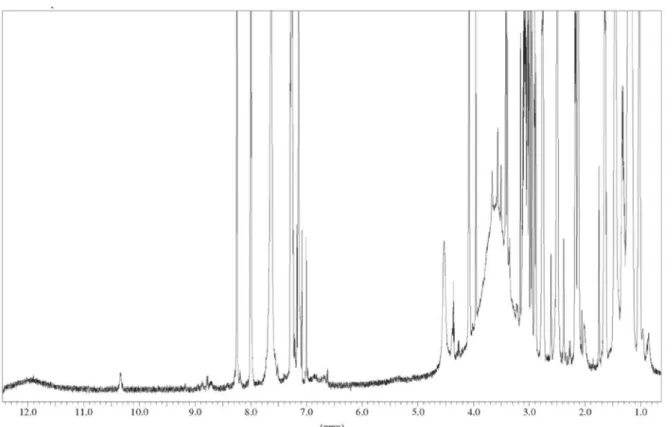
Figure S2. 1H NMR spectrum of rodriguesic acid (3) in DMSO-d
6at 600 MHz.
Pereira et al. S3 Vol. 25, No. 4, 2014
Rodriguesic Acids, Modified Diketopiperazines from the Gastropod Mollusc Pleurobranchus areolatus J. Braz. Chem. Soc.
S4
Pereira et al. S5 Vol. 25, No. 4, 2014
Figure S6. 1H NMR spectrum of the rodriguesic acid hydroxamate (4) in DMSO-d6 at 600 MHz.
Rodriguesic Acids, Modified Diketopiperazines from the Gastropod Mollusc Pleurobranchus areolatus J. Braz. Chem. Soc.
S6
Pereira et al. S7 Vol. 25, No. 4, 2014
Figure S9. 1H NMR spectrum of rodriguesic acid methyl ester (5) in DMSO-d
6at 600 MHz.
Rodriguesic Acids, Modified Diketopiperazines from the Gastropod Mollusc Pleurobranchus areolatus J. Braz. Chem. Soc.
S8
Pereira et al. S9 Vol. 25, No. 4, 2014
Figure S12. 1H NMR spectrum of the methyl ester of the rodriguesic acid hydroxamate (6) in DMSO-d
6at 600 MHz.
Rodriguesic Acids, Modified Diketopiperazines from the Gastropod Mollusc Pleurobranchus areolatus J. Braz. Chem. Soc.
S10
Pereira et al. S11 Vol. 25, No. 4, 2014
Figure S15. HRFTMS/MS analysis of methyl ester of the rodriguesic acid hydroxamate (6) [M+H]+ ion at m/z 519.31476.
Rodriguesic Acids, Modified Diketopiperazines from the Gastropod Mollusc Pleurobranchus areolatus J. Braz. Chem. Soc.
S12
Figure S17. Circular dichroism spectrum of rodriguesic acid methyl ester (5) in MeOH (0.033 mg mL-1).
![Table 1. 1 H NMR data for compounds 3-6 in DMSO-d 6 [δ, multiplicity (J in Hz)] at 600 MHz](https://thumb-eu.123doks.com/thumbv2/123dok_br/18998646.463003/4.892.94.817.148.722/table-nmr-data-compounds-dmso-multiplicity-hz-mhz.webp)