Article
0103 - 5053 $6.00+0.00*e-mail: [email protected]
#Present address: Fleury Medicina e Saúde, Av. General Valdomiro de
Lima 508, 04344-070 São Paulo-SP, Brazil
A Fragmentation Study of Di-Acidic Mycosporine-like Amino Acids
in Electrospray and Nanospray Mass Spectrometry
Karina H. M. Cardozo,a,b,# Ricardo Vessecchi,c Sérgio E. Galembeck,c Thais Guaratini,a,b Paul J. Gates,b Ernani Pinto,d Norberto P. Lopes*,e and Pio Colepicoloa
aInstituto de Química de São Paulo, Universidade de São Paulo, 05508-900 São Paulo-SP, Brazil
bSchool of Chemistry, University of Bristol, Cantock’s Close, Bristol, BS8 1TS, United Kingdom
cDepartamento de Química, Faculdade de Filosoia Ciências e Letras de Ribeirão Preto, Universidade de São Paulo, 14040-901 Ribeirão Preto-SP, Brazil
dDepartamento de Análises Clínicas e Toxicológicas, Faculdade de Ciências Farmacêuticas, Universidade de São Paulo, 05508-900 São Paulo-SP, Brazil
eDepartamento de Física e Química, Faculdade de Ciências Farmacêuticas de Ribeirão Preto, Universidade de São Paulo, 14040-903 Ribeirão Preto-SP, Brazil
No presente estudo, duas micosporinas (MAAs) contendo um segundo ácido carboxílico foram submetidas à fragmentação em eletrospray e nanospray em diferentes equipamentos. Em contraste com resultados anteriores, a eliminação de radical metila no modo positivo de análise foi um processo minoritário de fragmentação. Neste trabalho apresentamos também a via de fragmentação destas substâncias em modo negativo e cálculos teóricos para caracterizar os sítios de protonação.
Two mycosporine (MAAs), containing an extra acid function, were analyzed by nanospray and electrospray ionization tandem mass spectrometry. In contrast to the previous studies it is demonstrated that no signiicant characteristic methyl radical loss occurred in positive mode. The fragmentation pathway in negative mode was also proposed in this work, along with theoretical calculations to characterize the site of protonation.
Keywords: mycosporine, nanoESI-MS/MS, ESI-MS/MS, natural products, algae
Introduction
Mycosporine-like amino acids (MAAs) are a group of chemically related, water soluble compounds responsible for UV photoprotection in a diverse range of organisms including invertebrates, fish, bacteria, cyanobacteria, micro- and macroalgae.1-3 These compounds are chemically
characterised by the presence of either a cyclohexenone or cyclohexenimine chromophore conjugated with a substituent nitrogen of an amino acid, amino alcohol or
amino group (Figure 1).1 MAAs normally show a strong
UV absorption between 310 and 360 nm with high molar extinction coeficients. These characteristics indicate a possible photoprotective role that has been demonstrated
in a number of studies4-7 and for the economic interest of
pharmaceutical companies. In addition to a UV protective function, it has been suggested that MAAs also have antioxidant activity,8 osmotic functions,9 and regulatory
role in reproduction.10 More than 20 MAAs have so far
been characterised and identiied in aquatic environments and this number is increasing with the development and application of more eficient and sensitive methodologies such as high performance liquid chromatography (HPLC) and mass spectrometry.11-13
Tandem mass spectrometry (MS/MS) fragmentation studies in positive ion mode along with UV spectra are the normal tools for the structure elucidation for the new derivatives of MAAs.12-18 The increased application of mass
spectrometry to the analysis of natural products is related to the improved quality of the structural information produced by elucidation of fragmentation patterns, mechanisms and pathways.19-20 Previously, several studies have focussed
on the fragmentation of some MAAs showing a highly characteristic loss of mass 15 when analysed by positive mode ESI-MS/MS, in addition to a complex fragmentation pathway.13,15,17 Recently, a theoretical and FT-ICR-MS mass
spectrometry study showed that the elimination of mass 15 is due to a radical processes taking place at the methoxyl group link to a unsaturated carbon.18 Attempts to obtain
spectra in negative mode were not successful.17
In the last few years, nanospray ionisation (nanoESI) is beginning to increase in importance, especially with the development of automated systems using ‘chips’ (arrays of uniform nanospray needles that are used only once to avoid contamination).21 NanoESI offers the possibility of
improved sensitivity and lower sample consumption over conventional ESI for the analysis of natural products.22
This is especially important for the study of extracts from biological and medicinal sources when often only a very small amount of material is available.
In ‘chip based’ nanoESI, the analyte solution is sprayed from a conducting pipette tip pressed against the rear of a chip using a small gas pressure and much lower voltages to create the spray. Recently, analysis of some natural antioxidants (retinoids23,24 and carotenoids25,26)using ‘chip
based’ technology showed differences in the ionisation behavior when compared with conventional ESI. This increases the possibilities for structure elucidation at low sample amounts as new fragmentation routes are made available by controlling the types of ions being formed. Based upon these previous studies, the purpose of this work is to compare the ionisation of MAAs in negative and positive mode nanoESI and establish, for the irst time, a sound basis for the mechanism of the fragmentation of MAA anions.
Experimental
Chemicals
All solvents used were HPLC grade (Tedia, J. Baker and Fisher). Water was puriied using a Milli-Q system (Millipore, Bedford, MA, USA). Trifluoroacetic acid (99.9%) was purchased from Aldrich. Galena Química e Farmacêutica Ltda/Brazil kindly supplied the standards
of shinorine and porphyra-334 (product Helioguard® 365-
Porphyra umbilicalis extracts)
Instrumentation
Nanospray ionisation analyses were performed on two quadrupole-time of light hybrid instruments: (a) an UltrOTOF-Q (Bruker Daltonics, Billerica, MA) using Tip™ Emittek (glass tip capillaries working with 500 V) or (b) a QStar-XL (Applied Biosystems, Warrington, UK) using a Nanomate HD automatic ‘chip based’ nanospray system (Advion Biosciences, Norwich, UK). The Nanomate was set for 5 µL of solution to be aspirated and sprayed through a Nanomate 400 chip at 1.45 kV, with a nitrogen back pressure of 0.4 psi. On both instruments, the ion source gas and curtain gas were nitrogen.
Electrospray ionisation analyses were performed on ive instruments: (a) an Apex 4 7.0 Tesla Fourier-transform ion-cyclotron resonance mass spectrometer (Bruker Daltonics, Billerica, MA, USA). Samples were directly infused into the Apollo electrospray source from a syringe pump at 100 µL h-1. Analyses were performed at
a capillary voltage, of 4600V and capillary exit potential of 200 V (except were indicated otherwise). The N2 drying gas temperature was 200 °C. A mixture of PEG grades was used as an external calibrant for accurate-mass ESI analysis; (b) on a quadrupole-time of light instrument (UltrOTOF-Q, Bruker Daltonics, Billerica, MA). The analyses were performed in positive ion ESI mode at a capillary voltage of 3400 V and N2 drying gas temperature of 180 °C. NaTFA 10 mmol L-1 was used as a standard
for internal and external calibration; (c) on a QStar-XL quadrupole-time-of-light instrument (Applied Biosystems, Warrington, UK); (d) on a Quattro-LC triple quadrupole
mass spectrometer (Micromass, Manchester, UK); (e)on
an Esquire HCT ion trap instrument (Bruker Daltonics, Billerica, MA, USA) using a syringe pump (Cole-Parmer, Vernon Hills, IL, USA). Ion trap analyses were performed using nitrogen as the nebulising and drying gas and helium as the bath gas (4×10⁻⁶ mbar).
Theoretical calculations
All calculations were performed in Gaussian 0327 suite
of programs using the B3LYP/6-31+G(d,p) model.28,29
Results and Discussion
Mycosporine-like amino acids with one acidic function were previously analysed by positive mode ESI sequential mass spectrometry (at high-resolution and accurate-mass). The loss of a methyl radical by the homolytic cleavage of the O-C bond was observed to be the preferred fragmentation pathway.13,17,18 This characteristic loss was thoroughly studied
by a range theoretical calculations and the homolytic cleavage was found to be dependent on the weakening of the O-C bond. This cleavage is directly affected by the protonation site with the radical elimination occurring from the most unstable conformer.18 In this study, we have analysed the
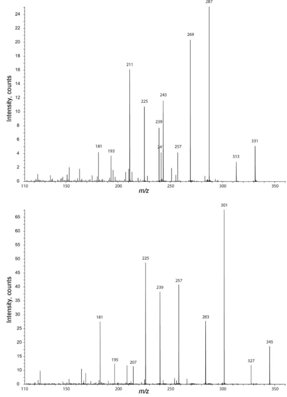
effect that the presence of a second acidic function in the side chain has on the preferred fragmentation pathways and to pinpoint any differences observed. Figure 2 shows the product ion spectrum of porphyra-334 (precursor ion
m/z 347) obtained from a range of instruments. The spectra clearly show the occurrence of the radical fragmentation route (to product ion m/z 332), but at very low intensity in all instruments used (with a range of different ESI source designs, including nanoESI). Shinorine exhibits the same result as porphyra-334, which differs to those previously obtained for the mono acidic MAAs where the radical elimination mechanism is the major fragmentation pathway. However, the MS3 spectra (ion trap) of the di-acidic MAA
(fragmentation of [M + H-CO2]+) shows an intense loss
of the methyl radical (Figure 2, spectrum A’). This result indicates that the presence of the two acidic functions changes the driving force for the fragmentation with a neutral elimination of CO2 being the predominant irst step with all instrument set-ups. To conirm this theory, MAAs with two carboxylic acid functions were submitted to theoretical computational analysis to deine the site of protonation. As described, our previous results were based on topologic analysis of electronic density, gas-phase basicity and bond order for each protonated conformer. The imine nitrogen was indicated as the most basic site. However, the most reactive ion occurs by protonation on the vinyl carbon. In order to know the protonation site of shinorine, we have calculated its gas-phase basicity and proton afinity. These results are in agreement with our previous studies; where the less energetic ion occurs by protonation on the imine nitrogen whose gas-phase basicity is 247.32 kcal mol-1
(Figure 3). This data indicates that the presence of the second carboxylic acid effects the protonation site leading to an increased CO2 elimination. This elimination reduces the internal ion energy and results in the observed intensity of the methyl radical elimination route. These observations suggest that the analytical methodology (such as quantiication) based on protonated molecules and radical fragmentation route is not always a good choice for all MAAs. However, the selection of MS3 from [M + H-15]+ is still a good option
for the screening of extracts which may contain MAAs. A
Figure 2. Positive ion ESI spectra of Porphyra-334 (m/z 347). A) ion trap MS2 spectra of ion m/z 347; A’) ion trap MS3 spectra of ion m/z 303; B) Q-Q
Figure 4. Negative ion nanoESI spectra of Shinorine and Porphyra-334.
previous study with the ionophore monensin A, showed signiicantly different fragmentation intensities between MS/MS with ESI and nanoESI,31 but all of the experiments
with the MAAs show similar behaviour in both nanoESI and ESI analysis with no signiicant differences being observed.
As expected, analysis in negative ion mode (Figure 4) showed no radical elimination. Considering that in the positive ion mode, the bond cleavage was observed to be dependent of the protonation site that weakens the C-O bond, another driving force must govern the fragmentation pathways in negative ion mode. Detailed analyses suggest that the carboxylic acid function may lose the proton, which causes an increase of electron density that is stabilised by
resonance. Scheme 1 represents the proposed fragmentation pathway for shinorine (R= H) and porphyra-334 (R= CH3). Product ion spectra of both shinorine and porphyra-334 produced initial CO2 eliminations followed by various combinations of H2O and/or CO2 losses. After the elimination of H2O and CO2 from the side chains, all the resultant ions must keep the negative charge on the more acidic positions. Based on this observation we expected to have the charge on the alkoxy groups, and these two possibilities must produce different terminal ions. It is proposed that the shinorine product ions (m/z 225 and 243) can go on to lose methanol (producing the ions m/z 193 and 211, respectively) in opposition to the isomeric structure with the charge on
N NH HO HO OCH3 C O O HO O O H N NH HO HO OCH3 R HO H - CO2
- H2O
N NH HO HO OCH3 R C O O O O N NH HO HO OCH3 - H2O
- CO2
N NH O O OCH3 R HO N NH O HO OCH3 - CO2
N
NH O
O
OCH3
- CO2
N
NH O
HO
OCH3 - H2O
OH R H H H N NH O OCH3 R HO H N NH O OCH3 - CH3OH
or H N NH HO OCH3 C O OH HO O O H
- CO2
N NH HO OCH3 HO N NH O OCH3 HO R N NH O OCH3 H C HO O OH
m/z 181
R
R= H m/z 331 R= CH3m/z 345
R
R R
H H
R
R
- CH3OH
- H2O R
R
R= H m/z 313 R= CH3m/z 327
R= H m/z 313 R= CH3m/z 327
R= H m/z 287 R= CH3m/z 301
R= H m/z 287 R= CH3m/z 301
R= H m/z 269 R= CH3m/z 283 R= H m/z 269
R= CH3m/z 283
R= H m/z 243 R= CH3m/z 257
R= H m/z 225 R= CH3m/z 239
R= H m/z 269 R= CH3m/z 283
R= H m/z 269 R= CH3m/z 283
R= H m/z 211 R= CH3m/z 225
R= H m/z 193 R= CH3m/z 207
H H O OH O O O O O OH C O O C O OH H
the other neighbouring oxygen atom that will produce the ion m/z 181. The same loses occur for porphyra-334. Careful analyses of the porphyra-334 and shinorine spectra indicate the presence of an additional fragmentation pathway for shinorine (Scheme 2). After the loss of a proton from the carboxylic acid function, the charge can be stabilised through assistance of the neighboring hydroxyl group. The non-substitution on the carbinolic carbon allows the loss of a formaldehyde neutral followed by water on one side or loss of a formic acid neutral on the other side of the bridge. These simple neutral losses may provide some further information about the side chain for theses MAAs with carboxylic acid on the side chain.
Conclusions
These results indicate that the presence of a second carboxylic acid function signiicantly reduces the intensity of the observed product ions from the radical methyl cleavage in positive mode MS/MS. As expected, in the negative ion mode, the radical fragmentation pathway does not occur. Taken together, these results conirm the importance of careful selection of the product ions used for analytical protocols for the analysis of crude extracts containing MAAs where the presence of the second acid function may change the fragmentation behavior and the classical analysis of loss of methyl radical may lead to the wrong conclusions during screening for MAAs.
Acknowledgments
The authors thank FAPESP (Fundação de Amparo à Pesquisa do Estado de São Paulo, 05/01572-1,
01/13482-6 and 03/08735-8), CAPES (Coordenação de Aperfeiçoamento de Pessoal de Nível Superior), CNPq and CNPq-Millenium (Conselho Nacional de Desenvolvimento Cientíico e Tecnológico) for research funding and inancial support.
References
1. Bandaranayake, W. M.; Nat. Prod. Rep.1998, 15, 159. 2. Dunlap, W. C.; Shick, J. M.; J. Phycol.1998,34, 418. 3. Groniger, A.; Sinha, R. P.; Klisch, M.; Hader, D. P.;
J. Photochem. Photobiol., B 2000,58, 115.
4. Carreto, J. I.; Carignan, M. O.; Daleo, G.; De Marco, S. G.;
J. Plankton Res. 1990, 12, 909.
5. Neale, P. J.; Banaszak, A. T.; Jarriel, C. R.; J. Phycol. 1998,34, 928.
6. Shick, J. M.; Dunlap, W. C.; Annu. Rev. Physiol. 2002, 64, 223. 7. Klisch, M.; Häder, D. P.; J. Photochem. Photobiol., B2000,55,
178.
8. Dunlap, W. C.; Yamamoto, Y.; Comp. Biochem. Physiol., Part B: Biochem. Mol. Biol. 1995, 112, 105.
9. Oren, A.; Geomicrobiol. J.1997, 14, 231.
10. Bandaranayake, W. M.; Des Rocher, A.; Mar. Biol.1999, 133, 163.
11. Carreto, J. I.; Carignan, M. O.; Montoya, N. G.; Mar. Biol. 2005,
146, 237.
12. Volkmann, M.; Gorbushina, A. A.; Kedar, L.; Microbiol. Lett. 2006,258, 50.
13. Cardozo, K. H. M.; Carvalho, V. M.; Pinto, E.; Colepicolo, P.;
Rapid Commun. Mass Spectrom. 2006, 20, 253.
14. Whitehead, K.; Karentz, D.; Hedges, J. I.; Mar. Biol. 2001, 139, 1013.
15. Whitehead, K.; Hedges, J. I.; Mar. Chem. 2002, 80, 27. N NH HO HO OCH3 C HO O O H H H N NH HO HO OCH3 C O O O H H H H H H H O H N NH HO HO OCH3 C HO O H H N NH HO HO OCH3 C O O H H
m/z = 287 m/z = 257
H N NH HO HO OCH3 C O O O H H H H H
m/z = 287
H2O
-N NH HO HO OCH3 O H Hm/z = 241 O
H
OH
-m/z = 239
16. Volkmann, M.; Whitehead, K.; Rütters, H.; Rullkötter, J.; Gorbushina, A. A.; Rapid Commun. Mass Spectrom. 2003, 17, 897.
17. Whitehead, K.; Hedges, J. I.; Rapid Commun. Mass Spectrom. 2003, 17, 2133.
18. Cardozo, K. H. M.; Vessecchi, R.; Carvalho, V. M.; Pinto, E.; Gates, P. J.; Colepicolo, P.; Galembeck, S. E.; Lopes, N. P.;
Int. J. Mass Spectrom. 2008, 273, 11.
19. Lopes, N. P.; Stark, C. B. W.; Hong, H.; Gates, P. J.; Staunton, J.; Rapid Commun. Mass Spectrom. 2002, 16, 414.
20. Fonseca, T.; Lopes, N. P.; Gates, P. J.; Staunton, J.; J. Am. Soc. Mass Spectrom.2004, 15, 325.
21. Schultz, G. A.; Corso, T. N.; Prosser, S. J.; Zhang, S.; Anal. Chem. 2000, 72, 4058.
22. Wilm, M.; Mann, M.; Anal. Chem. 1996,68, 1.
23. Guaratini, T.; Vessecchi, R. L.; Lavarda, F. C.; Campos, P. M. B. G. M.; Naal, Z.; Gates, P. J.; Lopes, N. P.; Analyst2004, 129, 1223.
24. Guaratini, T.; Gates, P. J.; Cardozo, K. H. M.; Campos, P. M. B. G. M.; Colepicolo, P.; Lopes, N. P. Eur. J. Mass Spectrom. 2006,12, 71.
25. Guaratini, T.; Vessecchi, R.; Pinto, E.; Colepicolo, P.; Lopes, N. P.; J. Mass Spectrom. 2005,40, 963.
26. Guaratini, T.; Gates, P. J.; Pinto, E.; Colepicolo, P.; Lopes, N. P.; Rapid Commun. Mass Spectrom. 2007,21, 3842.
27. Gaussian 03, Revision C.02; Frisch, M. J.; Trucks, G. W.; Schlegel, H. B.; Scuseria, G. E.; Robb, M. A.; Cheeseman, J. R.; Montgomery, Jr., J. A.; Vreven, T.; Kudin, K. N.; Burant, J. C.; Millam, J. M.; Iyengar, S. S.; Tomasi, J.; Barone, V.;
Mennucci, B.; Cossi, M.; Scalmani, G.; Rega, N.; Petersson, G. A.; Nakatsuji, H.; Hada, M.; Ehara, M.; Toyota, K.; Fukuda, R.; Hasegawa, J.; Ishida, M.; Nakajima, T.; Honda, Y.; Kitao, O.; Nakai, H.; Klene, M.; Li, X.; Knox, J. E.; Hratchian, H. P.; Cross, J. B.; Bakken, V.; Adamo, C.; Jaramillo, J.; Gomperts, R.; Stratmann, R. E.; Yazyev, O.; Austin, A. J.; Cammi, R.; Pomelli, C.; Ochterski, J. W.; Ayala, P. Y.; Morokuma, K.; Voth, G. A.; Salvador, P.; Dannenberg, J. J.; Zakrzewski, V. G.; Dapprich, S.; Daniels, A. D.; Strain, M. C.; Farkas, O.; Malick, D. K.; Rabuck, A. D.; Raghavachari, K.; Foresman, J. B.; Ortiz, J. V.; Cui, Q.; Baboul, A. G.; Clifford, S.; Cioslowski, J.; Stefanov, B. B.; Liu, G.; Liashenko, A.; Piskorz, P.; Komaromi, I.; Martin, R. L.; Fox, D. J.; Keith, T.; Al-Laham, M. A.; Peng, C. Y.; Nanayakkara, A.; Challacombe, M.; Gill, P. M. W.; Johnson, B.; Chen, W.; Wong, M. W.; Gonzalez, C.; and Pople, J. A.; Gaussian, Inc., Wallingford CT, 2004, 2866.
28. Becke, A. D.; J. Chem. Phys.1993, 98, 5648.
29. Lee, C.; Yang, W.; Parr, R. G.; Phys. Rev. B: Condens. Matter Mater. Phys. 1988, 37, 785.
30. Vessecchi, R.; Galembeck, S. E.; Lopes, N. P.; Nascimento, P. G. B. D.; Crotti, A. E. M.; Quim. Nova2008, 31, 840. 31. Souza-Junior, J. N.; Lopes, N. P.; Gates, P. J.; Eur. J. Mass
Spectrom. 2007, 13, 191.
Received: November 26, 2008
Web Release Date: September 4, 2009