Copyright
© ABE&M todos os dir
eitos r
eser
vados.
Relationship between insulin
and hypogonadism in men
with metabolic syndrome
Relação entre insulina e hipogonadismo em homens com síndrome metabólica
Amanda D. A. Caldas1,Adriana Lofrano Porto2,
Lucilia Domingues Casulari da Motta3,Luiz Augusto Casulari4
ABSTRACT
Objective: To evaluate the incidence of hypogonadism in men with metabolic syndrome and its correlation with serum insulin levels. Methods: Observational, transversal study with 80 men with metabolic syndrome. The individuals were divided into two groups: Group 1: 56 patients (70%) with total testosterone ≥ 300 ng/dL (normal gonadal function); Group 2: 24 patients (30%) with total testosterone < 300 ng/dL (hypogonadic). Results: The subjects from Group 2 com-pared to Group 1 presented higher body mass index (BMI), waist and hip circumferences, insu-lin, homeostasis model assessment insulin resistance index (Homa-IR) and beta cell (Homa-β), and triglycerides, but lower SHBG and free testosterone values. Inverse correlations between insulin levels and total testosterone and SHBG, as well as between Homa-IR and total testoster-one were observed. Conclusion: In this series of men with metabolic syndrome, hypogonadism was associated with insulin resistance and may be a marker of metabolic abnormalities. Arq Bras Endocrinol Metab. 2009;53(8):1005-11
Keywords
Metabolic syndrome; insulin resistance; hypogonadism; male; testosterone
RESUMO
Objetivo: Avaliar a frequência de hipogonadismo em homens portadores da síndrome metabó-lica e a sua correlação com a concentração sérica de insulina. Métodos: Estudo observacional e transversal com 80 homens portadores da síndrome metabólica. Os sujeitos foram estratifi-cados em dois grupos: Grupo 1: 56 pacientes (70%) com testosterona total ≥ 300 ng/dL (função gonadal normal); Grupo 2: 24 pacientes (30%) com testosterona < 300 ng/dL (hipogonádicos). Resultados: Os sujeitos do Grupo 2 comparados ao Grupo 1 tinham maior índice de massa corporal (IMC), de circunferências do quadril e da cintura, insulina, Homa-IR, Homa-β e trigli-cerídeos, mas tinham valores menores de SHBG e testosterona livre. Observou-se correlação inversa da concentração de insulina com a de testosterona total e SHBG, e do Homa-IR com a concentração de testosterona total. Conclusão: Nos indivíduos estudados, a presença de hipo-gonadismo esteve associada à resistência à insulina, podendo ser um marcador de alterações metabólicas. Arq Bras Endocrinol Metab. 2009;53(8):1005-11
Descritores
Síndrome metabólica; resistência à insulina; hipogonadismo; masculino; testosterona
Correspondence to:
Luiz Augusto Casulari SCN quadra 1, bloco F, Edifício América Office Tower, sala 1105 70711-905 – Brasília, DF, Brasil [email protected]
Received on Oct/29/2008 Accepted on May/21/2009 1 Departamento de Endocrinologia, Hospital das Forças Armadas, Brasília, DF, Brasil
2 Unidade de Endocrinologia, Centro de Clínicas Médicas, Hospital Universitário de Brasília e Laboratório de Farmacologia Molecular, Faculdade de Ciências da Saúde, Universidade de Brasília (UnB), Brasília, DF, Brasil 3 Departamento de Ginecologia e Obstetrícia, UnB, Brasília, DF, Brasil 4 Unidade de Endocrinologia, Centro de Clínicas Médicas, Hospital Universitário de Brasília, Brasília, DF, Brasil
INTRODUCTION
M
etabolic syndrome is currently one the mostcom-mon metabolic disorders, considered as the most important risk factor for the occurrence of cardiovascu-lar diseases. It is characterized by alterations in
carbo-hydrate metabolism, obesity, systemic arterial hyperten-sion and dyslipidemia (1).
gonado-Copyright
© ABE&M todos os dir
eitos r
eser
vados.
trophic axis, metabolic syndrome is associated with a state of male hypogonadism, whose origin and precise physiopathological mechanisms are still unknown (1,4).
The hyperinsulinemic state inherent to the metabolic syndrome leads to inhibition of the hepatic production of steroid hormone binding globulin (SHBG), with a consequent decrease in the total testosterone levels. Ho-wever, the free testosterone levels may remain normal or reduced, depending on the severity of obesity (1-3). Several theories have been proposed to explain the de-crease in the total and free testosterone levels, including the direct inhibition of testicular production by insulin and leptin, elevation of estrogen concentrations and al-teration in the secretion of gonadotrophins (1,4-7).
Alternatively, there are evidences that testosterone is an important regulator of insulin sensitivity in men; low concentrations of androgens are associated with several components of metabolic syndrome, including coronary artery disease, hypertension, dyslipidemia, a prothrombotic state and visceral obesity (5-9).
The diagnosis and treatment of hypogonadism in males are important issues, considering that testostero-ne deficit is related to sexual dysfunction, depressed hu-mor, irritability, concentration disturbance, weakness, fatigue, decrease in well-being and quality of life, os-teoporosis and changes in corporal composition (6-9).
In this paper, we have systematically evaluated the gonadal function from metabolic syndrome male car-riers, with emphasis on determining the frequency of hypogonadism and its relation to serum insulin levels.
METHODS
The study consisted of an observational and transver-sal evaluation of 80 male patients with metabolic syn-drome, who was under treatment at the University Hospital of Brasília, in the period from January 2003 to March 2007. The study protocol was approved by the Research Ethics Committee of the Faculty of Medi-cine of the University of Brasília. All participants have signed an Informed Consent Form. There was no con-flict of interests.
The diagnosis of metabolic syndrome was based on the presence of three or more of the following crite-ria (10,11): 1) waist circumference > 94 cm; 2) altered
fasting glucose levels (venous fasting glucose levels ≥
100 mg/dL) or intolerance to oral glucose (glycemia > 140 and < 200 mg/dL 120 min after the ingestion of
75 g of dextrosol) or diabetes mellitus (two fasting
glu-cose levels ≥ 126 mg/dL, or one randomly performed
glycemia > 200 mg/dL associated with typical diabetic symptoms, or glycemia 120 min after the ingestion of 75 g of dextrosol > 200 mg/dL); 3) triglycerides > 150 mg/dL; 4) HDL-cholesterol < 40 mg/dL; 5) arterial blood pressure > 130/85 mmHg.
Patients with metabolic syndrome were excluded from the study when they had: 1) low levels of free and total testosterone associated with high concentrations of FSH and LH, characterizing andropause or primary go-nadal insufficiency; 2) altered prolactin and/or TSH lev-els; and/or 3) Cushing’s syndrome (that was excluded when plasma cortisol levels were lower than 1.8 µg/dL at 8.00 p.m. after the intake of 1.0 mg of dexametha-sone at 11.00 p.m. on the previous day) (12).
Selected patients were divided into two groups ac-cording to the levels of total testosterone: Group 1:
≥ 300 ng/dL (preserved gonadal function); Group 2:
< 300 ng/dL (hypogonadic).
The following clinical parameters were assessed: body mass index (BMI), calculated by the formula
weight (kg)/height (m)2; measurements of the waist
and hip circumferences, taken, respectively, at the um-bilical scar and trochanters levels, with the use of a mea-suring tape; arterial blood pressure taken from left arm extended at the level of the precordium, using a mer-cury sphygmomanometer.
Blood samples were collected between 7 and 9 a.m. after a 12-hour night-fasting. The blood was centrifuged at 3,000 rpm for 10 minutes and the serum was stored at -20ºC until assayed. Triglycerides and glucose assays were performed immediately after blood was drawn, so that no degradation would occur from the freezing process.
Copyright
© ABE&M todos os dir
eitos r
eser
vados.
The Homeostasis Model Assessment Insulin Resis-tance Index (Homa-IR) was calculated using the
fol-lowing formula (13): Homa-IR = [fasting insulin (µU/
mL) x glucose (mmol/L)]/22.5. For the Brazilian population, this index defines the degree of insulin
re-sistance, as abnormal if > 2.7 (14). The Homa-β index,
which defines the function of the beta cell, was
calcu-lated by the formula (13): Homa-β = 20 x insulin (µU/
ml)/(glucose (mmol/L). Values between 81 and 227 are considered normal.
Statistical analysis
Student’s t-test was used for comparison between groups for the aspects presenting Gaussian distribution: age, weight, BMI, hip and waist circumferences. Mann-Whit-ney’s non-parametric test was used for the data which did not present Gaussian distribution: glucose, insulin,
Homa-IR, Homa-β, cholesterol, triglycerides,
HDL-cholesterol, FSH, LH, SHBG, total and free testosterone. A multiple linear regression model was adjusted to assess how the presence or absence of
hypogonad-ism, BMI, hips, waist, insulin, Homa-IR, Homa-β,
cholesterol, triglycerides, HDL-cholesterol, FSH, LH and total testosterone may influence in the behavior of SHBG, total and free testosterone.
Statistical analysis was performed using the SAS software, version 9.1.3. The results are presented as av-erage values and standard deviation.
RESULTS
Eighty patients, aged 42 ± 12 years old on average
(range, 18 to 65 years), entered the study. Group 1 was composed of 56 patients (70%) and Group 2 had 24 patients (30%).
The clinical profiles of both groups are shown in table 1. Hypogonadic patients (Group 2) had signifi-cantly higher BMI, waist and hip circumferences values than those of Group 1, but no significant differences in weight and age were found.
As shown in table 2, patients from Group 2 had SHBG, free and total testosterone levels significantly lower than those from Group 1, but no statistically sig-nificant differences on the FSH and LH levels were found between groups. However, insulin and triglycerides
lev-els, Homa-IR, and Homa-β were significantly higher in
Group 2 than in Group 1, but no statistically significant differences in the glucose, total cholesterol, and HDL-cholesterol levels were observed between the groups.
Table 1. Clinical proile of 80 men with metabolic syndrome, and normal (group 1) or low (group 2) testosterone levels
Variable Groups p
1 (n = 56) 2 (n = 24)
Age (year) 40.4 ± 13 42.4 ± 9.2 0.5
Weight (kg) 98.3 ± 16.2 108.2 ± 25.9 0.09
BMI (kg/m2) 31.9 ± 4.9 34.7 ± 6 0.03
Hip (cm) 108.2 ± 11.7 118.6 ± 18.9 0.01
Waist (cm) 107 ± 12.1 117.5 ± 16.8 0.009
p < 0.05; BMI: body mass index; Student´s t test.
Table 2. Laboratorial characteristics of 80 men with metabolic syndrome, and normal (group 1) or low (group 2) testosterone levels
Variable Groups p
1 (n = 56) 2 (n = 24)
Total testosterone (ng/dL) 455.7 ± 131.9 227.7 ± 38.3 0.001
Free testosterone (pg/mL) 19.1 ± 10.1 15.5 ± 17 0.004
SHBG (nmol/L) 18.6 ± 10.1 7.9 ± 4.2 0.001
FSH (mUI/mL) 3.8 ± 2.5 5.5 ± 5.6 0.34
LH (mUI/mL) 2.9 ± 1.6 3.6 ± 2.2 0.16
Glucose (mg/dL) 100.7 ± 30.6 99.5 ± 16.1 0.1
Insulin (µUI/mL) 12 ± 8.3 21.5 ± 10 0.001
Homa-IR 3.1 ± 2.6 5.3 ± 2.6 0.003
Homa-β 145.6 ± 110.3 230.9 ± 114.6 0.001
Total cholesterol (mg/dL) 191.4 ± 34.1 196.9 ± 34.1 0.6
HDL-cholesterol (mg/dL) 44.5 ± 11.4 42.1 ± 6.7 0.4
Triglycerides (mg/dL) 165.7 ± 100.7 206.6 ± 94.4 0.02
p < 0.05; SHBG: sex hormone-binding globulin; FSH: follicle-stimulating hormone; LH: luteinizing hormone; Mann-Whitney test.
In the adjustment of the multiple linear regression model, insulin had a significant and inversely propor-tional effect on total testosterone (p = 0.04) and SHBG levels (p = 0.03), but not on free testosterone levels (p = 0.65); Homa-IR had a significant and inversely proportional effect on total testosterone levels (p = 0.02), but not on free testosterone (p = 0.50) and
SHBG levels (p = 0.06); and Homa-β did not correlate
with any of them: total (p = 0.13) and free (p = 0.82) testosterone, and SHBG levels (p = 0.10).
As presented in figure 1, the variables insulin
Copyright
© ABE&M todos os dir
eitos r
eser
vados.
Group 2 had an average decay in total testosterone that was 5.64 units superior to that of Group 1 patients, with a 95% confidence interval ranging from -11.26 to -0.02.
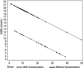
As shown in Figure 2, the SHBG correlated, in-versely and significantly, with insulin levels (p = 0.03) and group (p = 0.01) (test F = 15.1; p < 0.01), as fol-lows: for the increment of each unit in the insulin lev-els, the average levels of SHBG decreased 0.25 units, for both groups: Group 1, without hypogonadism, pre-sented average SHBG concentrations 8.2 units higher than those of the group with hypogonadism, with a 95% confidence interval ranging from 3.46 to 13.05.
ly characterized by low concentrations of total testoster-one (< 300 ng/dL). Most investigators consider 300 mg/ dL as the lower limit for normality, since inferior values are associated with symptoms and clinical alterations compatible with hypogonadism (9).
A frequency of 33% of hypogonadism was found in another systematic evaluation of 103 men with diabe-tes, a rate similar to that found in the present study. However, a variation of 20% to 64% in the frequency of low androgen levels was found when assessing men
with diabetes mellitus and metabolic syndrome (9).
Previous data has shown that the age of the study population may influence the frequency of testoster-one deficiency. For example, in testoster-one series, testostertestoster-one levels lower than 300 ng/dL were observed in 64% of the diabetic patients, and 38% of the non-diabetic ones, when the subject’s age was higher than 73 years old (15). On the other hand, when the age of the patients ranged from 40 to 79 years old, low levels of testos-terone were observed in 21% of the diabetic men, and 13% of the non-diabetic (16). However, in the pres-ent study, the patipres-ents pertaining to both groups had similar average ages (40 and 42, for individuals without and with hypogonadism, respectively). Such relatively young ages are not commonly associated with a high frequency of hypogonadism (17,18), which suggests that age had not specifically interfered on the results.
The decrease in testosterone levels associated with age can be precluded in healthy men, that is, in those men with no evidence of chronic diseases and/or who have a healthy life style (17). Indeed, a significant num-ber of men remain eugonadic even at advanced ages (18). Nevertheless, patients with chronic diseases have a higher risk of having their testosterone levels dimin-ished. A prevalence of 38.7% of hypogonadism, defined as testosterone levels under 300 ng/dL, was found in 2,162 patients with 45 or more years of age who re-ceived attention in primary care clinics in the United States (19). The authors of the cited paper observed that chronic diseases such as arterial hypertension, hy-perlipidemia, prostate disease, asthma or chronic ob-structive pulmonary disease, and, mainly, obesity and
type 2 diabetes mellitus were associated with a higher
risk of hypogonadism (19).
We observed a significantly higher BMI, hip and waist circumferences, and a tendency, albeit not statis-tically significant, to a higher weight in hypogonadic patients (Group 2), as compared to the group of pa-tients without hypogonadism (Group 1). These
find-Figure 2. Multiple linear regression for SHBG in relation to insulin and groups with and without hypogonadism, showing that SHBG levels are inversely and significantly correlated to insulin levels (p = 0.03), in both groups.
Figure 1. Multivariate analysis, showing the inverse significant correlation of the variables insulin concentration and group with total testosterone levels, in both groups of patients (p = 0.04).
DISCUSSION
In this series of men with metabolic syndrome, we found a high frequency (30%) of hypogonadism, which is
main-22 21 20 19 18 17 16 15 14 13 12 11 10 9 8 7 6 5 4 3
SHBG (nmol/L)
Group With hipogonadism Without hipogonadism
10
0 20 30 40 50
600
500
400
300
200
Total testosterone (ng/dL)
0 10 20 30 40 50
Insulin (μUI/mL)
Copyright
© ABE&M todos os dir
eitos r
eser
vados.
ings suggest that a larger accumulation of abdominal fat is associated with a higher frequency of hypogonad-ism. These results are in accordance to those found by other authors who have also demonstrated that higher levels of BMI and a centripetal distribution of body fat are associated with low concentrations of testosterone (7,20-24). Besides that, in a study about the prevalence of low testosterone levels (inferior to 300 ng/dL) in men, obesity was associated with the highest odds ra-tio (2.38) for hypogonadism, as compared to other co-morbid conditions evaluated (19).
The reasons for the association between metabolic syndrome and hypogonadism are not completely clear. It has been suggested that insulin resistance, acting through signs mediated by the adipose tissue, includ-ing increased levels of leptin, would have a direct role in the regulation of gonadal function, thus decreasing the concentrations of testosterone (1,4,25,26). At the same time, pro-inflammatory cytokines, such as TNF alpha, IL-1 and IL-6, produced by visceral adipocytes, are also associated with insulin resistance (27,28). It was demonstrated that there is a strong correlation be-tween insulin sensitivity and the function of Leydig’s cell (29), but other studies have not confirmed such findings (23,30).
Our results point to a possible association between insulin resistance and hypogonadism, since the con-centrations of basal insulin, the insulin resistance index (Homa-IR) and the index that measures the pancreatic
beta cell function (Homa-β) were significantly higher in
the hypogonadic group as compared to the eugonadic group. These results are in accordance to the previous observation of an inverse correlation between fasting insulin and testosterone levels (22,24). Moreover, male patients with insulin resistance-associated diseases, such
as obesity (20-24) and type 2 diabetes mellitus (31), have
levels of testosterone lower than control patients with normal weight who are not diabetic. Transversal studies have also demonstrated an inverse correlation between testosterone and fasting insulin levels in men, regardless of age, obesity and fat distribution (20,21,25,31).
We have also found significantly lower SHBG lev-els in hypogonadic patients and an inverse correlation between insulin and SHBG. It means that high levels of insulin corresponded to low levels of SHBG, inas-much as that every one-unit increase in the levels of insulin resulted in a 0.25 unit decrease in the average SHBG levels, in both groups. A similar decrease in the SHBG levels in hypogonadic patients has been
previ-ously described in individuals with metabolic syndrome and is associated with insulin resistance, carbohydrate intolerance and central obesity (23,32-36). It has been suggested that SHBG concentration could be used as a specific marker of insulin resistance as well as one of the components of the metabolic syndrome (37,38).
The increase in insulin levels observed in metabolic syndrome carriers is probably secondary to the insu-lin resistance action, and may develop as a means to overcome this defect. Our results are in compliance with this hypothesis, since we noted an increase in both the insulin resistance index (Homa-IR) and the index
that measures pancreatic beta cell function (Homa-β)
in hypogonadic patients. The pathological increase in
Homa-β possibly represents an attempt to overcome
the insulin resistance action by enhancing the function of the pancreatic beta cell. This hypothesis is also re-inforced by the fact that increased testosterone levels were observed during the hyperinsulinemic state of the clamp glucose test (29,39).
However, the precise mechanisms involved in in-sulin action that result in low testosterone and SHBG levels are not clear yet, but might be due to a functional defect at one or more levels of the hypothalamus-pitu-itary-gonadal axis (29).
It is known that insulin plays a relevant role in the central regulatory systems involved in reproduc-tive function, acting synergistically to various factors to mediate the secretion of gonadotrophins and the control of body weight (40). Insulin is also involved in the pathophysiology of obesity following lesions to the hypothalamus-pituitary region (41). It has been sug-gested that obese patients with insulin resistance have a diminished sensitivity to insulin action in the hypo-thalamus-pituitary-gonadal axis due to the production of cytokines and hormones by the adipose tissue (29). However, in the present study, the gonadotrophins lev-els were similar in both groups, suggesting that the hy-pothalamus and pituitary had no influence on the low-er levels of testostlow-erone obslow-erved in the hypogonadic group. Similarly, other authors have found no correla-tion between insulin sensitivity and basal, GnRH- or clomiphene-stimulated LH secretion (29,30). Never-theless, the concentrations and pulse amplitude of LH may be abnormal in morbid obesity patients (41-43).
Copyright
© ABE&M todos os dir
eitos r
eser
vados.
testosterone into estradiol. This would inhibit gonado-trophin production centrally, and further decrease tes-tosterone levels. Subsequently, the decrease in testos-terone would facilitate a higher deposition of body fat and a more severe degree of hypogonadism (1,7,44).
On the other hand, there is evidence that a low tes-tosterone level in men would result in insulin resistance and abdominal obesity, as well as be a predictive factor for
the development of type 2 diabetes mellitus (6,45-49). It
has been proposed that the hypogonadism resultant from prostate cancer treatment, by the blockage of gonadotro-phins by GnRH-agonists (50,51) or by castration (52), could be associated with an increase in insulin levels.
Moreover, multiple interventional studies have shown that exogenous testosterone has a favorable impact on body mass index, insulin secretion and sensitivity, lipid profile and blood pressure, which are the parameters that are often disturbed in metabolic syndrome. Testos-terone supplementation was able to reduce abdominal circumference and waist-hip ratio, and to improve in-sulin sensitivity and glycemic control in men with
hy-pogonadism and type 2 diabetes mellitus (6,8,53,54).
Testosterone replacement was also able to decrease the levels of pro-inflammatory cytokines (TNF-alpha, IL-1 and IL-6) produced by visceral adipocytes and associa-ted with insulin resistance (27,55). Although the im-pact of these findings on an individual’s cardiovascular risk have not been systematically investigated, concomi-tant improvements in glycemic control, insulin resistan-ce, lipid profile and visceral adiposity probably represent an overall reduction in cardiovascular risk. Prospective long-term clinical trials are necessary to clarify this issue.
The observation of low levels of HDL-cholesterol is one of the diagnostic criteria for metabolic syndrome. We have not found a significant difference in HDL-cholesterol levels nor in total HDL-cholesterol levels in either group of patients. These results might be interpreted based on the previous observations that testosterone supplementation can decrease total cholesterol levels without affecting HDL-cholesterol levels (53,55).
In summary, there is sufficient evidence that reduc-tions in SHBG and total and free testosterone levels mi-ght be related to high concentrations of insulin, secon-dary to insulin resistance, as demonstrated in the present study (5,21,29). On the other hand, the state of hy-pogonadism would favor, by itself, the development of
insulin resistance, central obesity, type 2 diabetes mellitus
and increased cardiovascular risk (8,37,38,54,56). In conclusion, our results demonstrate a high fre-quency of hypogonadism in male patients with
metabo-lic syndrome. Moreover, they suggest that insulin might be related to the findings of low SHBG, total and free testosterone levels, in this series of patients. We also hi-ghlight the importance of the precocious identification of hypogonadism in men with metabolic syndrome.
Disclosure: no potential conflict of interest relevant to this article was reported.
REFERENCES
1. Matos AFG, Moreira RO, Guedes EP. Aspectos neuroendó-crinos da síndrome metabólica. Arq Bras Endocrinol Metab. 2003;47(4):410-21.
2. Carvalheira JBC, Saad MJA. Doenças associadas à resistência à insulina/hiperinsulinemia não incluídas na síndrome metabólica. Arq Bras Endocrinol Metab. 2006;50(2):360-7.
3. Livingstone C, Collison M. Sex steroids and insulin resistance. Clin Sci (London). 2002;102(2):151-66.
4. Lordelo RA, Mancini MC, Cercato C, Halpern A. Eixos hormo-nais na obesidade: causa ou efeito? Arq Bras Endocrinol Metab. 2007;51(1):34-41.
5. Pasquali R. Obesity and androgens: facts and perspectives. Fertil Steril. 2006;85(5):1319-37.
6. Kapoor D, Jones TH. Androgen deficiency as a predictor of me-tabolic syndrome in aging men: an opportunity for intervention? Drugs Aging. 2008;25(5):357-69.
7. Kapoor D, Malkin CJ, Channer KS, Jones TH. Androgens, insu-lin resistance and vascular disease in men. Cinsu-lin Endocrinol. 2005;63(3):239-50.
8. Shabsigh R, Arver S, Channer KS, Eardley I, Fabbri A, Gooren L, et al. The triad of erectile dysfunction, hypogonadism and the me-tabolic syndrome. Int J Clin Pract. 2008;62(5):791-8.
9. Kalyani RR, Dobs AS. Androgen deficiency, diabetes, and the me-tabolic syndrome in men. Curr Opin Endocrinol Diabetes Obes. 2007;14(3):226-34.
10. Eckel RH, Grundy SM, Zimmet PZ. The metabolic syndrome. Lan-cet. 2005;365(9468):1414-28.
11. Picon PX, Zanatta CM, Gerchman F, Zelmanovitz T, Gross JL, Ca-nani LH. Análise dos critérios de definição da síndrome metabóli-ca em pacientes com diabetes melitos tipo 2. Arq Bras Endocrinol Metab. 2006;50(2):264-70.
12. Vilar L, Freitas MC, Albuquerque JL, Botelho CA, Egito CS, Arruda MJ, et al. The role of non-invasive dynamic tests in the diagnosis of Cushing’s syndrome. J Endocrinol Invest. 2008;31(11):1008-13. 13. Matthews DR, Hosker JP, Rudenski AS, Naylor BA, Treacher DF, Turner RC. Homeostasis model assessment: insulin resistance and beta-cell function from plasma glucose and insulin concen-trations in man. Diabetologia. 1985;28(7):412-9.
14. Geloneze B, Repetto EM, Geloneze SR, Tambascia MA, Ermetice MN. The threshold value for insulin resistance (HOMA-IR) in an admixtured population IR in the Brazilian Metabolic Syndrome Study. Diabetes Res Clin Pract. 2006;72(2):219-20.
15. Tan RS, Pu SJ. Impact of obesity on hypogonadism in the andro-pause. Int J Androl. 2002;25(4):195-201.
16. Barrett-Connor E, Khaw KT, Yen SS. Endogenous sex hormo-ne levels in older men with diabetes mellitus. Am J Epidemiol. 1990;132(5):895-901.
Copyright
© ABE&M todos os dir
eitos r
eser
vados.
18. Feldman HA, Longcope C, Derby CA, Johannes CB, Araujo AB, Coviello AD, et al. Age trends in the level of serum testosterone and other hormones in middle-aged men: longitudinal results from the Massachusetts male aging study. J Clin Endocrinol Me-tab. 2002;87(2):589-98.
19. Mulligan T, Frick MF, Zuraw QC, Stemhagen A, McWhirter C. Pre-valence of hypogonadism in males aged at least 45 years: the HIM study. Int J Clin Pract. 2006;60(7):762-9.
20. Abate N, Haffner SM, Garg A, Peshock RM, Grundy SM. Sex ste-roid hormones, upper body obesity and insulin resistance. J Clin Endocrinol Metab. 2002;87(10):4522-7.
21. Tsai EC, Matsumoto AM, Fujimoto WY, Boyko EJ. Association of bioavailable, free, and total testosterone with insulin resistance: influence of sex hormone-binding globulin and body fat. Diabe-tes Care. 2004;27(4):861-8.
22. Zumoff B, Strain GW, Miller LK, Rosner W, Senie R, Seres DS, et al. Plasma free and non-sex-hormone-binding-globulin bound testosterone are decrease in obese men in proportion to their de-gree of obesity. J Clin Endocrinol Metab. 1990;71(4):929-31. 23. Glass AR, Swerdloff RS, Bray GA, Dahms W, Atkinson RL. Low
se-rum testosterone and sex hormone binding globulinin massively obese men. J Clin Endocrinol Metab. 1977;45(6):1211-9.
24. Pasquali R, Casimirri F, Cantobelli S, Melchionda N, Morselli La-bate AM, et al. Effect of obesity and body fat distribution on sex hormones and insulin in men. Metabolism. 1991;40(1):101-4. 25. Isidori AM, Caprio M, Strollo F, Moretti C, Frajese G, Isidori A, et
al. Leptin and androgens in male obesity: evidence for leptin con-tribution to reduced androgen levels. J Clin Endocrinol Metab. 1999;84(10):3673-80.
26. Magni P, Motta M, Martini L. Leptin: a possible link between food intake, energy expenditure, and reproductive function. Regul Pept. 2000;92(1-3):51-6.
27. Corrales JJ, Almeida M, Burgo R, Mories MT, Miralles JM, Orfao A. Androgen-replacement therapy depresses the ex-vivo production of inflammatory cytokines by circulating antigen-presenting cells in aging type-2 diabetic men with partial androgen deficiency. J Endocrinol. 2006(3);189:595-604.
28. Pittas AG, Joseph NA, Greenberg AS. Adipocytokines and insulin resistance. J Clin Endocrinol Metab. 2004;89(2):447-52.
29. Pitteloud N, Hardin M, Dwyer AA, Valassi E, Yialamas M, Elahi D, et al. Increase insulin resistance is associated with a decrease in Leydig cell testosterone secretion in men. J Clin Endocrinol Me-tab. 2005;90(5):2636-46.
30. Amatruda JM, Hochstein M, Hsu TH, Lockwood DH. Hypotha-lamic and pituitary dysfunction in obese males. Int J Obes. 1982;6(2):183-9.
31. Barrett-Connor E. Lower endogenous androgen levels and dys-lipidemia in men with non-insulin-dependent diabetes mellitus. Ann Intern Med. 1992;117(10):807-11.
32. Lima N, Cavaliere H, Halpern A, Medeiros GN. A função gonadal do homem obeso. Arq Bras Endocrinol Metab. 2000;44(1):31-7. 33. Pasquali R, Casimiri F, de Iasio R, Mesini P, Boschi S, Chierici R,
et al. Insulin regulates testosterone and sex hormone-binding globulin concentrations in adult normal and obese men. J Clin Endocrinol Metab. 1995;80(2):654-8.
34. Birkeland KI, Hanssen KF, Torjesen PA, Vaaler S. Level of sex hormone-binding globulin is positively correlated with insulin sensitivity in men with type 2 diabetes. J Clin Endocrinol Metab. 1993;76(2):275-8.
35. Muller M, Grobbee DE, den Tonkelaar I, Lamberts SW, van der Schouw YT. Endogenous sex hormones and metabolic syndrome in aging men. J Clin Endocrinol Metab. 2005;90(5):2618-23. 36. Chen RY, Wittert GA, Andrews GR. Relative androgen deficiency
in relation to obesity and metabolic status in older men. Diabetes Obes Metab. 2006;8(4):429-35.
37. Nestler JE. Sex hormone-binding globulin: a marker for hype-rinsulinemia and/or insulin resistance? J Clin Endocrinol Metab. 1993;76(2):273-4.
38. Hautanen A. Synthesis and regulation of sex hormone-binding glo-bulin in obesity. Int J Relat Metab Disord. 2000;24(Suppl 2):S64-70. 39. Pasquali R, Macor C, Vicennati V, Novo F, De Lasio R, Mesini P, et
al. Effects of acute hyperinsulinemia on testosterone serum con-centrations in adult obese and normal-weight men. Metabolism. 1997;46(5):526-9.
40. Bruning JC, Gautam D, Burks DJ, Gillette J, Schubert M, Orban PC, et al. Role of brain insulin receptor in control of body weight and reproduction. Science. 2000;289(5487):2122-5.
41. Papadia C, Naves LA, Costa SSS, Vaz JAR, Domingues L, Casulari LA. Incidence of obesity does not appear to be increased after tre-atment of acute lymphoblastic leukaemia in Brazilian childhood: role of leptin, insulin, and IGF-1. Horm Res. 2007;68(4):164-70. 42. Vermeulen A, Kaufman JM, Deslypere JP, Thomas G. Attenuated
luteinizing (LH) pulse amplitude but normal LH pulse frequency, and its relation to plasma androgens in hypogonadism of obese men. J Clin Endocrinol Metab. 1993;76(5):1140-6.
43. Giagulli VA, Kaufman JM, Vermeulen A. Pathogenesis of the de-creased androgen levels in obese men. J Clin Endocrinol Metab. 1994;79(4):997-1000.
44. De Ronde W. Therapeutic uses of aromatase inhibitors in men. Curr Opin Endocrinol Diabetes Obes. 2007;14(3):235-40. 45. Tsai EC, Boyko EJ, Leonetti DL, Fujimoto WY. Low testosterone
le-vel as a predictor of increased visceral fat in Japanese-American. Int J Obes Relat Metab Disord. 2000;24(4):485-91.
46. Spark RF. Testosterone, diabetes mellitus, and the metabolic syn-drome. Curr Urol Rep. 2007;8(6):467-71.
47. Fukui M, Kitagawa Y, Ose H, Hasegawa G, Yoshikawa T, Nakamura N. Role of endogenous androgen against insulin resistance and atherosclerosis in men with type 2 diabetes. Curr Diabetes Rev. 2007;3(1):25-31.
48. Goulis DG, Tarlatzis BC. Metabolic syndrome and reproduction: I. testicular function. Gynecol Endocrinol. 2008;24(1):33-9. 49. Gould DC, Kirby RS, Amoroso P. Hypoandrogen-metabolic
syn-drome: a potentially common and underdiagnosed condition in men. Int J Clin Pract. 2007;61(2):341-4.
50. Dockery F, Bulpitt CJ, Agarwal S, Donaldson M, Rajkumar C. Tes-tosterone suppression in men with prostate cancer leads to an increase in arterial stiffness and hyperinsulinaemia. Clin Sci (Lon-don). 2003;104(2):195-201.
51. Smith JC, Bennett S, Evans LM, Kynaston HG, Parmar M, Mason MD, et al. The effects of induced hypogonadism on arterial stiff-ness, body composition and metabolic parameters in males with prostate cancer. J Clin Endocrinol Metab. 2001;86(9):4261-7. 52. Xu T, Wang X, Hou S, Zhu J, Zhang X, Huang X. Effect of
surgi-cal castration on risk factors for arteriosclerosis of patients with prostate cancer. Chin Med J. 2002;115(9):1336-40.
53. Kapoor D, Goodwin E, Channer KS, Jones TH. Testosterone repla-cement therapy improves insulin resistance, glycaemic control, visceral adiposity and hypercholesterolaemia in hypoganadal men with type 2 diabetes. Eur J Endocrinol. 2006;154(6):899-906. 54. Simon D, Charles M, Nahoul K, Orssaud G, Kremski J, Hully V, et al. Association between plasma total testosterone and cardiovas-cular risk factors in healthy adult men: the Telecom study. J Clin Endocrinol Metab. 1997;82(2):682-5.
55. Malkin CJ, Pugh PJ, Kapoor D, Jones RD, Channer KS, Jones TH. The effect of testosterone replacement on endogenous inflam-matory cytokines and lipid profiles in hypogonadal men. J Clin Endocrinol Metab. 2004;89(7):3313-8.