CASE REPORT
Journal of Evolution of Medical and Dental Sciences/ Volume 2/ Issue 42/ October 21, 2013 Page 8119
SUBLINGUAL DERMOID CYST A RARE CASE
–
CASE REPORT
Prasant M C1, Fareedi Mukram Ali2, Anuroop Singhai3, Kishor Patil4
HOW TO CITE THIS ARTICLE:
Prasant MC, Fareedi Mukram Ali, Anuroop Singhai, Kishor Patil. Sublingual dermoid cyst a rare case – case report. Journal of Evolution of Medical and Dental Sciences 2013; Vol. 2, Issue 42, October 21; Page: 8119-8122.
ABSTRACT: Presenting a case of 16 years old male with a sub lingual swelling causing discomfort in
deglutition and speech is reported in this case report. The swelling had displaced the tongue cranially Sublingual dermoid cyst is a very rare lesion occurring in oral and maxillofacial area. Epidermoid and dermoid cysts represent less than 0.01% of all oral cavity cysts. The pathology was diagnosed as sub lingual dermoid cyst by histopathological examination. It was completely excised through an intra-oral approach. Post-operatively normal functions of tongue were restored.
KEY WORDS: Sublingual swelling, Dermoid cyst, Cystic enucleation, Dyspnea.
INTRODUCTION: Development of face, head and neck is a very balanced and co-ordinated process
during which the process from pharyngeal arches fuses in midline. A small piece of epidermis may get entrapped deep in the mid line, during the mandibular processes from both sides fuses together. This residual entrapped epidermis later in life may proliferate under some stimulus to form a cystic enlargement either above or below the tongue. The term sublingual dermoid cysts was first used Roser (1859) maintained that many cases of ranula and sebaceous cysts on the base of the tongue were dermoid cysts.1
The cysts can be defined as epidermoid when the lining presents only epithelium, dermoid cysts when skin adnexa are found, and teratoid cysts when other tissue such as muscle, cartilage, and bone are present. Midline cyst in the floor of mouth pathogenesis is not well understood and dysontogenetic, traumatic, and thyroglossal anomaly theories have been suggested.2 Meyer had
histologically, divided these types of cysts into three groups: epidermoid, dermoid, and teratoid. Although dermoid cysts represent a separate entity, the term "dermoid" is typically used to indicate all three categories. In fact, dermoid cysts occur primarily in the testes and ovaries, and the most common location in the head and neck is the external third of the eyebrow.3
Dermoid cyst is a very rare anomaly seen in the oral cavity in early period of life. These should be differentially diagnosed from common pathologies like ranula, thyroglossal cysts, bilateral and unilateral blockade of Wharton’s duct and cystic hygroma.
CASE REPORT: A 16 years old male, reported to SMBT Dental College and Hospital; Sangamner, with
CASE REPORT
Journal of Evolution of Medical and Dental Sciences/ Volume 2/ Issue 42/ October 21, 2013 Page 8120 consistency, fluctuant and immobile, with yellowish discoloration of overlying mucosa, which was quite easy to be pinched off. The regional lymphadenopathy was not present. The aspiration of cystic contents showed a thick viscous yellow colour cheesy material. On radiological assessment with Orthopantomogram no change in mandible were seen. On clinical assessment of this lesion provisional diagnosis of sublingual dermoid cyst was made. Patient was admitted in the wards with all routine blood and urine investigations done and preanesthetic assessment done for oral intubation as it may decrease the view and access to the glottis. The swelling was excised via the oral route by an incision, which extended from the tip of under surface of tongue upto the floor of mouth. As the swelling was superficial to mylohyoid muscle, it was possible to enucleate the cyst completely via this approach. The wound was closed in layers using 3/0 vicryl. The histopathology report of biopsy confirmed the provisional diagnosis of dermoid cyst by showing the presence of cheesy keratin material in the cavity. The cyst wall was lined with keratinized stratified squamous epithelium. The post-operative recovery was uneventful and patient was discharged on third postoperative day without any residual illness. Patient was followed up till 1 year with no recurrence of lesion.
DISCUSSION: Dermoid cyst is considered as a congenital anomaly appearing due to inclusion of the
epidermis in the midline. The term dermoid cyst does not appear to be restricted to a single kind of lesion as dermoid cysts appear to be a common lesion in various parts of body like cranium, ovaries, neck and very rarely in oral cavity. The incidence mentioned in the international literature is 1.6% to 6.5%, and they represent less than 0.01% of all oral cavity cysts.4 Intra-orally the most common
site of occurrence is floor of mouth and other less common sites are tongue, mandible and parotid gland. There are again two sites of occurrence in the sublingual region above mylohyoid and below the mylohyoid muscle. Dermoid cysts occur primarily in the testes and ovaries, and the most common location in the head and neck is the external third of the eyebrow.3 Dermoid cyst can occur
at any age but more commonly in second and third decade of life with its occurrence in infants rarely. It is thus very important to differentiate between various other congenital anomalies like ranula, cystic hygroma, thyroglossal cyst from dermoid cyst. Dermoid cyst is surrounded by a thin capsule of fibrous connective tissue and lined internally by a stratified squamous epithelium. Dermoid cyst may contain hair, hair follicle, sebaceous glands sweat glands nails and teeth. Dermoid cyst has a potential to grow and attain such a size that it may obstruct the oral cavity completely.5,6
There are chances of occurrence of squamous cell carcinoma in lining of dermoid cyst as reported by Devine JC7.
These sublingual dermoid cysts are generally approached in two ways either intraoral or extraoral. Those with the swelling above mylohyoid muscle are approached intraorally as seen in this case and those below the mylohyoid are approached extraorally.8 The extra oral approach is
also used in emergency situations in which breathing is hampered. But extraoral approach gives an unaesthetic scar while intraoral approach is esthetic and healing is faster.9,10
REFERENCES:
1. B. D. Gold, D. E. Sheinkopf, and B. Levy, Dermoid, epidermoid and teratomatous cysts of
the tongue and the floor of the mouth, Journal of Oral Surgery, vol. 32, no. 2, pp. 107–
CASE REPORT
Journal of Evolution of Medical and Dental Sciences/ Volume 2/ Issue 42/ October 21, 2013 Page 8121 2. Calderon S, Kaplan I. Concomitant sublingual and submental epidermoid cysts: a case
report. J Oral Maxillofac Surg. 1993; 51:790–792.
3. Howell CJT. The sublingual dermoid cyst: Report of five cases and review of the literature. Oral Surg Oral Med Oral Pathol. 1985; 59:578. doi: 10.1016/0030-4220(85)90184-7.
4. Bonet-coloma C, Minguez-Martinez I, Palma-Carrio C; Orofacial dermoid cysts in pediatric patients: a review of 8 cases. Med Oral Pathol Oral Cir Bucal.2011 Mar 1; 16(2):e200-3.
5. Walstad WR, Solomon JM, Schow SR, Ochs MW. Midline cystic lesion of the floor of the mouth. J Oral Maxillofac Surg. 1998; 56:70–74.
6. RefKoca H, Seckin T, Sipahi A, Kazanc A. Epidermoid cyst in the floor of the mouth: Report of a case. Quintessence Int. 2007; 38:473–477.
7. Devine JC, Jones DC. Carcinomatous transformation of a sublingual dermoid cyst. A case report. Int J Oral Maxillofac Surg. 2000 Apr; 29(2):126-7.
8. El-Hakim IE, Alyamani A; Alternative surgical approaches for excision of dermoid cyst of the floor of mouth. Int J Oral Maxillofac Surg. 2008 May; 37(5):497-9. Epub 2008 Feb 12. 9. Di Francesco A, Chiapasco M, Biglioli F, Ancona D Intraoral approach to large dermoid
cysts of the floor of the mouth: a technical note. Int J Oral Maxillofac Sur. 1995 Jun;24(3):233-5
10.Armstrong JE, Darling MR, Bohay RN, Cobb G, Lawen D, Daley TD. Trans-geniohyoid dermoid cyst: considerations on a combined oral and dermal surgical approach and on histogenesis. J Oral Maxillofac Surg.2006 Dec; 64(12):1825-30.
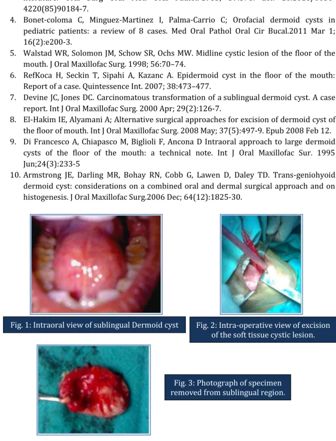
Fig. 1: Intraoral view of sublingual Dermoid cyst Fig. 2: Intra-operative view of excision
of the soft tissue cystic lesion.
CASE REPORT
Journal of Evolution of Medical and Dental Sciences/ Volume 2/ Issue 42/ October 21, 2013 Page 8122
AUTHORS:
1. Prasant M.C. 2. Fareedi Mukram Ali 3. Anuroop Singhai 4. Kishor Patil
PARTICULARS OF CONTRIBUTORS:
1. Dean & HOD, Department of Oral & Maxillofacial Surgery, RKDF Dental College, Bhopal.
2. Reader, Department of Oral & Maxillofacial Surgery, SMBT Dental College, Sangamner Taluka, Maharashtra State.
3. Senior Lecturer, Department of Oral & Maxillofacial Surgery, RKDF Dental College and Research Centre, NH-12, Hoshangabad Road, Misrod, Bhopal.
4. Post Graduate Student, Department of Oral Pathology & Microbiology, SMBT Dental College, Sangamner Taluka, Maharashtra State.
NAME ADDRESS EMAIL ID OF THE
CORRESPONDING AUTHOR:
Dr. Fareedi Mukram Ali,
Department of Oral & Maxillofacial Surgery, SMBT Dental College, Sangamner Taluka, Maharashtra State.
Email – [email protected]