204 Rev. odonto ciênc. 2010;25(2):204-207 Epidermoid cyst
Epidermoid cyst: highlights on diagnosis and
magnetic resonance imaging features
Cisto epidermóide: processo de diagnóstico e características das
imagens por ressonância magnética
Larissa Santana Arantes Elias a
Angélica Ferreira Oton-Leite a
Clóvis Martins Silva b
Rejane Faria Ribeiro-Rotta a
Aline Carvalho Batista a
Elismauro Francisco de Mendonça a
a Department of Oral Medicine (Oral Pathology),
Dental School, Federal University of Goiás, Goiânia, GO, Brazil
b Araújo Jorge Hospital, Association of Cancer
Combat of Goiás, Goiânia, GO, Brazil
Correspondence:
Elismauro Francisco de Mendonça
Faculdade de Odontologia da Universidade Federal de Goiás
Primeira Avenida Número 1964, Setor Universitário Goiânia, GO – Brasil
74605-020
E-mail: [email protected]
Received: September 22, 2009 Accepted: December 10, 2009
Conflict of Interest Statement: The authors state that there are no financial and personal conflicts of interest that could have inappropriately influenced their work.
Copyright: © 2010 Elias et al.; licensee EDIPUCRS. This is an Open Access article distributed under the terms of the Creative Commons Attribution-Noncommercial-No Derivative Works 3.0 Unported License.
Abstract
Purpose: Epidermoid and dermoid cysts are extremely rare developmental cysts of a benign nature, which may occur anywhere in the body, although about 7% are found in the head and neck. This article reports a clinical case of a patient who had an epidermoid cyst and its magnetic resonance imaging (MRI) features.
Case report: This case discusses an epidermoid cyst in a 36-year-old woman complaining about speech difficulty. Clinical examination revealed an extensive swelling on the floor of the mouth. MRI findings showed a cystic homogenous lesion located underneath the mylohyoid muscle which was removed by surgery. Histological examination of the mass confirmed the diagnosis of an epidermoid cyst.
Conclusion: We concluded that MRI was considered useful for a more accurate diagnosis prior to treatment.
Key words: Epidermoid cyst; dermoid cyst; magnetic resonance images; differential diagnosis
Resumo
Proposta: Cistos epidermóide e dermóide são cistos de desenvolvimento extremamente raros, de natureza benigna, que podem ocorrer em qualquer região do corpo e somente 7% são encontrados na região de cabeça e pescoço. Este artigo apresenta o caso clínico de uma paciente que possuía um cisto epidermóide juntamente com as características das imagens por ressonância magnética.
Descrição do caso: Paciente do gênero feminino, 36 anos, apresentou-se com queixa de dificuldade de fonação e deglutição. O exame clínico intrabucal revelou uma extensa tumefação em soalho bucal. As imagens por ressonância magnética indicaram uma lesão de natureza cística, homogênea, de conteúdo sólido e localizada abaixo do músculo milohiódeo. A lesão foi enucleada e encaminhada para exame anatomopatológico, cujo diagnóstico foi compatível com cisto epidermóide.
Conclusão: A imagem por ressonância magnética foi essencial para determinar as características da lesão de acordo com a sua localização anatômica.
Palavras-chave: Cisto epidermóide; cisto dermóide; imagens por ressonância magnética; diagnóstico diferencial.
Rev. odonto ciênc. 2010;25(2):204-207 205 Elias et al.
Introduction
Epidermoid and dermoid cysts are extremely rare developmental cysts of a benign nature, which may occur anywhere in the body, but most predominantly in the ovary and scrotal regions (1-3). Only about 7% are found in the head and neck (1,2,4). The occurrence of such cysts in the oral cavity is extremely rare, with approximately
1.6% located in this area (1,2,4). The loor of the mouth
is the most commonly affected area, however, these cysts can also be found in the tongue, lips, buccal mucosa and
jaw and mandible bones (2-5). Because of the dificulty
of making a correct diagnosis of these lesions when using clinical examinations and conventional radiological images, it is recommended that specialized imaging examinations should be used, such as ultrasonography (US), computed tomography (CT) and Magnetic Resonance Imaging (MRI) (1,2,4-10).
We describe a case of a patient who had an epidermoid cyst and discuss features such as age of occurence, anatomic location, differential diagnosis and imaging indings.
Case report
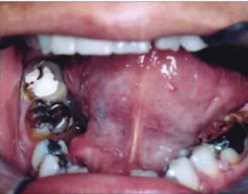
A 36-year-old female patient was referred to the Head and Neck Division of the Araújo Jorge Hospital (Association
of Cancer Combat of Goiás), Goiânia-GO, complaining mainly about “a growth in her mouth that had affected her speech”. The patient stated that the lesion had appeared two years ago, was painless, but was causing progressive
speech dificulty. During the intrabucal clinical examination,
good dental conditions were noted as well as a swelling
measuring 5 x 4 cm, located in the loor of the mouth
(Fig. 1a). The initial diagnosis was the possibility of a salivary gland lesion.
To assist with the diagnosis of the lesion, it was decided to request basic complementary examinations including conventional images such as panoramic and total occlusal images of the jaw. MRI was also requested. The results of the hematological and biochemical examinations were normal. Neither the panoramic nor occlusal images made any further contribution to the diagnosis. However, a lesion was detected in the MRI of the maxillofacial regions (Fig. 1d and 1e). It was an ovoid lesion of regular margins with a slightly hyperintense signal on the T1 sequence and a markedly hyperintense signal on the T2 sequence with no attenuation when fat suppression was used (Fig. 1b and 1c). The lesion
measuring 4.4 x 3.6 x 2.9 cm was located in the loor of the
mouth and, after using paramagnetic contrast, presented well
deined borders (Fig. 1c). The right submandibular salivary
duct presented with sialectasia before its opening. The MR
indings were compatible with an epidermoid cyst.
206 Rev. odonto ciênc. 2010;25(2):204-207 Epidermoid cyst
On the basis of the clinical and radiographic data the patient underwent general anesthesia to remove the lesion. Aspiratory puncture of the lesion was carried out followed by incision and tecidual divulsion with total enucleation of the cyst. The specimen was sent for anatomopathological
examination in which the microscopic aspects conirmed a cystic-type lesion lined with stratiied squamous epithelium and illed with keratin (Fig. 2a and 2b). The postoperative
period was uneventful (Fig. 3).
Discussion
The terminology and nomenclature of the epidermoid and dermoid cysts are controversial. The term dermoid is a distinct entity that is normally used to indicate three different types of cyst: the real dermoid, the epidermoid and the teratoma (3,4,7). All dermoid cysts have an epithelial covering, but contain cutaneous annexes, whereas epidermoid cysts do not contain dermic annexes and the covering of the teratoma contains structures whose origins are in the three germinative layers (1,3,4,5,7). Dermoid cysts occur more frequently than epidermoid cysts, in a ratio of 2:1 (2). The current case was considered an epidermoid cyst because it did not present the necessary characteristics to classify it as a dermoid cyst.
The etiology of epidermoid cysts is unknown, but is associated with the presence of remnants of embryonic tissue
(ectoderm) during the fusion of the irst (mandible) and
second (hyoid) branchial arches during the third and fourth intrauterine weeks (1,4).
With regard to location, these cysts can be classiied as being
located in a median or lateral position (2,3,4,7). Median cysts develop below the lingual brake and between the genioglossal muscle while lateral cysts are located between the genioglossal and mylohyoid muscles (4). Cysts may
also be classiied according to their position within the muscle, since this inluences the therapeutic approach to be
adopted. Thus, epidermoid cysts can be located above the geniohyoid and below the genioglossal muscles, curving
with the loor of the mouth; they can also be present below
the geniohyoid muscle and above the mylohyoid muscle, arching in the submental region, or can be located laterally in the submandibular space above the mylohyoid muscle and
to the side of the base of the tongue (2-4,7). In this speciic
case, the magnetic resonance image showed a median cyst located above the geniohyoid and below the genioglossal
muscles, clinically presented as a swelling in the loor of
the mouth.
Epidermoid cysts are generally diagnosed between the ages of 15 and 50, on average at 35, occurring with the same frequency in both males and females (3,4). Our patient was a 36-year-old, thereby being consistent with the literature. In general, these cysts grow slowly and painlessly and,
depending on their size and location, can make it dificult
to chewing, swallowing and speaking (1,3,4). Such reports are consistent with the present case since the patient only sought treatment when her speech was impaired.
Fig. 2. Epidermoid cyst: histopathological characteristics. (a) Cystic-type lesion lined with stratified squamous epithelium and filled with keratin (red arrow) (hematoxylin-eosin stain; original magnification 5x (a) and 10x (b)).
Rev. odonto ciênc. 2010;25(2):204-207 207 Elias et al.
In a differential diagnosis of lesions located in the loor
of the mouth, the dermoid, epidermoid, linfoepithelial, and tireoglossac duct cysts must be included as well as neoplasms of the sublingual or minor salivary glands, cystic
hygroma and ranula (1,2,4,5,6,10). Because of the dificulty
encountered in making a correct diagnosis of these lesions it is recommended that specialized imaging examinations should be used (1,2,4-10).
US is the irst choice because of its feasibility, low cost and eficacy (4). Together with MRI it presents better tecidual
resolution when compared with computed tomography (8,9).
In general, these cysts can be visualized as a well-deined
mass with homogenous internal structures and a well-outlined cystic wall (7,8).
Some authors consider MRI superior to other methods, since it shows the exact position, extension and demarcation of dermoid cysts (2,4,10). With this exam, different slices and plans of the region in question can be obtained since the sagittal image is extremely important for the positioning of medial tumors and the diagnosis of benign cystic lesions (2,4,10). With MRI, the epidermoid cyst is described as a cystic mass, which is hypointense in T1-weighted images and hyperintense in T2-weighted images (5,8,10). The intensity of the signal varies according to the ratio of keratin (protein) to cholesterol (lipid) (8). A lipid material
shows high intensity on both T1 and T2-weighted images, whereas a material with keratin shows a low to intermediate intensity on T1-weighted images and intermediate to high intensity on T2-weighted images (8). Considering the large amount of keratin in our case, the signal was low intensity on T1-weighted images and high intensity on T2-weighted images. An epidermoid cyst is typically seen on CT as a
well-deined, lobulated mass with hypoattenuation similar to that of lesions containing luid (8,11).
The treatment to be given to these lesions varies according to the clinical presentation of the cyst and its location (11). It could involve an intraoral or extraoral procedure or a combination of both. Various authors have suggested that an intraoral procedure be undertaken in cases of a sublingual cyst or small cysts while an extraoral procedure would be indicated for those cysts located in the submental, supra-hyoid region, that is, when the submandibular and submental spaces are involved or in cases of large lesions (2-4,11).
Therefore, we conclude that, in the current case of an epidermoid cyst, the MRI exam was of utmost importance in complementing the clinical diagnosis and it could be
correlated with the microscopic indings of the lesion, since
the epidermoid cyst contained a large amount of keratin providing consistent images in MRI.
References
1. De Ponte FS, Brunelli A, Marchetti E, Bottini DJ. Sublingual epidermoid cyst. J Craniofac Surg 2002;13:308-10.
2. Fuchshuber S, Grevers G, Issing WJ. Dermoid cyst of the floor of the mouth – a case report. Eur Arch Otorhinolaryngol 2002; 259:60-2.
3. Bataineh AB, Mansour MJ. Extraoral epidermoid cysts. Br J Oral Maxillofac Surg 1997;35:49-51.
4. Kandogan T, Koç M, Vardar E, Selek E, Sezgin O. Sublingual epidermoid cyst: a case report. J Med Case Reports 2007;1: 1-4.
5. Kim SH, Han MH, Park SW, Chang KH. Radiologic – pathologic correlation of unusual lingual masses: Part I: Congenital lesions/ Part II: Benign and malignant tumors. Korean J Radiol 2001; 2:37-51.
6. Anavi Y, Kaplan I, Calderon S. Lateral neck mass. Oral Surg Oral Med Oral Pathol Oral Radiol Endod 2002;94:536-39.
7. Ariyoshi Y, Shimahara M. Magnetic resonance imaging of a submental dermoid cyst: Report of a case. J Oral Maxillofacial Surg 2003;61:507-10.
8. Ariji Y, Gotoh M, Naitoh M, Izumi M, Shimozato K, Kurita K, et al. Magnetic resonance imaging assessment of tumorous lesions in the floor of the mouth: case reports and review of the literature. Oral Radiol 2006;22:18-26.
9. Chikui T, Shimizu M, Goto TK, Nakayama E, Yoshiura K, Kanda S, et al. Interpretation of the origin of a submandibular mass by CT and MRI imaging. Oral Surg Oral Med Oral Pathol Oral Radiol Endod 2004;98:721-29.
10. Potts M, Macleod RI, McLean NR, Chippindale AJ. The value of a magnetic resonance imaging in the assessment of a sublingual epidermoid cyst. Dentomaxillofac Radiol 1992;21:102-4. 11. Fung HS, Wong HN, Lau S, Wong WK, Tang KW. A 50-year-old man