ABSTRACT - Congenital dermoid inclusion cyst over the anterior fontanel (CDIC) is an uncommon cystic lesion
located over the anterior fontanel. It is a benign and curative lesion and most of the time, can be diagnosed
at birth. From 1994 to 2001, three patients were operated with this kind of lesion and after reviewing the
literature we found 229 cases and only 6 cases described in Brazil. Our objective in this study is to report three
more cases.
KEY WORDS: congenital inclusion, dermoid cyst, epidermoid cyst, anterior fontanel.
Cisto dermóide de inclusão congênita sobre a fontanela anterior: relato de três casos
Cisto dermóide de inclusão congênita sobre a fontanela anterior: relato de três casos
Cisto dermóide de inclusão congênita sobre a fontanela anterior: relato de três casos
Cisto dermóide de inclusão congênita sobre a fontanela anterior: relato de três casos
Cisto dermóide de inclusão congênita sobre a fontanela anterior: relato de três casos
RESUMO - Cisto dermóide de inclusão congênita sobre a fontanela anterior (CDIC) é lesão rara localizada na
região da fontanela anterior. Trata-se de lesão benigna e curável que, na maioria das vezes, é diagnosticada
no nascimento. De 1994 a 2001, três pacientes foram operados com este tipo de lesão e, através dos dados
disponíveis na literatura, verificamos somente 229 casos descritos, apenas 6 descritos no Brasil, o que nos
motivou a registrar mais três casos.
PALAVRAS-CHAVE: inclusão congênita, cisto dermóide, cisto epidermóide, fontanela anterior.
1
Neurosurgery Service of Hospital Municipal Dr. José de Carvalho Florence, São José dos Campos, SP, Brazil
1;
2Discipline of Neurosurgery,
Department of Neurology, Faculty of Medical Science, State University of Campinas UNICAMP, Campinas SP, Brazil
Received 12 April 2002, received in final form 28 January 2003. Accepted 30 January 2003.
Dr. Humberto Belem de Aquino - Rua Jaime Spinelli 5 - 12282-436 Caçapava SP – Brasil. E-mail: [email protected]
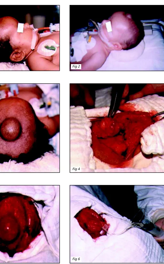
Many different types of lesions over the children’s
skull exist and some are commonly diagnosed in daily
practice. Congenital dermoid inclusion cyst over the
anterior fontanel (CDIC) is a rare and benign lesion
located over the anterior fontanel. Many children
are examinated with lesions over the skull in our
hos-pital. In a recent review of these children, our
atten-tion was drawn to three patients with a cystic
roun-ded mass over the anterior fontanel. These three
pa-tients showed no neurological abnormality and the
diagnosis of CDIC were confirmed by surgery and
histological examination. Our literature search found
229 other cases (Table 1) of CDIC worldwide.
CASES
From 1994 to 2001, three patients were operated with
a cystic mass over the anterior fontanel, being of different
Fig 5
Fig 6
Fig 4
Fig 2
Fig 1
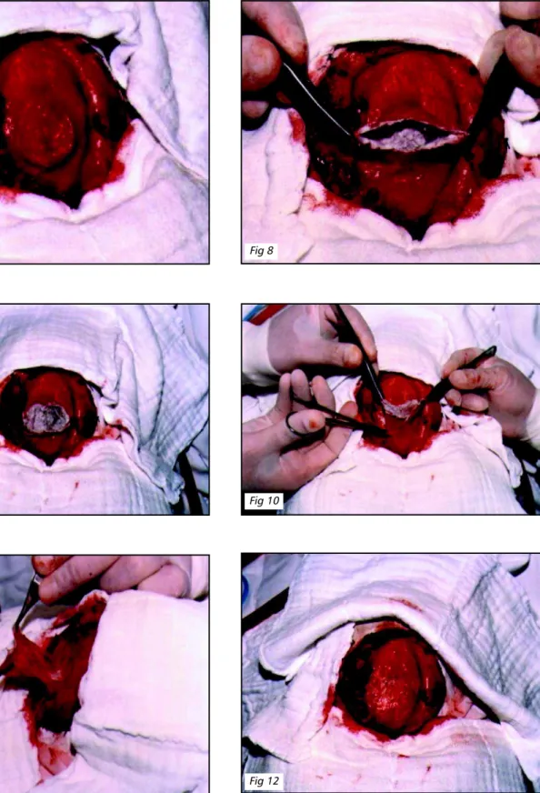
DISCUSSION
Congenital dermoid inclusion cyst over the
ante-rior fontanel is reported as an uncommon cystic
le-sion, located over the anterior fontanel. Adeloye
and Odeku (1971)
1were the first to publish a clear
and complete description about this lesion, having
treated eighteen patients. CDIC is a cystic mass
co-vered by normal skin. It is soft, mobile and it does
not cause discomfort, pain or throbbing. There is no
communication between the cyst and intracranial
cavity. Several cystic sizes have been reported
1-4, this
depending on the age of patient at the time of
diag-nosis. Most of the time, the diagnosis can be made
at birth, although some authors have reported adult
cases
4-6. CDIC is a developmental tumor due to
in-clusion of dermal elements within the neuroaxis
bet-ween the third and fifth week of the embryogenesis
when the ectoderm folds into the neural tube
6,7.
Based on its pathogenesis, they can be classified as:
1) congenital dermoid cyst of the teratoma type that
Fig 7
Fig 8
Fig 10
Fig 9
Fig 13
Fig 14
Table 1.CDIC registered cases*.
Hayat S et al.
41989
1 case
Oliveira HA et al.
61989
1 case
Macedo NTL et al.
71985
1 case
Wong TT et al.
211986
8 cases
Pereira CU et al.
132000
2 cases
Tateshima S et al.
142000
1 case
Parizek J et al.
111989
13 cases
Hibaut-Macarde P et al.
161991
1 case
Tan EC et al.
301993
4 cases
Sinclair RD et.al
311992
3 cases
Martinez LS et al.
321992
3 cases
Mlay SM et al.
331993
6 cases
Stannard MW et al.
341990
6 cases
Saito M et al.
351988
2 cases
Isozumi T et al.
361995
1 case
Nicolau A et al.
371986
6 cases
Peter JC et al.
381992
35 cases
Total
94 cases
*Review of the liteture by Macedo NTL et al. until 1984 – 135 cases reported
is derived from the embryogenic epithelium,
confi-ned to the ovaries and testis; 2) acquired
implanta-tion dermoid cyst formed by cells implanted
trauma-tically into deeper structures; 3) congenital dermoid
inclusion cyst resulting from the inclusion of
displa-ced dermal cells along the embryonic fusion line
7.
Histologically, the cyst wall is lined by squamous
epi-thelium and inside the cyst exists adnexial appendage
structures including hair follicles, sebaceous and
sweat glands
8-13. There are factors that change other
lesions over the anterior fontanel, such as epidermoid
cyst. The fluid content can be clean or yellow,
de-pending on the size and age of the lesion and
exo-crine sweat gland content and some authors
disco-vered sodium, potassium, chloride and glucose
con-centrations within their cases, has been low
inci-dence
14,15. A skull X-ray will show the shadow of the
swelling in the extracranial space and can reveal
changes which include flattening of the outer table
underneath the swelling or some depression
16,17. In
the past, some authors injected air or contrast
me-dium (Pantopaque), others used ventriculography,
in order to show interconnection between the cyst
and intracranial cavity
18. Currently, CT and MRI are
considered the best examination methods, to
con-firm its extracranial position
19-21. Encephalocele,
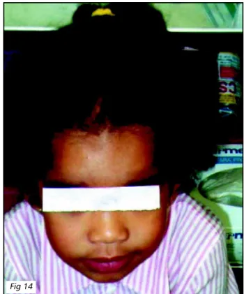
The-because it is considered to be a simple lesion. In the
cases reported in the literature, we did not find any
description addressing the long-term outcomes of
patients who were treated. Therefore, we are
inclu-ding our cases in order to provide a long-term view
of this condition. We predict our results will prove
that CDIC is a benign and curative lesion.
Acknowledgments
Acknowledgments
Acknowledgments
Acknowledgments
-Acknowledgments - We thank Lynn Falkner from Salt
Lake City (Utah – USA), Carolina Brom from Caçapava (São
Paulo) and Andy Shepherd from England, for English
support.
REFERENCES
1. Adeloye A, Odeku EL. Congenital subgaleal cysts over the anterior fontanelle in Nigerians. Arch Dis Child 1971;46:95-96.
2. Glasauser FE, Levy LF, Auchterlonie WC. Congenital inclusion dermoid cyst of the anterior fontanel. J Neurosurg 1978;48:274-278.
3. Sonntag VKH, Waggener JD. Congenital dermoid cyst of the anterior fontanel in a mexican-american. Surg Neurol 1980;13:371-373. 4. Hayath S, Seetharam W, Kumari G, Dinakar I, Nightingale F. Congenital
dermoid cyst over the anterior fontanelle. Br J Clin Pract 1989;43:119- 120. 5. Ojikutu NA, Mordi VPN. Congenital inclusion dermoid cyst located
over the region of the anterior fontanel in adult Nigerians. J Neurosurg 1980; 52:724-727.
6. Oliveira HA. Cisto dermóide de inclusão localizado na região da fontanela anterior no adulto. Arq Neuropsiquiatr 1989;47:375-377. 7. Macedo NTL, Ramos VP, Lins C. Cisto dermóide de inclusão da
fontanela anterior. Arq Neuropsiquiatr 1985;43:407-412.
8. Zülch KJ. Brain tumor The biology and pathology Epidermoid and dermoid cyst. 3.Ed. New York: Springer Verlag, 1986:433-437. 9. Brownstein MH, Helwig EB. Subcutaneous dermoid cysts. Arch
Dermatol 1973;107:237-239.
10. Naidich TP. Dermoids of the anterior fontanelle. Neuro Image Quiz, Answers 1988:278-279.
11. Parízek J, Nìmecek S, Nemecková J, Cernoch Z, Šercl M. Congenital dermoid cyst over the anterior fontanel: report on 13 cases in Czechoslovak children. Child’s Nerv Syst 1989;5:234-237.
12. Chaudhari AB, Ladapo F, Mordi VPN, Choudhury KJ, Naseem A, Obe JA.Congenital inclusion cyst of the subgaleal space. J Neurosurg 1982;56:540-544.
19. Stokes RB, Saunders CJ, Thaller SR. Bregmatic epidermoid inclusion cyst eroding both calvarial tables. J Craniof Surg 1996;7:148-150. 20. Kriss TC, Kriss VM, Warf BC. Recurrent meningitidis: the search for
the dermoid or epidermoid tumor. Pediatr Infect Dis 1995;14:697-700. 21. Wong TT, Wann SL, Lee LS. Congenital dermoid cyst of the anterior
fontanelle in Chinese children. Child’s Nerv Syst 1986;2:175-178. 22. Mohanty S, Clezy JKA, Adeloye A. Dermoid cyst of the anterior
fontanel. J Neurosurg 1978;48:627-628.
23. Pereira WC, Andrade AF, Lopes PG. Cisto dermóide na região do bregma: relato de dois casos. Arq Neuropsiquiatr 1969;27:349-352. 24. Fujimaki T, Miyazaki S, Fukushima T, Sato Y, Fujimaki W, FujitaY.
Dermoid cyst of the frontal bone away the anterior fontanel. Child’s Nerv Syst 1995;11:424-427.
25. Yuasa H, Tokito S, Izumi K, Oyama M. Congenital inclusion dermoid cyst of the anterior fontanel in a Japanese infant: case report. Neurosurgery 1981;9:67-69.
26. Chaudhari AB, Rosenthal AD. Congenital inclusion cysts of the subgaleal space. Surg Neurol 1984;21:61-66.
27. Stella L, Spaziante R, Maiuri F, Gangemi M, Divìtììs E. Congenital dermoid cysts at the anterior fontanelle. Neurochirurgia 1984;27:186-189. 28. Borges A, Carelli EF, Maciel Jr. JA, Alvarenga M, Castro R, Bonilha L. Pilonidal
cyst on the vault:case report. Arq Neuropsiquiatr 1999;20:273-276. 29. Mehta MH, Patel RV. Congenital midline subgaleal cyst. Indian
Pediatrcs 1990;27:403-404.
30. Tan EC, Takagi T. Congenital inclusion cyst over the anterior fontanel in Japanese children: a study of five cases. Child’s Nerv Syst 1993;9:81-83. 31. Sinclair RD, Darley C, Dawber RP. Congenital inclusion dermoid cyst
of the scalp. Australas J Dermatol 1992;33:135-140.
32. Martinez-Lage Sanches JF, Almargro Navarro MJ, Poza Poza M, Puche Mira A, Sola Perez J. Dermoide cyst of the anterior fontanelle in children: clinical significance and differentiation from encephalocele. An Esp Pediatr 1992;36:355-358.
33. Mlay SM, Sayi EN. Anterior fontanelle scalp cysts in infancy. East Afr Med J 1993;70:578-579.
34. Stannard MW, Currarino G. Subgaleal dermoid cyst of the anterior fontanelle: diagnosis with sonography. AJNR Am J Neuroradiol 1990;11:349-352.
35. Saito M, Takagi T, Ishikawa T. Dermoid cyst of the anterior fontanel: advantage of MRI for the diagnosis. Brain Dev 1988;10:252-255. 36. Isozumi T, Tsuji A, Nakasu M, Handa J. Congenital dermoid cyst over
the anterior fontanelle: case report. No Shinkei Geka 1995;23:423-427. 37. Nicolau A, Daney I, Diard F, Risch M, Kind M. Midline subepicranial (subgaleal) dermoid cysts in children: report of 6 cases and review of the literature. Ann Radiol (Paris) 1986;29:511-518.