Abstract
Submitted: September 1st, 2016
Modiication: November 30, 2016 Accepted: January 12, 2017
Bacterial endotoxin adhesion to
different types of orthodontic
adhesives
Bacterial endotoxin (LPS) adhesion to orthodontic brackets is a known contributing factor to inlammation of the adjacent gingival tissues. Objective: The aim of this study was to assess whether LPS adheres to orthodontic adhesive systems, comparing two commercial brands. Material and Methods: Forty specimens were fabricated from Transbond XT and Light Bond composite and bonding agent components (n=10/component), then contaminated by immersion in a bacterial endotoxin solution. Contaminated and non-contaminated acrylic resin samples were used as positive and negative control groups, respectively. LPS quantiication was performed by the Limulus Amebocyte Lysate QCL-1000™ test. Data obtained were scored and subjected to the Chi-square test using a signiicance level of 5%. Results: There was endotoxin adhesion to all materials (p<0.05). No statistically signiicant difference was found between composites/bonding agents and acrylic resin (p>0.05). There was no signiicant difference (p>0.05) among commercial brands. Afinity of endotoxin was signiicantly greater for the bonding agents (p=0.0025). Conclusions: LPS adhered to both orthodontic adhesive systems. Regardless of the brand, the endotoxin had higher afinity for the bonding agents than for the composites. There is no previous study assessing the afinity of LPS for orthodontic adhesive systems. This study revealed that LPS adheres to orthodontic adhesive systems. Therefore, additional care is recommended to orthodontic applications of these materials.
Keywords: Corrective orthodontics. Composite resins. Endotoxins.
Priscilla Coutinho ROMUALDO1
Thaís Rodrigues GUERRA1
Fábio Lourenço ROMANO1
Raquel Assed Bezerra da SILVA1
Izaíra Tincani BRANDÃO2
Célio Lopes SILVA2
Lea Assed Bezerra da SILVA1
Paulo NELSON-FILHO1
http://dx.doi.org/10.1590/1678-7757-2016-0434
1Universidade de São Paulo, Faculdade de Odontologia de Ribeirão Preto, Departamento de Clínica
Infantil, Ribeirão Preto, SP, Brasil.
2Universidade de São Paulo, Faculdade de Medicina de Ribeirão Preto, Departamento de Bioquímica
e Imunologia, Ribeirão Preto, SP, Brasil.
Corresponding address: Priscilla Coutinho Romualdo Departamento de Clínica Infantil Faculdade de Odontologia de Ribeirão Preto -Universidade de São Paulo. Avenida do Café, S/N, Monte Alegre -Ribeirão Preto - SP - 14040-904 - Brazil. Phone: +55-16-3315-4099 - Fax: +55-16-3315-4102 -
Introduction
Orthodontic appliances are composed of different materials and accessories with irregular surfaces
like brackets, ligatures, bands and wires that create
additional sites that harbor dental plaque and oral
microorganisms21, changing chemical properties of the oral medium10. Fixed orthodontic therapy inevitably
predisposes patients to an increased risk of dental
problems, as ixed appliances make an effective oral
hygiene challenging and limit the mechanical cleansing
of saliva low, tongue and oral muscles24.
The use of orthodontic appliances can also increase
the levels of periodontal pathogens in the supragingival
and subgingival, associated with gingival inlammation
that can occur during orthodontic treatment14.
Periodontopathogenic microbiota is predominantly
composed of anaerobic microorganisms9, especially
Gram-negative bacteria18, which contain endotoxin in their cell wall23. Bacterial endotoxin, also referred to
as LPS due to its lipopolysaccharide nature, is released
during bacterial multiplication or death, causing a
series of important biological effects23 that lead to
inlammatory reaction and bone resorption in the
periapical region25.
Endotoxin has a high afinity for different materials,
e.g., metals13, silica, zirconium7, acrylic resins4, ceramics13 and even titanium and titanium alloys1.
In vitro and in vivo studies11,19 have shown that bacterial endotoxin adheres to metal brackets and
such afinity affects endotoxin concentration in the gingival sulcus, contributing to inlammation of tissues
adjacent to the brackets. By analogy, a similar process
could occur on the surface of adhesive systems used
for ixation of orthodontic brackets to the dental
enamel. To the best of our knowledge there is not a
previous study assessing bacterial endotoxin afinity
for orthodontic adhesive systems. Therefore, the aim
of this study was to assess whether LPS adheres to the components of orthodontic adhesive systems
(bonding agent and composite resin), comparing two
commercial brands.
Material and methods
Fabrication of specimens
In order to obtain the test specimens, it was used
a circular Telon matrix, manufactured at the Precision
Workshop of the University of São Paulo, Ribeirão
Preto, SP, Brazil. The matrix consisted of two nested parts: an outer portion and an inner portion in the form
of a 3-mm-diameter plunger. Accompanying the matrix
there was a 2-mm-thick spacer, which was engaged
in the plunger between the two portions so that the outer potion was 2 mm higher than the inner portion,
providing adequate thickness to the specimen.
Therefore, forty disc-shaped specimens (3 mm
diameter and 2 mm thick) were fabricated from each component (composite or bonding agent) of two largely
used orthodontic adhesive systems (Transbond XT; 3M
Unitek, Monrovia, CA, USA and Light Bond; Reliance
Orthodontic Products, Inc., Itasca, IL, USA). Groups were created with 10 specimens of each component
(test groups). As bacterial endotoxin is known to
have a high afinity for acrylic resin4, 10 additional specimens of a self-curing acrylic resin (JET Classic; Art. Odontológicos Ltda, Campo Limpo Paulista, SP,
Brazil) served as positive (n=5) and negative (n=5)
controls; the positive control was contaminated with
the endotoxin solution and negative control was not contaminated.
Each component was inserted into the matrix in
increments followed by pressure with a glass plate
until excess low. All components were activated
with a halogen light device for 40 seconds, with light
intensity of 400 mW/cm2. Then, specimens were
removed from the matrix and their size checked with
a precision caliper.
All specimens were sterilized with ethylene oxide
(Oximed, São José do Rio Preto, SP, Brazil) and then
contaminated by immersion in a bacterial endotoxin
solution, except for the negative control group.
Endotoxin (LPS) solution preparation
In a laminar low chamber, 350 mg of lyophilized
endotoxin from Escherichia coli (Lipopolysaccharide B E.coli 055:B5 – Sigma Aldrich Corporation, St. Louis, MO, USA) was suspended into 4.7 mL of pyrogen-free
water, resulting in a 25 ng/mL concentration endotoxin
solution. For contamination, the specimens were
immersed in the solution in glass tubes placed under agitation (126 rpm) in an incubator at 37°C for 24 h.
The negative control specimens were not immersed
Quantiication of bacterial endotoxin (LPS) by
the Limulus Amebocyte Lysate QCL-1000™ test
After contamination with LPS, the specimens were
individually placed in new nonpyrogenic glass tubes
with lids (BioWhittaker; Cambrex Corporation, East
Rutherford, NJ, USA) containing 1 mL of pyrogen-free water (recovery solution) and taken to an ultrasonic
cleaner (Ultracleaner USC 1600ª; Unique Indústria e
Comércio de Produtos Eletrônicos Ltda., Indaiatuba,
SP, Brazil) for 15 min to release endotoxin from the material.
Endotoxin quantiication in the bonding agent,
composite and acrylic specimens was performed using
the 1000™ test (Limulus Amebocyte Lysate QCL-1000™; Lonza, Walkersville, MD, USA) following the
manufacturer’s instructions. LAL is a quantitative test
for detection of endotoxin with a sensitivity range
of 0.1 - 1.0 EU/ml (endotoxin units per milliliter). A standard curve of known endotoxin levels was
used to determine the amount of endotoxin in the
samples. Fifty µL of solutions of each known standard
concentration (1.0 EU/mL, 0.5 EU/mL, 0.25 EU/mL and 0.1 EU/mL) and 50 µL of the negative control
(pyrogen-free water) was dripped in duplicate in the
wells of a non-pyrogenic 96-well polystyrene plate
(Corning Incorporated, Corning, NY, USA). Fifty µl of the samples diluted in pyrogen-free water at a ratio
of 1:1 were added to the remaining wells and after
that 50 µL of LAL solution were added to all wells
containing samples or standards, the microplate was then incubated at 37°C for 10 min. After that, 100
µL of chromogenic substrate that was preheated to
37°C was added to the wells, stirred and incubated
at 37°C for 6 min in the dark, following the same dripping protocol and maintaining a constant dripping
rate. Subsequently, 100 µL of the blocking reagent
(25% v/v glacial acetic acid in water) was added to
stop the reaction.
The absorbance of each sample was determined
using an ELISA (enzyme-linked immunosorbent assay)
reader (Ultramark; Bio-Rad Laboratories, Hercules, CA,
USA) at 405 nm. Absorbance was considered directly
proportional to endotoxin levels in the wells and it correlated directly to the endotoxin concentration
in the range from 0.1 to 1.0 EU/mL. The amount of
endotoxin in each sample was expressed in EU/mL
and calculated from the solution absorbance values with known endotoxin levels (standard) multiplied by
the dilution factor.
Statistical analysis
For statistical analysis, values of endotoxin
concentrations were classiied into three scores: score 1 (concentration ≤0.5 EU/mL); score 2 (0.51 to 1.0
EU/mL); and score 3 (>1.0 EU/mL). Comparisons of
scores between composites and bonding agents and between the two brands of both types of materials
were performed with the Chi-square test, using the
GraphPad 5.0a Software (Graphpad Software Inc., San
Diego, CA, USA). The signiicance level was set at 5%.
Results
All experimental groups differed significantly
(p<0.05) from the negative control group
(non-contaminated acrylic resin), demonstrating bacterial
endotoxin adhesion to all tested materials. No
statistically signiicant difference (p>0.05) was found
between experimental groups and positive control
group (acrylic resin contaminated with endotoxin).
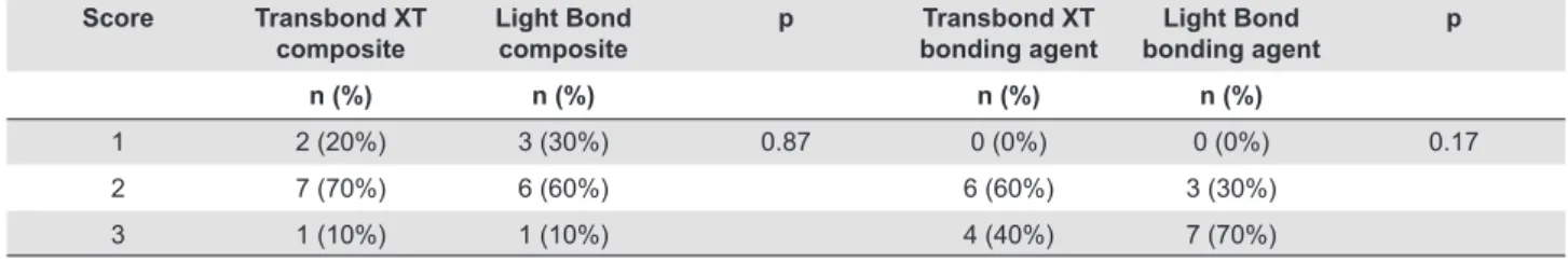
Table 1 shows the distribution of relative and absolute endotoxin levels (by scores) in experimental
groups. Since no statistically signiicant difference
(p>0.05) was found between the two composites
or the two bonding agents, the materials were compared regardless of their brand. Figure 1 is a
graphical representation of scores distribution between
composites and bonding agents, and it shows a
signiicantly higher endotoxin adhesion to bonding
agents than to composites (p=0.0025).
Score Transbond XT composite
Light Bond composite
p Transbond XT bonding agent
Light Bond bonding agent
p
n (%) n (%) n (%) n (%)
1 2 (20%) 3 (30%) 0.87 0 (0%) 0 (0%) 0.17
2 7 (70%) 6 (60%) 6 (60%) 3 (30%)
3 1 (10%) 1 (10%) 4 (40%) 7 (70%)
Discussion
The present study quantiied in vitro bacterial endotoxin adhered to the components (composite and
bonding agent) of two orthodontic adhesives. The use
of tests derived from the aqueous extractof Limulus polyphemus crab blood cells, known as Limulus Amebocyte Lysate (LAL) tests, is recommended
to assess the presence or absence of endotoxin in
solutions or instruments. In the presence of endotoxin,
LAL components are activated by a protein cascade, which results in the cleavage of a substrate present
in the test reagent, with the release of yellowish
p-nitroaniline (pNA). The release of pNA is measured
spectrophotometrically at 405-410 nm, after disruption of the reaction by a stop reagent. The LAL test has
been widely used for endotoxin detection in different
areas of Dentistry.
Bacterial endotoxin (LPS) is a major virulence factor of the surface of Gram-negative microorganisms, playing
a key role in triggering periodontal inlammation26. It is
a bacterial antigen present in the subgingival bioilm,
acting directly on the innate immune system at the site of infection27. Acting as a powerful stimulus for a
variety of host cells, LPS stimulates the expression of
important pro-inlammatory cytokines, such as IL-1 and TNF-α5, which increase the expression and release
of other pro-inlammatory cytokines and induce the
release of cell adhesion molecules8. LPS also stimulates
the production of reactive oxygen species and the
phosphorylation of protein kinases in the cells2. In this
way, LPS contributes to the recruitment of immune
cells, a major component of the innate immune response17, causing a series of biological effects that
trigger an inlammatory response with subsequent
bone resorption25.
Endotoxin has high afinity for a variety of dental
materials1,7,13, including acrylic resin commonly used as
a temporary material4, and a high afinity for titanium
(present in dental implants) with a signiicant decrease
in titanium corrosion resistance30. Also in Dentistry, endotoxin is present in necrotic root canals15 where
its presence has been associated with periapical
inlammation and bone resorption6.
Our option of using endotoxin derived from E. coli
in this study was based on its broad indication, based
on its proven toxicity, to evaluate the biological activity
of LPS at different research levels12,25. In addition, the
molecular structure of E. coli, according to Mattison, et al.16 (1987), is representative of most endotoxins.
Moreover, this endotoxin is easier to obtain and
cheaper.
The results of this in vitro study showed that bacterial endotoxin has affinity for adhesives
frequently used in orthodontics. Previous studies
have demonstrated that LPS also adheres to metallic
brackets, contributing to the inlammation of tissues
adjacent to the brackets11,19. However, the lack
of studies assessing endotoxin adhesion to other
orthodontic adhesive systems does not allow to
compare our indings.
Numerous orthodontic adhesive systems are
available for bonding orthodontic brackets. The choice
for Transbond XT is because it is often referred to a “gold standard” in a number of studies28. Light Bond
adhesive system is also widely used in orthodontic
practice and it was selected due to its luoride releasing
property. Fluoride-containing orthodontic adhesives
have gained attention due to thebeneicial role of luoride in inhibiting enamel demineralization around
orthodontic brackets20.
Previous studies evaluating the same adhesive systems showed differences among them regarding
shear bond strength22, degree of monomer conversion
and cytotoxicity3. In the present study, however, the
afinity of bacterial endotoxin for both materials was
similar.
An important inding of the present study was the
occurrence of greater bacterial endotoxin adhesion to
bonding agents than to composites, which could be explained by differences in their composition. Although
these materials have a similar composition, it is known
that composites must contain higher amounts of
inorganic iller particles, which are not always present
in bonding agents29.
According to the manufacturers, the bonding
agents evaluated in this study do not contain inorganic
fillers, while both composites have over 80% of inorganic particles by volume.
Considering the higher affinity of endotoxin
for orthodontic bonding agents, additional care is
recommended to orthodontists in the sense of avoiding “overwetting” and limiting the application of these
materials to the bracket base. Excess material on
dental enamel should be carefully removed to avoid
leaving areas of bonding agent/composite exposed to oral medium, which could favor the adhesion of
LPS to the materials and stimulate the occurrence of
inlammation in the gingival tissues adjacent to the
brackets.
Further laboratory research and clinical studies are
necessary to compare and substantiate these indings.
Conclusion
The results of this study revealed that bacterial
endotoxin (LPS) adhered to orthodontic adhesive systems. The bonding agents of both systems
presented greater afinity for endotoxin than for
composites.
Acknowledgments
This work was supported by Fundação de Amparo
à Pesquisa do Estado de São Paulo (2013/26611-6).
References
1- Barão VA, Mathew MT, Yuan JC, Knoernschild KL, Assunção WG,
Wimmer MA, et al. Inluence of corrosion on lipopolysaccharide afinity
for two different titanium materials. J Prosthet Dent. 2013;110:462-70. 2- Bhattarai G, Poudel SB, Kook SH, Lee JC. Resveratrol prevents
alveolar bone loss in an experimental rat model of periodontitis. Acta Biomater. 2016;29:398-408.
3- Cörekçi B, Irgin C, Halicioğlu K, Dursun S, Yavuz MZ. Effects of
plasma-emulating light-emitting diode (LED) versus conventional
LED on cytotoxic effects and polymerization capacity of orthodontic composites. Hum Exp Toxicol. 2014;33:1000-7.
4- Silva LA, Silva RA, Branco LG, Navarro VP, Nelson-Filho P. Quantitative radiographic evaluation of periapical bone resorption in
dog's teeth contaminated with bacterial endotoxin (LPS) associated or not with calcium hydroxide. Braz Dent J. 2008;19:296-300. 25
5- Gagnon F, Knoernschild KL, Payant L, Tompkins GR, Litaker MS,
Schuster GS. Endotoxin afinity for provisional restorative resins. J
Prosthodont. 1994;3:228-36.
6- Glauser MP. The inlammatory cytokines. New developments in the
pathophysiology and treatment of septic shock. Drugs. 1996;52:9-17. 7- Grundling GL, Melo TA, Montagner F, Scarparo RK, Vier-Pelisser FV.
QMix(R) irrigant reduces lipopolysacharide (LPS) levels in an in vitro model. J Appl Oral Sci. 2015;23:431-5.
8- Harder S, Quabius ES, Ossenkop L, Mehl C, Kern M. Surface contamination of dental implants assessed by gene expression analysis
in a whole-blood in vitro assay: a preliminary study. J Clin Periodontol. 2012;39:987-94.
9- Hopkins SJ. The pathophysiological role of cytokines. Leg Med (Tokyo). 2003;5:S45-57.
10- Jenkins WM, Papapanou PN. Epidemiology of periodontal disease in children and adolescents. Periodontol 2000. 2001;26:16-32.
11- Jung WS, Kim H, Park SY, Cho EJ, Ahn SJ. Quantitative analysis of changes in salivary mutans streptococci after orthodontic treatment.
Am J Orthod Dentofacial Orthop. 2014;145:603-9.
12- Knoernschild KL, Rogers HM, Lefebvre CA, Fortson WM, Schuster
GS. Endotoxin afinity for orthodontic brackets. Am J Orthod Dentofacial
Orthop. 1999;115:634-9.
13- Li D, Fu L, Zhang Y, Yu Q, Ma F, Wang Z, et al. The effects of LPS on adhesion and migration of human dental pulp stem cells in vitro. J
Dent. 2014;42:1327-34.
14- Lieder R, Petersen PH, Sigurjónsson ÓE. Endotoxins - the
invisible companion in biomaterials research. Tissue Eng Part B Rev. 2013;19:391-402.
15- Liu H, Sun J, Dong Y, Lu H, Zhou H, Hansen BF, et al. Periodontal health and relative quantity of subgingival Porphyromonas gingivalis
during orthodontic treatment. Angle Orthod. 2011;81:609-15. 16- Marinho AC, Martinho FC, Zaia AA, Ferraz CC, Gomes BP. Monitoring
the effectiveness of root canal procedures on endotoxin levels found in teeth with chronic apical periodontitis. J Appl Oral Sci. 2014;22:490-5.
17- Mattison GD, Haddix JE, Kehoe JC, Progulske-Fox A. The effect
of Eikenella corrodens endotoxin on periapical bone. J Endod.
1987;13:559-65.
18- Mizgerd JP, Spieker MR, Doerschuk CM. Early response cytokines
and innate immunity: essential roles for TNF receptor 1 and type I IL-1 receptor during Escherichia coli pneumonia in mice. J Immunol.
19- Mombelli A, Décaillet F. The characteristics of bioilms in
peri-implant disease. J Clin Periodontol. 2011;38:203-13.
20- Nelson-Filho P, Valdez RM, Andrucioli MC, Saraiva MC, Feres M, Sorgi
CA, et al. Gram-negative periodontal pathogens and bacterial endotoxin in metallic orthodontic brackets with or without an antimicrobial agent:
an in-vivo study. Am J Orthod Dentofacial Orthop. 2011;140:e281-7. 21- Paschos E, Kurochkina N, Huth KC, Hansson CS, Rudzki-Janson
I. Failure rate of brackets bonded with antimicrobial and
luoride-releasing, self-etching primer and the effect on prevention of enamel demineralization. Am J Orthod Dentofacial Orthop. 2009;135:613-20.
22- Peixoto IT, Enoki C, Ito IY, Matsumoto MA, Nelson-Filho P. Evaluation of home disinfection protocols for acrylic baseplates of removable
orthodontic appliances: a randomized clinical investigation. Am J Orthod Dentofacial Orthop. 2011;140:51-7.
23- Reimann S, Mezey J, Daratsianos N, Jäger A, Bourauel C. The
inluence of adhesives and the base structure of metal brackets on
shear bond strength. J Orofac Orthop. 2012;73:184-93.
24- Rietschel ET, Brade H. Bacterial endotoxins. Sci Am.
1992;267:54-61.
25- Rosenbloom RG, Tinanoff N. Salivary Streptococcus mutans levels
in patients before, during, and after orthodontic treatment. Am J Orthod Dentofacial Orthop. 1991;100:35-7.
26- Sun Y, Shu R, Li CL, Zhang MZ. Gram-negative periodontal bacteria induce the activation of Toll-like receptors 2 and 4, and
cytokine production in human periodontal ligament cells. J Periodontol. 2010;81:1488-96.
27- To TT, Gumus P, Nizam N, Buduneli N, Darveau RP. Subgingival plaque in periodontal health antagonizes at toll-like receptor 4 and
inhibits e-selectin expression on endothelial cells. Infect Immun. 2015;84:120-6.
28- Tüfekçi E, Pennella DR, Mitchell JC, Best AM, Lindauer SJ. Eficacy of a luoride-releasing orthodontic primer in reducing demineralization
around brackets: an in-vivo study. Am J Orthod Dentofacial Orthop. 2014;146:207-14.
29- Van Landuyt KL, Snauwaert J, De Munck J, Peumans M, Yoshida Y, Poitevin A, et al. Systematic review of the chemical composition
of contemporary dental adhesives. Biomaterials. 2007;28:3757-85. 30- Yu F, Addison O, Baker SJ, Davenport AJ. Lipopolysaccharide