Edivaldo de Morais*, Fábio Lourenço Romano**, Lourenço Correr Sobrinho***, Américo Bortolazzo Correr****, Maria Beatriz Borges de Araújo Magnani*****
Shear bond strength of composites using an
adhesion booster
Objective: To evaluate the shear strength of two composites (Transbond XT and Concise) using an adhesion booster (Ortho Primer). Methods: The sample consisted of 90 bovine incisors divided into six groups (n=15). All teeth were subjected to prophylaxis with pum-ice and enamel etching with phosphoric acid. Transbond XT was used conventionally in Group I. Group II was handled similarly to Group I, except that Ortho Primer was applied instead of XT Primer. After etching, the enamel in Group III was contaminated with saliva, Ortho Primer was then applied and bonding performed using Transbond XT. In Group IV, Concise was used conventionally. Group V was handled similarly to Group IV, except that Ortho Primer was applied instead of the fluid resin. After etching, the enamel in Group VI was contaminated with saliva, Ortho Primer was then applied and bonding performed using Concise. The specimens were stored in distilled water at 37 ºC for 24 hours and subjected to shear strength testing. Data were analyzed with ANOVA and Tukey’s test (5%). Results:
Bond strength in Group IV was statistically higher than in Groups II, III and VI (p<0.05). No statistically significant differences were found among Groups I, IV and V, and between I, II, III and VI (p>0.05). When used conventionally, both Transbond XT and Concise yielded the highest bond strength values. When applied to dry enamel, Ortho Primer acted effec-tively as a bonding agent for the composites under evaluation. When applied to contami-nated enamel, Concise yielded low bond strength.
Abstract
Keywords: Orthodontic brackets. Shear strength. Orthodontics.
* Specialist in Orthodontics, Piracicaba Dental School, University of Campinas - UNICAMP.
** PhD and Professor of Orthodontics, Department of Pediatric Clinic, Preventive and Community Dentistry, Ribeirão Preto Dental School, University of São Paulo - USP.
*** Head Professor of Dental Materials, Department of Restorative Dentistry, Piracicaba Dental School, University of Campinas - UNICAMP. **** PhD in Dental Materials, Piracicaba Dental School, University of Campinas - UNICAMP.
***** PhD and Professor of Orthodontics, Department of Child Dentistry, Piracicaba Dental School, University of Campinas - UNICAMP.
» The authors report no commercial, proprietary, or inancial interest in the products or companies described in this article.
How to cite this article: Morais E, Romano FL, Correr Sobrinho L, Correr
intROductiOn
For many years orthodontic treatment was performed with the aid of orthodontic bands placed on all teeth. Not only was this procedure rather laborious, it also caused considerable dis-comfort to patients, compromised oral hygiene, presented unfavorable esthetics and remaining spaces between the teeth after removal of the orthodontic appliance.3,30
Changing from banding teeth to bonding brackets directly to tooth enamel was one of the most significant advances in orthodontic appli-ance placement procedures of all times. This pro-cedure was only possible thanks to Buonocore8
(1955), who introduced the technique for etch-ing dental enamel with phosphoric acid, and to Newman19 (1965) who spearheaded the bonding
of orthodontic attachments.
Currently, a broad range of orthodontic attach-ments such as brackets, hooks and tubes are avail-able to orthodontists as well as a variety of bonding materials, among which composites are undoubt-edly the most widespread. The procedure for bonding brackets to enamel with these materials needs to be conducted in an orderly and cautious manner in order to prevent bond failures when applying orthodontic mechanics, which might un-dermine treatment effectiveness. This procedure is time consuming and requires a dry surgical field free from any type of contamination.4 The
bond-ing of accessories with composites in the presence of contamination or moisture may cause a higher number of bracket bond failures, delaying treat-ment and increasing costs to orthodontists.2,28
With the purpose of finding a solution to this critical issue in orthodontic bonding, manufac-turers have developed materials that can be used in moist or contaminated environments without compromising bond strength.11,12,14,18,29
Addition-ally, these products save consultation time, de-creasing the number of bond failures and conse-quently affording greater ease and effectiveness to orthodontists.21,24,25
A new product was recently introduced in the market known by its commercial name as Or-tho Primer (Morelli, Sorocaba, Brazil). Accord-ing to the manufacturer it is an adhesion booster with hydrophilic properties suitable for bonding brackets in adverse clinical situations, including cases where saliva or moisture contamination oc-curs after enamel etching. This material acts as a bonding agent with the function of chemically activating orthodontic composite adhesion and is suitable for all types of brackets.
The mere introduction of a new product in the market, however, is not enough to ensure that such material is suitable for clinical use. Product effectiveness and suitability must be verified in laboratory experiments and clinical trials. The aim of this study was to examine the shear strength of metal brackets bonded with different composites onto dry surfaces contaminated with saliva using Ortho Primer as an adhesion enhancing agent.
MAtERiAL And MEtHOdS
The study made use of 90 right and left man-dibular permanent bovine22 incisors, freshly
ex-tracted, properly cleaned with periodontal cu-rettes (Duflex, Juiz de Fora, Brazil), which were stored for one week in an aqueous solution of 0.1% thymol and subsequently stored in distilled water in the refrigerator at 4 °C. Criteria for tooth selection required intact crowns, no decalcifica-tion (softening), cracks or fractures.
tube in the sandy phase under vibration (Fig 1), and the excess removed from the bottom of the die with a LeCron spatula (Duflex, Juiz de Fora, Brazil). Then the tooth crowns were removed from the wax and cleaned (Fig 2).
To verify the correct positioning of the crown in the PVC tube a glass square ruler was used with the 90-degree angle resting on the upper portion of the die and on the buccal surface of each tooth (Figs 3A and B). This verification was necessary to ensure proper shear testing. Badly positioned teeth were excluded from the experi-ment. The entire tooth-PVC tube set was filled with resin, numbered for better identification and stored again in distilled water under refrigeration. All buccal surfaces were subjected to prophy-laxis with rubber cup, non-fluoridated pumice and water for 10 seconds, then washed and dried for the same period of time. After five prophy-laxis procedures the rubber cup was replaced to ensure standardization. Subsequently, all teeth had their enamel etched with phosphoric acid gel at 37% for 30 seconds, followed by washing and drying for about 20 seconds.
The specimens were divided into six groups (n=15), as described in Table 1.
In Groups II, III, V and VI, after Ortho Primer bonding agent had been applied, light-curing was performed for 10 seconds.
Ninety maxillary central incisor metal brackets Dyna-lock Standard, with mesh base, no torque or angulation (code 018-501, 3M Unitek, Monrovia, USA) (Fig 4), were positioned on the buccal sur-face of each tooth with the aid of bracket placing tweezers (Orthoply, Philadelphia, USA). All bond-ing procedures were performed by the same exam-iner after properly positioning each attachment and pressing them against the tooth surface in order to reduce composite thickness between bracket and enamel. At this time, all bonding material excess was removed with an explorer probe (Fig 5).
In the groups using Transbond XT, bonding was light cured for 40 seconds, i.e., 10 seconds on
FIGURE 1 - Steps taken to insert bovine teeth in PVC tube.
FIGURE 2 - Bovine tooth inserted in PVC tube.
each surface (mesial, distal, incisal and gingival) as close as possible to the base of the bracket with a halogen light unit XL 2500 (3M/ESPE, St. Paul, USA) with 500 mW/cm2 power. This light
A B
FIGURE 3 - Proper tooth positioning verification. A) Lateral view. B) Front view. FIGURE 4 - Bracket used in the experiment.
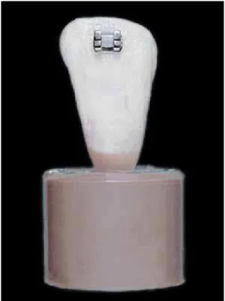
FIGURE 5 - Specimen with bracket bonded to buccal surface.
In the groups using Concise composite no light cur-ing was performed as this is a self-curcur-ing material.
After bracket bonding, the specimens were once again stored in distilled water in an oven (Odontobrás, Ribeirão Preto, Brazil) for 24 hours at 37 ºC to simulate oral conditions. Thereafter,
all samples were subjected to shear bond strength testing on an Instron machine, model 44.11, (Canton, USA) with a speed of 0.5 mm/minute with a chisel-shaped tip resting on the upper enamel/bracket interface.
Shear bond strength values were obtained in Kgf (kilograms-force), transformed into N (New-ton) and divided by the area of the bracket base (15.64 mm2), yielding values in MPa.
Statistical treatment
Data on shear strength were analyzed using multifactorial analysis of variance and Tukey’s test at 5% level of significance for comparison be-tween groups.
Group Enamel condition
Bonding
agent Composite
I Dry XT Primer Transbond XT
II Dry Ortho Primer Transbond XT
III Contaminated
with human saliva Ortho Primer Transbond XT
IV Dry Fluid resin
A and B Concise*
V Dry Ortho
Primer Concise*
VI Contaminated with human saliva
Ortho
Primer Concise* TABLE 1 - Groups used in the experiment.
RESuLtS
Mean shear strength values obtained for the six groups as well as statistical comparisons be-tween them are shown in Table 2.
Group IV showed a mean shear strength value statistically higher than Groups II, III and VI (p<0.05). Groups I, II and V were cally higher than Group VI (p<0.05). No statisti-cally significant difference was observed between Groups I, IV and V, between I, II and III, nor be-tween Groups III and VI (p>0.05).
diScuSSiOn
In orthodontic bonding, tooth surface condi-tions and type of bonding material greatly affect bond strength. In some situations, enamel prepa-ration is not adequate to ensure bracket adhe-sion during orthodontic mechanotherapy, causing treatment delays and damage to tooth structure due to the need for repeated rebonding.5,9,10,13,17
Groups I, II and III were bonded with Trans-bond XT light-cured composite, a material used as control in several studies available in the
lit-erature.1,4,6,7,11,12,15,24,25,26 There were no statistically
significant differences between them, regardless of surface treatment. In Group I bracket bonding was performed with Transbond XT in a conven-tional manner, i.e., according to the manufactur-er’s directions, yielding a shear strength value of 11.35 MPa. This is close to values found by other authors6,7,11,15 and higher than other studies.1,25
The differences found between the results of this and other studies are probably due to the differ-ent methodologies used during the mechanical tests as well as the different substrates.
Between Groups II (dry enamel, Ortho Primer and bonding with Transbond XT) and III (enamel contaminated with saliva, Ortho Primer and bonding with Transbond XT), the mean shear strength values were very close and therefore not statistically significant. This was not the case with Concise since Group V, where Ortho Primer was used on dry enamel, yielded better adhesion than on saliva-contaminated enamel (Group VI), and was therefore statistically significant.
Self-curing composites – Concise in particular – have been used to bond orthodontic accessories for several decades primarily thanks to their adequate bond strength in a dry environment.21,25 Table 2
shows that Group IV, which used Concise as bond-ing material accordbond-ing to the manufacturer’s rec-ommendations, achieved the highest shear strength mean value in this study (16.34 MPa). This is close to values found by other authors15,22,27 and higher
than other studies cited in the literature.1,24
In situations where maintenance of a dry surgi-cal field becomes challenging, the literature shows inadequate or even absence of bond strength of this type of material (composite) to enamel.16 Group
VI, which was bonded with Concise after appli-cation of Ortho Primer to saliva-contaminated enamel, showed the lowest shear strength in this study (5.62 MPa). This result is lower than the val-ue proposed by Reynolds20 for laboratory trials and
confirms that saliva contamination after acid etch-ing is a key factor underminetch-ing bracket adhesion to TABLE 2 - Mean shear strength (MPa) and statistical analysis of
experi-mental groups.
Means followed by different letters differ by Tukey’s test at 5% level. Standard deviation in parentheses.
Groups Shear Strength
(MPa)
IV 16.34 (4.76)a
V 12.43 (3.83)ab
I 11.35 (3.62)ab
II 9.85 (3.12)b
III 7.68 (4.52)bc
enamel using composites.10,12 The bond strength of
12.43 MPa found in Group V (dry enamel, Ortho Primer and bonding with Concise) was statistically higher than in Group VI, which reinforces the ar-gument that bonding with composites is signifi-cantly enhanced in a dry environment.
Groups V and VI used a light-cured mate-rial (Ortho Primer) with a self-curing composite (Concise). Potential lack of compatibility between curingmethods23 was not investigated in this work.
It is worth mentioning the work of Grandhi et al15 in 2001, who found that the low effectiveness
of a self-curing composite (Concise) associated with a hydrophilic primer (Transbond MIP) in a contaminated environment could be related to a failure to activate the hydrophilic primer, since in this experiment the primer was not light-cured prior to bonding. In the present study another hydrophilic bonding agent was utilized (Ortho Primer) which was cured after application. Giv-en these methodological differGiv-ences one cannot state that light-curing Ortho Primer enhanced bond strength in the infected groups.
In a comparison between bonding materials (Transbond XT and Concise), both showed simi-lar bond strength values in light of a variety of surface treatment approaches and different types of bonding agents (Tables 1 and 2). When the composites were bonded to dry enamel using their respective bonding agents the highest bond strength values were obtained, followed by the condition of dry enamel and saliva-contaminated enamel associated with Ortho Primer, which ex-hibited lower bond strength values.
cOncLuSiOnS
1) Composites Transbond XT and Concise™ Orthodontic Bonding System, when used con-ventionally, achieved the highest bond strength values.
2) When applied to dry enamel, Ortho Primer acted effectively as a bonding agent for the com-posites mentioned above.
contact address
Fábio Lourenço Romano
Av. Engenheiro José Hebert Faleiros, 600 Casa 78 CEP: 14.098-780 - Ribeirão Preto/SP, Brazil E-mail: [email protected]
1. Aasrum E, Ng’ang’a PM, Dahm S, Ogaard B. Tensile bond
strength of orthodontic brackets bonded with a
luoride-releasing light-curing adhesive. An in vitro comparative study. Am J Orthod Dentofacial Orthop. 1993;104(1):48-50. 2. Arnold RW, Combe EC, Warford JH Jr. Bonding of stainless steel brackets to enamel with a new self-etching primer. Am J Orthod Dentofacial Orthop. 2002;122(3):274-6.
3. Bishara SE, Khowassah MA, Oesterle LJ. Effect of humidity and temperature changes on orthodontic direct-bonding adhesive systems. J Dent Res. 1975;54(4):751-8. 4. Bishara SE, Olsen ME, Damon P, Jakobsen JR. Evaluation
of a new light-cured orthodontic bonding adhesive. Am J Orthod Dentofacial Orthop. 1998;114(1):80-7.
5. Bishara SE, Gordan VV, VonWald L, Jakobsen JR. Shear bond strength of composite, glass ionomer, and acidic primer adhesive systems. Am J Orthod Dentofacial Orthop. 1999;115(1):24-8.
6. Bishara SE, VonWald L, Laffoon JF, Warren JJ. Effect of a self-etch primer/adhesive on the shear bond strength of orthodontic brackets. Am J Orthod Dentofacial Orthop. 2001;119(6):621-4.
7. Bishara SE, Laffoon JF, Vonwald L, Warren JJ. The effect of repeated bonding on the shear bond strength of different orthodontic adhesives. Am J Orthod Dentofacial Orthop. 2002;121(5):521-5.
8. Buonocore MG. A simple method of increasing the adhesion
of acrylic illing materials to enamel surfaces. J Dent Res.
1955;34(6):849-53.
9. Buyukyilmaz T, Usumez S, Karaman AI. Effect of self-etching primers on bond strength: are they reliable? Angle Orthod. 2003;73(1):64-70.
10. Cacciafesta V, Sfondrini MF, De Angelis M, Scribante A, Klersy C. Effect of water and saliva contamination on shear bond strength of brackets bonded with conventional, hydrophilic, and self-etching primers. Am J Orthod Dentofacial Orthop. 2003;123(6):633-40.
11. Caputo MC, Nouer PRA, Arsati YBOL, Almeida RC, Almeida MHZ. Avaliação in vitro da resistência ao cisalhamento na colagem de bráquetes com adesivos resinosos e um potencializador de adesão. Rev Dental Press Ortod Ortop Facial. 2004;9(2 Supl):45-56.
12. Crane MD. Effect of moisture contamination on bracket bond strength of hydrophilic bond materials [abstract 3237]. J Dent Res. 2000;548.
13. David VA, Staley RN, Bigelow HF, Jakobsen JR. Remnant amount and cleanup for 3 adhesives after debracketing. Am J Orthod Dentofacial Orthop. 2002;121(3):291-6.
14. Domingues-Rodrigues GC, Carvalho PAL, Horliana RF,
Bonim RA, Vigorito JW. Avaliação “in vitro” da resistência
à tração de braquetes metálicos colados com o novo
sistema adesivo “Self etching primer” (SEP). Ortodontia.
2002;35(2):28-34.
15. Grandhi RK, Combe EC, Speidel TM. Shear bond strength of stainless steel orthodontic brackets with a moisture-insensitive primer. Am J Orthod Dentofacial Orthop. 2001;119(3):251-5.
REfEREncES
16. Kula KS, Nash TD, Purk JH. Shear-peel bond strength of orthodontic primers in wet conditions. Orthod Craniofacial Res. 2003;6(2):96-100.
17. Meehan MP, Foley TF, Mamandras AH. A comparison of the shear bond strengths of two glass ionomer cements. Am J Orthod Dentofacial Orthop. 1999;115(2):125-32.
18. Miller RA. Laboratory and clinical evaluation of a self-etching primer. J Clin Orthod. 2001;35(1):42-5.
19. Newman G. Epoxy adhesives for orthodontics attachments: progress report. Am J Orthod. 1965;51(12):901-12. 20. Reynolds IR. A review of direct orthodontic bonding. Br J
Orthod. 1975;2(3):171-8.
21. Romano FL, Ruellas ACO. Estudo comparativo in vitro da resistência ao cisalhamento da colagem e do índice de remanescente resinoso entre os compósitos Concise e Superbond. Rev Dental Press Ortod Ortop Facial. 2003;8(1):69-75.
22. Romano FL, Tavares SW, Ramalli EL, Magnani MBBA, Nouer DF. Análise in vitro da resistência ao cisalhamento de bráquetes metálicos colados em incisivos bovinos e humanos. Rev Dental Press Ortod Ortop Facial. 2004;9(6):63-9.
23. Romano FL, Tavares SW, Nouer DF, Consani S, Magnani MBBA. Shear bond strength of metallic orthodontic brackets bonded to enamel prepared with self-etching primer. Angle Orthod. 2005;75(5):849-53.
24. Romano FL. Shear bond strength of metallic brackets bonded under various enamel conditions. Braz Oral Res. 2006;20(1):28-33.
25. Santos PCF, Santos JFF, Chaves CM Junior, Campos BGP, Santos HMG. Colagem em ambiente úmido: avaliação da capacidade de resistência à tração de braquetes metálicos. Rev Dental Press Ortod Ortop Facial. 2000;5(6):33-43. 26. Schaneveldt S, Foley TF. Bond strength comparison of
moisture-insensitive primers. Am J Orthod Dentofacial Orthop. 2002;122(3):267-73.
27. Surmont P, Dermaut L, Martens L, Moors M. Comparison in shear bond strength of orthodontic brackets between
ive bonding systems related to different etching times:
an in vitro study. Am J Orthod Dentofacial Orthop. 1992;101(5):414-9.
28. Tortamano A, Vigorito JW, Nauff F, Garone GM, Santos RSC. Avaliação da resistência à tração de agentes cimentantes para bráquetes ortodônticos. Rev Assoc Paul Cir Dent. 2002;56(4):259-63.
29. Webster MJ, Nanda RS, Duncanson MG Jr, Khajotia SS, Sinha PK. The effect of saliva on shear bond strengths of hydrophilic bonding systems. Am J Orthod Dentofacial Orthop. 2001;119(1):54-8.
30. Zachrisson BU, Buyukyilmaz T. Recent advances in bonding to gold, amalgam, and porcelain. J Clin Orthod. 1993;27(2):75-81.