CLINICAL SCIENCE
Renal macrophage infiltration is associated with a
poor outcome in IgA nephropathy
Gyl Eanes Barros Silva,IRoberto Silva Costa,IRoberto Cuan Ravinal,ILeandra Naira Zambelli Ramalho,I Marlene Antonia dos Reis,IIMiguel Moyses-Neto,III Elen Almeida Romao,III Terezila Machado Coimbra,IV Ma´rcio DantasIII
IUniversity of Sa˜o Paulo, Faculty of Medicine of Ribeira˜o Preto, Department of Pathology, Ribeira˜o Preto/SP, Brazil.IIFaculty of Medicine of Triaˆngulo Mineiro, Department of General Pathology, Uberaba/MG, Brazil.IIIUniversity of Sa˜o Paulo, Faculty of Medicine of Ribeira˜o Preto, Division of Nephrology, Ribeira˜o Preto/SP, Brazil.IVUniversity of Sa˜o Paulo, Faculty of Medicine of Ribeira˜o Preto, Department of Physiology, Ribeira˜o Preto/SP, Brazil.
OBJECTIVES: The objectives of our study were as follows: 1) to analyze the prognostic value of macrophage infiltration in primary IgA nephropathy (IgAN) and 2) to study the relationship between macrophages and other factors associated with the development of renal fibrosis, including mast cells, TGF-b1,a-SMA and NF-kB.
METHODS: We analyzed 62 patients who had been diagnosed with IgAN between 1987 and 2003.
Immunohistochemical staining was performed with monoclonal antibodies against CD68 and mast cell tryptase and polyclonal antibodies against TGF-b1,a-SMA and NF-kB p65. We also used Southwestern histochemistry for the
in situdetection of activated NF-kB.
RESULTS: The infiltration of macrophages into the tubulointerstitial compartment correlated with unfavorable clinical and histological parameters, and a worse clinical course of IgAN was significantly associated with the number of tubulointerstitial macrophages. Kaplan-Meier curves demonstrated that increased macrophage infiltration was associated with decreased renal survival. Moreover, the presence of macrophages was associated with mast cells, tubulointerstitiala-SMA expression and NF-kB activation (IH and Southwestern histochemistry). In the multivariate analysis, the two parameters that correlated with macrophage infiltration, proteinuria and tubulointerstitial injury, were independently associated with an unfavorable clinical course.
CONCLUSION:An increased number of macrophages in the tubulointerstitial area may serve as a predictive factor for poor prognosis in patients with IgAN, and these cells were also associated with the expression of pro-fibrotic factors.
KEYWORDS: IgA nephropathy; Macrophage; Renal fibrosis; Glomerulonephritis; Southwestern immunohistochemistry. Silva GEB, Costa RS, Ravinal RC, Ramalho LNZ, Reis MA, Moyses-Neto M, et al. Renal macrophage infiltration is associated with a poor outcome in IgA nephropathy. Clinics. 2012;67(7):697-703.
Received for publication onFebruary 17, 2012;First review completed onFebruary 29, 2012;Accepted for publication onMarch 5, 2012 E-mail: [email protected]
Tel.: 55 16 3602-0224
INTRODUCTION
IgA nephropathy (IgAN) is the most common type of primary glomerulonephritis (GN) worldwide and is char-acterized by a highly variable clinical course (1). Within 10 to 20 years of disease onset, these patients may progress to end-stage renal disease (ESRD) (2,3), and only 3.7% of IgAN patients develop long-standing spontaneous clinical remission (4). As a result of these findings, many potential prognostic factors have been extensively studied, and many
clinical and histological parameters have been implicated in the prognosis of IgAN, including hypertension, proteinuria, high levels of serum creatinine and injury to the tubuloin-terstitial, glomerular and vascular compartments of the kidney. The extent of tubulointerstitial fibrosis is believed to better predict a decline in renal function and poor prognosis than other known parameters (1,5), and this process has been shown to be associated with phenotypic changes in glomerular and interstitial cells and can be recognized by the expression of smooth muscle actin (a-SMA) (6). A large number of cytokines, inflammatory mediators and growth factors produced by macrophages and other cells are related to renal fibrosis (7,8), as macrophages orchestrate kidney tissue repair and the replacement of specialized renal cells following injury (9). Recently, many reports have described two different populations of macrophages that act during either the initiation or recovery phase of scarring (9,10). M1 Copyrightß2012CLINICS– This is an Open Access article distributed under
the terms of the Creative Commons Attribution Non-Commercial License (http:// creativecommons.org/licenses/by-nc/3.0/) which permits unrestricted non-commercial use, distribution, and reproduction in any medium, provided the original work is properly cited.
macrophages are recruited during the first 48 hours after an acute injury, whereas M2 macrophages predominate at later time points (11). Moreover, transcription factors such as nuclear factor-kappa B (NF-kB) have been shown to regulate many genes involved in the progression of renal disease (12).
Our objectives for this study were as follows: 1) to analyze whether the amount of macrophage infiltration in the kidney would be associated with clinical and histolo-gical parameters for the prediction of an unfavorable clinical course in patients with IgAN and 2) to study the relationship between macrophages and other factors associated with the development of renal fibrosis, such as mast cells, TGF-b1 (transforming growth factor beta 1), a -SMA and NF-kB.
PATIENTS AND METHODS
Patients and Clinical Evaluation
We studied the renal biopsies of 62 patients who had been diagnosed with primary IgAN between 1987 and 2003. The records survey covered a four-month period during 2008, and a review was performed in 2011. Patients with a clinical history of alcoholism, liver diseases or Henoch-Scho¨nlein purpura and any specimens with fewer than six identifiable glomeruli were excluded from the study. The biopsies were obtained from the files of the Department of Pathology at the Medical School of Ribeira˜o Preto, Brazil. Clinical and laboratory data were obtained from the medical records of the patients using a previously reported protocol (13).
The following classifications of IgAN outcomes were applied during the clinical follow-up period (13): (i) clinical remission (CR), which was defined as proteinuria below 200 mg/day with normal renal function; (ii) clinical improvement (CI), which was defined as regression of nephrotic or nephritic syndrome or reduced proteinuria with stable or improved renal function; (iii) unchanged (UC), which was defined as unchanged clinical and laboratory data during the course of the disease; (iv) clinical worsening (CW), which was defined as aggravation of nephrotic or nephritic syndrome or increased proteinuria with normal renal function; and (v) chronic kidney disease (CKD), which was defined as a serum creatinine level of 1.5 mg/dL or greater, a creatinine clearance of less than 90 mL/min per 1.73 m2, or the need for dialysis or renal transplant (ESRD). We considered remission, clinical improvement and unchanged conditions to indicate a favorable clinical outcome, whereas clinical worsening and CKD indicated an unfavorable clinical outcome.
Regardless of the schedule used, all patients were assumed to have been treated for IgAN when they received corticosteroids, regardless of whether these were adminis-tered in combination with immunosuppressive drugs. At the time of biopsy collection, 49 of the 62 patients had received angiotensin-converting enzyme (ACE) inhibitor therapy, and 6 of the 62 patients had been treated for IgAN.
Morphological Evaluation
The samples were analyzed using standard light and immunofluorescence microscopy. The tissues obtained by renal biopsy in the 62 IgAN patients and control sections were stained with hematoxylin and eosin, Masson’s trichrome and methenamine silver. Interstitial fibrosis was assessed using the following semiquantitative scoring
system: absent,,10% of the total area; mild, 10 to 25% of the total area; moderate, 26 to 50% of the total area; and severe, .50% of the total area. Frozen renal tissue speci-mens were prepared for immunofluorescence microscopy.
Immunohistochemical (IHC) Studies
These assays were performed as previously described (13). We used the following primary antibodies: a polyclonal antibody for NF-kB (1:200, Santa Cruz BiotechnologyH, Santa Cruz, CA, USA); a monoclonal mouse antibody for human macrophage CD68 (1:150, DAKO CorporationH, Glostrup, Denmark); a monoclonal antibody for human mast cell tryptase (1:30, NovocastraH, Newcastle Upon Tyne, UK); a polyclonal antibody for human TGF-b1 (1:250, Santa Cruz Biotechnology, Santa Cruz, CA, USA); and a monoclonal antibody for human a-SMA (1:1000). Chromogenic development was performed using 3,3’ -diaminobenzidine (DAB) (Sigma), and the samples were counterstained with methyl green prior to dehydration and mounting. Control kidney specimens were obtained from normal kidney tissues of ten patients with renal cell carcinoma who had no history of diabetes or hypertension and no detectable histological lesions.
Quantification of mast cells and macrophages was performed, and the number of cells/mm2 was counted using a light microscope with a 10x magnification objective and an automatic image analyzer (KS 300 Kontron Imaging System, Eching bei Mu¨nchen, Germany). Extracapillary cells, such as those present in crescents, were excluded from the count of intraglomerular immune cells. Glomerular expression of a-SMA and TGF-b1 was graded semi-quantitatively as follows: (13) 0, no staining; 1, trace mesangial staining; 2, weak segmental mesangial staining; 3, strong segmental mesangial staining; and 4, strong diffuse mesangial staining. Thea-SMA and TGF-b1 immunoreac-tion in the tubulointerstitium and tubular NF-kB expression in the renal cortex were scored as follows: 0, absent staining; 1, weak staining with focal distribution; 2, moderate staining with focal distribution; 3, strong staining with focal distribution or weak and diffuse staining; and 4, strong and diffuse staining.
Southwestern histochemical (SWH) analysis
Statistical Analysis
For normally distributed data, values were expressed as the means¡standard deviations. Proportions were tested using
the standard chi-squared test (x2) with a Yates correction or Fisher’s exact test. Associations between continuous variables were examined by calculating Pearson’s correlation coeffi-cient. Simple and multivariate logistic regression models were used to assess associations. A significant difference was accepted ifp,0.05.
Survival analyses were performed to test the association between renal failure and the number of macrophages infil-trating the glomerulus and tubulointerstitial area. Survival times from the first clinical assessment to the last follow-up visit were obtained for each patient. Univariate comparisons of renal survival rates were made using Kaplan-Meier curves and the log-rank test. The end-point for renal survival was defined as ESRD requiring dialysis or transplant.
RESULTS
Sixty-two patients were included in this study, including 32 men (51.6%) and 30 women (48.4%). Twenty-four patients were excluded from our study due to short follow-up periods (,12 months), specimens with fewer than 6 glomeruli and one case without hematuria. We found significant correla-tions between an unfavorable clinical outcome of IgAN and male gender, the absence of macroscopic hematuria, arterial hypertension, high levels of serum creatinine, severe protei-nuria, and elevated glomerular or tubulointerstitial injury scores (p,0.05). For the purposes of statistical analysis, we classified 28 (45%) patients as having a favorable clinical course and 34 (55%) patients as having an unfavorable clinical course. The average follow-up times were very similar between groups, as patients with an unfavorable course demonstrated an average follow-up time of 76.32¡47.84
months and those with a favorable course had an average follow-up time of 73.29¡50.85 months. The clinical and
laboratory data for these two groups are shown in Table 1. Macrophages were rarely detected in the interstitial compartment of normal control samples, although these cells were frequently detected in the glomerular compart-ment. In contrast, macrophages were often detected in the cortical interstitium and glomeruli of patients with IgAN (Figure 1). The average number of macrophages infiltrating the tubulointerstitial compartment was 7.14 cells/mm2for patients with IgAN and 3.0 cell/mm2for the controls, and
the average number of macrophages infiltrating the glo-merular compartment was 2.6 cells/mm2for patients with IgAN and 2.3 cell/mm2for the controls.
The number of tubulointerstitial macrophages was posi-tively correlated with the serum creatinine level at the time
of biopsy (p= 0.02; r = 0.28) and at the end of the follow-up period (p,0.01; r = 0.46). The number of tubulointerstitial macrophages was also correlated with the magnitude of urinary protein excretion at the time of biopsy (p= 0.01; r = 0.44) and at the end of the follow-up period (p,0.01; r = 0.33). These data are shown in Figure 2.
The presence of tubulointerstitial macrophages was not correlated with the glomerular (p= 0.50) or tubulointerstitial (p= 0.70) expression of TGF-b1 or the glomerular expression ofa-SMA (p= 0.52). Macrophage numbers had a tendency to correlate with NF-kB expression, as measured by SWH (p= 0.07) and shown in Figure 3. Interestingly, the numbers of tubulointerstitial macrophages showed positive correla-tions with tubulointerstitial a-SMA (p= 0.01) and NF-kB expression, as measured by IHC (p,0.01), as well as tubulointerstitial injury (p,0.01) and poor clinical outcome (p= 0.01). The correlation between the presence of mast cells and macrophages infiltrating the tubulointerstitium of the kidney was also statically significant (p,0.01).
As shown in Figure 4, we observed a significant correlation between the infiltration of macrophages in the tubulointerstitial compartment and the clinical disease course (p= 0.01). Infiltrating macrophages in the tubuloin-terstitial compartment were also correlated with the degree of tubulointerstitial lesions (p,0.01). As shown in Figure 5, a significant association was also detected between macro-phages and the annual decrement of estimated glomerular filtration rate (eGFR) during the first three years following renal biopsy (r = 0.531,p,0.01). Because a minimum of three years was required for the calculation of the annual decrement of eGFR, only 47 of the 62 cases were included in this analysis. For the other 15 patients, the follow-up duration was between one and three years or there was insufficient data for the eGFR calculation.
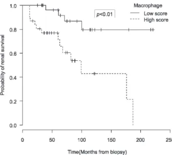
The Kaplan-Meier curve shown in Figure 6 demonstrates the correlation between the presence of macrophages and survival over the course of several months, whereby a higher number of macrophages infiltrating the tubulointer-stitium of the renal cortex predicted a lower survival rate for renal function (p,0.05). There were no correlations between macrophage infiltration into the glomerular compartment and various clinical or laboratory parameters, unfavorable clinical course or renal survival (p.0.05).
In the multivariate analysis (Table 2), the number of macrophages was not independently associated with an unfavorable clinical course (OR = 1.0, CI = 0.91-1.09,p= 0.93). However, the degree of tubulointerstitial injury and the levels of proteinuria were independently associated with progressive disease (OR = 10.5, CI = 212-425, p= 0.01 and OR = 7.65, CI = 1.39-42.3,p= 0.02).
Table 1 -Clinical parameters of IgAN patients with favorable vs. unfavorable clinical disease courses.
Favorable course (n = 28)
Unfavorable course (n = 34)
Age at onset (years) 27.7¡9.2 27.2¡13.0
Gender (male - female) 10:18* 22:12
Urinary protein excretion at the time of biopsy (g/24 h) 1.9¡0.8* 2.9¡1.0
Urinary protein excretion at the end of follow-up (g/24 h) 1.1¡0.8 1.0¡0.5
Serum creatinine at the time of biopsy (mg/dL) 1.4¡0.5* 1.9¡0.4
Serum creatinine at the end of follow-up (mg/dL) Time of follow-up (months)
2.2¡0.7* 76¡47.32
3.8¡1.3 73¡50.85
Figure 1 -Renal macrophage infiltration. (A) The absence of macrophages in a preserved area of the cortex, and (B) macrophages within areas of interstitial fibrosis (A and B: objective 40x).
DISCUSSION
In the present study, high levels of serum creatinine (at the time of biopsy and at the end of the follow-up period), severe proteinuria (at the time of biopsy and at the end of the follow-up period), an elevated degree of tubulointer-stitial injury and increased tubulointertubulointer-stitial expression of NF-kB anda-SMA were all significantly associated with the number of macrophages in the tubulointerstitial compart-ment. Furthermore, macrophage infiltration served as a marker of the annual decrement of eGFR.
The infiltration of macrophages into the renal cortex has been reported in patients with progressive renal disease (17,18). In a study of 204 patients with IgAN, the presence of macrophages (CD68-positive cells) was positively correlated with the following: serum creatinine at the time of biopsy, proteinuria, progression of renal disease and a worse disease outcome (19). In addition, similar results have been observed in pediatric patients (20). These findings are similar to those of the present study, as we demonstrated a correlation between macrophage infiltration and protei-nuria and serum creatinine levels both at the time of biopsy and at the end of the follow-up period.
Studies have shown that macrophages can synthesize collagen I and fibronectin in vitro and can release several fibrogenic cytokines, including TGF-b, PDGF and IL-2 (21,22). In our sample, there was no relationship between TGF-bexpression and glomerular or interstitial infiltration by macrophages, which may have been due to the limited sensitivity of immunohistochemistry to detect TGF-b activ-ity in the kidney (8).
In a study of patients with chronic cyclosporine nephro-pathy, Young et al. (23) reported that macrophage infiltra-tion precedes the development of interstitial fibrosis. Moreover, Geleilete et al. (24) observed an increase in the diffuse distribution of a-SMA and ED-1 (monocytes/ macrophages) immunoreactivity in the renal cortex of animals treated with gentamicin. In the same study, the animals that were sacrificed 30 days after treatment demon-strated a focal distribution of these proteins that was pre-dominantly localized within damaged areas. Therefore, these authors concluded that macrophages, TGF-b, endothelin and myofibroblasts may contribute to renal injury. In our patients, there was a correlation between the interstitial expression ofa-SMA, but not the glomerular expression of a-SMA, and macrophage infiltration.
Figure 3 -Southwestern histochemistry for NF-kB expression. (A) The absence of staining and (B) moderate-to-intense staining in the tubular compartment (A and B: objective 40x).
One previous study found a correlation between the activation of NF-kB and macrophage interstitial infiltration in patients with lupus nephritis (25). In addition, recent studies have suggested that the NF-kB signaling pathway is involved in the activation of macrophages (15,26). In our study, we found a correlation between macrophage infiltra-tion and NF-kB expression, as measured by IHC, as well as a trending correlation between macrophage infiltration and NF-kB expression, as measured by SWH. Although the role of NF-kB in the activation of mast cells has been demon-strated (27), no study has examined this relationship in renal disease.
NF-kB participates in the regulation of gene transcription by binding to a specific sequence of nuclear DNA known as a response element. To better understand the operation of this transcriptional factor, an analysis of expression at the individual cell level is required. In our IHC analysis, NF-kB was detected whether or not it was bound to the response element, whereas in the SWH analysis, NF-kB was only detected when it was unbound or weakly bound to the response element. Because these methods provide comple-mentary results, they can be used together to characterize the physiological state of NF-kB (15). However, none of the techniques described in the literature provide a fully comprehensive description of NF-kB activation and func-tion (28,29).
In the current study, the number of macrophages was not independently associated with an unfavorable clinical course, although the degree of tubulointerstitial injury was found to be the variable most significantly associated with poor clinical course. In addition, the multivariate analyses identified a significant interaction between macrophage infiltration and levels of proteinuria and tubulointerstitial injury. In agreement with these findings, other reports have consistently shown a relationship between interstitial macrophage infiltration and the progression of renal fibrosis (11,16).
Recently, several studies have suggested that there are functionally distinct populations of macrophages that exist within the same tissues and have opposing pro- and anti-fibrotic actions (9,10). Despite the potential protective role of
macrophages in renal fibrosis, the number of macrophages in the tubulointerstitial area may serve as a predictive factor for poor prognosis in patients with IgAN, and their presence was associated with the expression of pro-fibrotic factors in the current study. Therefore, renal function and its prognosis are closely correlated with the extent of tubu-lointerstitial damage and, consequently, with the extent of mononuclear infiltration into this compartment. However, we observed no correlation between macrophage infiltration into the glomerular compartment and any of the examined clinical or histological parameters. This finding could likely be explained by the very high percentage of patients with non-proliferative histological forms (77.5% with focal and segmental sclerosis and mesangiopathic forms; 6.5% with mesangiopathic forms) in our study. In addition, the study by Nagata et al. demonstrated that the number of macro-phages in the glomerular compartment was significantly higher in patients who received pathological diagnoses of focal and diffuse proliferative glomerulonephritis than in patients with minor glomerular abnormalities or sclerosis (30).
However, certain limitations of our study should be noted. First, the investigation was performed casuistically using single-center, heterogeneous data regarding the use of angiotensin-converting enzyme (ACE) inhibitor therapy, the levels of renal function and immunosuppressive protocols. In addition, the study used retrospective data collection analysis.
In conclusion, an increased number of macrophages in the tubulointerstitial area may serve as a predictive factor for poor prognosis in patients with IgAN, and we found the presence of these cells to also be associated with the expression of pro-fibrotic factors.
Figure 5 -The correlation between the number of macrophages in the tubulointerstitial area and annual decrement of eGFR
AUTHOR CONTRIBUTIONS
Silva GEB participated in the conception and drafting of the manuscript as well as the revisions for critically important intellectual content. Costa RS was involved in the histopathological studies of the biopsies from selected patients as well as the revision of the manuscript for critically important intellectual content. Ravinal RC participated in the immunohistochemical studies. Reis MA participated in the statistical analysis. Ramalho LZ participated in the Southwestern histochemical studies. Moyses-Neto M participated in the morphometric analyses. Romao EA participated in the translation and editing of the manuscript. Coimbra TM participated in the conception, analysis of data, and in the revision of the manuscript for critically important intellectual content. Dantas M participated in the selection of patients, collection of biopsies, analysis of data, laboratory and radiological exams, and in the revision of the manuscript for critically important intellectual content.
REFERENCES
1. Berthoux FC, Mohey H, Afiani A. Natural history of primary IgA nephropathy. Semin Nephrol. 2008;28(1):4-9, http://dx.doi.org/10.1016/ j.semnephrol.2007.10.001.
2. Coppo R, D’Amico G. Factors predicting progression of IgA nephro-pathies. J Nephrol. 2005;18(5):503-12.
3. Manno C, Strippoli GF, D’Altri C, Torres D, Rossini M, Schena FP. A novel simpler histological classification for renal survival in IgA nephropathy: a retrospective study. Am J Kidney Dis. 2007;49(6):763-75, http://dx.doi.org/10.1053/j.ajkd.2007.03.013.
4. Costa RS, Droz D, Noel LH. Long-standing spontaneous clinical remission and glomerular improvement in primary IgA nephropathy (Berger’s disease). Am J Nephrol. 1987;7(6):440-4, http://dx.doi.org/ 10.1159/000167516.
5. D’Amico G. Natural history of idiopathic IgA nephropathy and factors predictive of disease outcome. Semin Nephrol. 2004;24(3):179-96, http:// dx.doi.org/10.1016/j.semnephrol.2004.01.001.
6. Alpers CE, Hudkins KL, Gown AM, Johnson RJ. Enhanced expression of ‘‘muscle-specific’’ actin in glomerulonephritis. Kidney Int. 1992;41(5):1134-42, http://dx.doi.org/10.1038/ki.1992.173.
7. Ehara T, Shigematsu H. Mast cells in the kidney. Nephrology (Carlton). 2003;8(3):130-8, http://dx.doi.org/10.1046/j.1440-1797.2003.00153.x. 8. Goumenos DS, Tsamandas AC, Oldroyd S, Sotsiou F, Tsakas S,
Petropoulou C, et al. Transforming growth factor-beta(1) and myofibro-blasts: a potential pathway towards renal scarring in human glomerular disease. Nephron. 2001;87(3):240-8, http://dx.doi.org/10.1159/ 000045921.
9. Ricardo SD, van Goor H, Eddy AA. Macrophage diversity in renal injury and repair. J Clin Invest. 2008;118(11):3522-30, http://dx.doi.org/ 10.1172/JCI36150.
10. Nishida M, Hamaoka K. Macrophage phenotype and renal fibrosis in obstructive nephropathy. Nephron Exp Nephrol. 2008;110(1):e31-6, http://dx.doi.org/10.1159/000151561.
11. Lee S, Huen S, Nishio H, Nishio S, Lee HK, Choi BS, et al. Distinct macrophage phenotypes contribute to kidney injury and repair. J Am Soc Nephrol. 22(2):317-26.
12. Harris RC, Neilson EG. Toward a unified theory of renal progression. Annu Rev Med. 2006;57:365-80, http://dx.doi.org/10.1146/annurev. med.57.121304.131342.
13. Ravinal RC, Costa RS, Coimbra TM, Dantas M, dos Reis MA. Mast cells, TGF-beta1 and myofibroblasts expression in lupus nephritis outcome. Lupus. 2005;14(10):814-21, http://dx.doi.org/10.1191/0961203305lu 2188oa.
14. Ashizawa M, Miyazaki M, Abe K, Furusu A, Isomoto H, Harada T, et al. Detection of nuclear factor-kappaB in IgA nephropathy using Southwestern histochemistry. Am J Kidney Dis. 2003;42(1):76-86, http://dx.doi.org/10.1016/S0272-6386(03)00411-6.
15. Koji T, Komuta K, Nozawa M, Yamada S, Nakane PK. Localization of cyclic adenosine 3’,5’-monophosphate-responsive element (CRE)-bind-ing proteins by southwestern histochemistry. J Histochem Cytochem. 1994;42(10):1399-405, http://dx.doi.org/10.1177/42.10.7930523. 16. Donadelli R, Abbate M, Zanchi C, Corna D, Tomasoni S, Benigni A, et al.
Protein traffic activates NF-kB gene signaling and promotes MCP-1-dependent interstitial inflammation. Am J Kidney Dis. 2000;36(6):1226-41, http://dx.doi.org/10.1053/ajkd.2000.19838.
17. Eddy A. Role of cellular infiltrates in response to proteinuria. Am J Kidney Dis. 2001;37(1 Suppl 2):S25-9, http://dx.doi.org/10.1053/ ajkd.2001.20735.
18. Forbes JM, Hewitson TD, Becker GJ, Jones CL. Ischemic acute renal failure: long-term histology of cell and matrix changes in the rat. Kidney Int. 2000;57(6):2375-85, http://dx.doi.org/10.1046/j.1523-1755.2000.00097.x. 19. Myllymaki JM, Honkanen TT, Syrjanen JT, Helin HJ, Rantala IS,
Pasternack AI, et al. Severity of tubulointerstitial inflammation and prognosis in immunoglobulin A nephropathy. Kidney Int. 2007;71(4): 343-8, http://dx.doi.org/10.1038/sj.ki.5002046.
20. Maruhashi Y, Nakajima M, Akazawa H, Shimoyama H, Nishiguchi M, Yamoto Y, et al. Analysis of macrophages in urine sediments in children with IgA nephropathy. Clin Nephrol. 2004;62(5):336-43.
21. Vaage J, Lindblad WJ. Production of collagen type I by mouse peritoneal macrophages. J Leukoc Biol. 1990;48(3):274-80.
22. Kliem V, Johnson RJ, Alpers CE, Yoshimura A, Couser WG, Koch KM, et al. Mechanisms involved in the pathogenesis of tubulointerstitial fibrosis in 5/6-nephrectomized rats. Kidney Int. 1996;49(3):666-78, http://dx.doi.org/10.1038/ki.1996.95.
23. Young BA, Burdmann EA, Johnson RJ, Andoh T, Bennett WM, Couser WG, et al. Kidney Int. 1995;48(2):431-8.
24. Geleilete TJ, Melo GC, Costa RS, Volpini RA, Soares TJ, Coimbra TM. Role of myofibroblasts, macrophages, transforming growth factor-beta endothelin, angiotensin-II, and fibronectin in the progression of tubulointerstitial nephritis induced by gentamicin. J Nephrol. 2002;15(6):633-42.
25. Zheng L, Sinniah R, Hsu SI. Renal cell apoptosis and proliferation may be linked to nuclear factor-kappaB activation and expression of inducible nitric oxide synthase in patients with lupus nephritis. Hum Pathol. 2006;37(6):637-47, http://dx.doi.org/10.1016/j.humpath.2006.01.002. 26. Wilson HM, Chettibi S, Jobin C, Walbaum D, Rees AJ, Kluth DC.
Inhibition of macrophage nuclear factor-kappaB leads to a dominant anti-inflammatory phenotype that attenuates glomerular inflammation in vivo. Am J Pathol. 2005;167(1):27-37, http://dx.doi.org/10.1016/ S0002-9440(10)62950-1.
27. Miyata N, Gon Y, Nunomura S, Yamashita K, Matsumoto K, Hashimoto S, et al. Inhibitory effects of parthenolide on antigen-induced microtubule formation and degranulation in mast cells. Int Immunopharmacol. 2008;8(6):874-80, http://dx.doi.org/10.1016/j.intimp.2008.02.002. 28. Panzer U, Steinmetz OM, Turner JE, Meyer-Schwesinger C, von Ruffer C,
Meyer TN, et al. Resolution of renal inflammation: a new role for NF-kappaB1 (p50) in inflammatory kidney diseases. Am J Physiol Renal Physiol. 2009;297(2):F429-39, http://dx.doi.org/10.1152/ajprenal. 90435.2008.
29. Sanz AB, Sanchez-Nino MD, Ramos AM, Moreno JA, Santamaria B, Ruiz-Ortega M, et al. NF-kappaB in renal inflammation. J Am Soc Nephrol. 21(8):1254-62.
30. Nagata M, Akioka Y, Tsunoda Y, Komatsu Y, Kawaguchi H, Yamaguchi Y, et al. Macrophages in childhood IgA nephropathy. Kidney Int. 1995;48(2):527-35, http://dx.doi.org/10.1038/ki.1995.323.
Table 2- Risk factors for unfavorable clinical course according to the multivariate analysis.
Risk Factors Gross Logistic Regression Adjusted Logistic Regression
Odds Ratio IC 95% p-value Adjusted Odds Ratio* IC 95% Adjustedp-value*
LI LS LI LS
Macrophages 1.10 1.01 1.19 0.04 1.00 0.91 1.09 0.93
Serum Creatinine at onset 6.94 1.94 24.82 ,0.01 3.17 0.81 12.44 0.10
Proteinuria at onset (261) 4.05 1.21 13.54 0.02 7.65 1.39 42.03 0.02
Proteinuria at onset (361) 5.36 1.24 23.21 0.02 6.98 0.88 55.61 0.07
Interstitial Fibrosis (260/1) 4.07 1.17 14.15 0.03 3.52 0.67 18.53 0.14
Interstitial Fibrosis (360/1) 40.70 4.50 367.91 ,0.01 30.05 2.12 425.69 0.01