Case Report
Relato de Caso
Juliana Fernandes Godoy1 Alcione Ghedini Brasolotto2 Giédre Berretin-Félix2 Adriano Yacubian Fernandes2
Descritores
Acidente vascular cerebral Distúrbios da voz Voz Testes de articulação da fala Técnicas de diagnóstico neurológico
Keywords
Stroke Voice disorders Voice Speech articulation tests Diagnostic techniques, neurological
Correspondence address:
Juliana Fernandes Godoy
Rua José Antônio Braga, 3-49, Bauru (SP), Brasil, CEP: 17018-540. E-mail: [email protected]
Received: 06/28/2013
Accepted: 03/10/2014
Study carried out at the School of Odontology of Bauru, Universidade de São Paulo – USP – Bauru (SP), Brazil. (1)School of Odontology of Bauru, Universidade de São Paulo – USP – Bauru (SP), Brazil.
(2) Department of Speech-Language Pathology and Audiology, School of Odontology of Bauru, Universidade de São Paulo – USP – Bauru (SP), Brazil.
Financial support: Coordination for the Improvement of Higher Education Personnel (CAPES).
Conlict of interests: nothing to declare.
Neuroradiology and voice indings in stroke
Achados de neurorradiologia e voz no acidente
vascular encefálico
ABSTRACT
Neurological dysphonias are vocal disorders followed by injuries or changes in the nervous system. Stroke is the second leading cause of death worldwide and the brain damage caused by it can affect communication in several aspects. The changes in the vocal features caused by these lesions are poorly described regarding the location and extent of cerebral involvement. The aim of this study was to describe vocal parameters of ten elderly patients affected by stroke according to the topography of the lesion at neuroimaging. We recorded from them: perceptual and auditory evaluation of voice and laryngeal diadochokinesis. Neuroimaging studies were classiied according to location, extent, laterality territory of brain injury. The results show a population of extensive middle cerebral artery strokes and strokes with varied location in the brain. The subjects’ voices showed predominantly the presence of roughness, breathiness, richeness and instability, and change in laryngeal motor control, demonstrated by reduced speed and instability in laryngeal diadochokinesis. These features presented in patients with extensive middle cerebral artery stroke and in patients with short strokes with varied location in the brain. The results suggest that the vocal features in the assessed cases do not depend only on the topography of the brain damage. Thus, it is important to consider the patient as unique in clinical evaluation.
RESUMO
INTRODUCTION
The newer methods of clinical research, such as structural and functional neuroimaging, as well as theoretical advances, make us ponder the concept of lesion–symptom relationships on language and speech. Thus, when considering the neural circuits responsible for language and speech, a lesion in the region of these circuits tends to affect the function as a whole, although in varying degrees and forms(1).
Vocal disorders resulting from neurological lesions may be present in cases of dysarthria, which is the abnormal speech due to neurological injury and affects the execution of the motor action. In dysarthria, changes occur in one or more of the following components: breathing, phonation, articulation, resonance, and prosody(2). Some authors have described the
following common symptoms in dysarthria: pneumophonoar-ticulatory incoordination, impaired vocal quality, changes in pitch and loudness, hypernasality, and changes in the articu-lation pattern(3).
There are few studies that relate the changes in phona-tion with neuroradiological images obtained in patients with neurological disorders. The evaluation of voice, especially in cases of stroke, is also poorly described, given its impor-tance in the restoration of the communication functions of these patients.
In general, studies in this ield observed the changes in patients during the acute phase of the onset, with the vocal and speech assessments conducted within the irst 72 hours(3-5),
but little is known about the changes in the stable phase of the onset. It is noteworthy that these studies evaluated the audi-tory-perception of the quality of patients’ voice and oral motor control through diadochokinesia (DDK) tasks. This assessment relects the adequacy of the individual’s neuromotor matura-tion and integramatura-tion, considering a test of neurological skills in which the subject must perform fast repetitions of the same syllable or vowel. In the case of the vowel, only the laryngeal motor control ability is evaluated; an understudied aspect(6),
but it provides important information.
Among the studies that observed the area of injury in the brain in cases after a stroke, one of them(3) reported that the
lesions responsible for dysarthria were located in the supra-tentorial region in 45.6% of the cases, and in the infrasupra-tentorial region in 54.4% of the cases; the supratentorial strokes were found more often on the left (74.2%) than on the right (25.8%) hemispheres. The same group of researchers, in another study(4),
reports that dysarthria in extracerebellar stroke was most often caused by lesions in the left cerebral hemisphere and its inten-sity was more pronounced when the lesion was in the same hemisphere, regardless of its topography.
Changes such as weakness, slowness, or incoordina-tion of the laryngeal muscles may be identiied by voice. Furthermore, the identiication of laryngeal signs and symp-toms can facilitate the early differential diagnosis of certain diseases(7). In more severe cases of neurological disorders,
examination of phonatory characteristics can reveal impor-tant information about the basic neuropathology. Aiming to
a better understanding of the interference of changes in the central nervous system (CNS) in voice emission and the pur-suit of more substrate for rehabilitation, the study of phona-tory characteristics in relation to the location and extent of injury is justiied.
Thus, the aim of this study was to describe the phonatory characteristics of 10 patients affected by stroke according to the topography of the lesion, observed by neuroimaging exams.
PRESENTATION OF CLINICAL CASES
This study was approved by the Research Ethics Committee (under protocol no. 139/2010), and the subjects signed a free and informed consent form.
Ten subjects with clinical diagnosis and who underwent imaging tests that conirmed brain injury from stroke were eval-uated. The sample consisted of individuals aged over 60 years, affected by stroke, conirmed by computed tomography (CT). Six subjects were female and four were male, and all of them were affected by stroke between 6 months and 3 years and 8 months before the time of the evaluation, with an average of 2 years and 1 month after the occurrence of ischemia. The average age for women was 77.3 years and that for men was 73 years. A laryngeal nasoendoscopy was performed to exclude indi-viduals who had laryngeal lesions not related to the stroke, such as polyps, nodules, cysts, leukoplakia, and sulcus vocalis.
The interpretation of CT scans of the skull was per-formed by two neuroradiologists, aiming to characterize brain lesions regarding their location, area of vascular irri-gation, extension, and laterality. Strokes with more than 3 cm in length and affecting more than one cerebral lobe were considered extensive.
Auditory-perceptual voice was assessed with Consensus Auditory-Perceptual Evaluation of Voice (CAPE-V), an instru-ment developed by a group of speech-language pathologists and voice specialists who are part of the Special Interest Division 3 — Voice as Voice Disorder of American Speech-Language and Hearing Association (SID-3 – ASHA).
CAPE-V assesses six predetermined parameters in three tasks: sustained vowel, speciic sentences from a protocol, and spontaneous speech. To indicate the degree of deviation observed, we used a linear analog scale, with 100 mm for each parameter. Evaluators indicated the degree of perception from normal to the deviation, for each parameter of the scale. The parameters analyzed in the protocol were the following: overall degree of change, roughness, breathiness, strain, pitch, and loudness. This protocol allows the inclusion of two extra parameters and, in this study, the additional parameters used were instability in the emission of sustained vowel and rich-ness in the two tests involving speech.
of this sample, the average value assigned by the three judges in the visual analog scale was considered for each parameter.
The DDK, which is the ability to perform rapid repetitions of relatively simple patterns comprising oppositional contrac-tions, was evaluated by repeating two distinct vowels separately, /a/ and /i/, uninterruptedly.
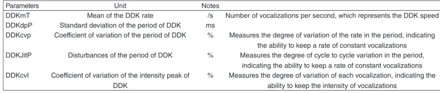
The analysis of the emissions of the vowels /a/ and /i/ was performed by the Motor Speech Proile – Advanced (MSP) software from KayPENTAX. The parameters of the DDK are provided automatically by the MSP software and are shown in Chart 1.
Data analysis was performed in a descriptive manner in relation to all subjects, as well as in relation to two groups of subjects according to the location and extent of injury: one with strokes with extensive lesions of the middle cerebral artery and the other with smaller-sized strokes with lesions in varied loca-tions of the brain.
RESULTS
In the analysis of skull CT scans, a large homogeneous group of strokes (example shown in Figure 1) were found, and in ive patients (three women and two men), the middle cerebral artery was found affected. These are indicated in the
tables, differentiating them from the rest of the group, which is formed by smaller strokes of varying locations in the brain (example shown in Figure 2). In addition, other indings of the examination have been described: ventriculomegaly, which corresponds to an increase in the size of the brain ventricles without increased intracranial pressure; calciications in the basal ganglia; mega cisterna magna, characterized by an increase and morphological alteration of the cisterna magna; and congenital alteration. Such indings were observed in the two groups (Table 1).
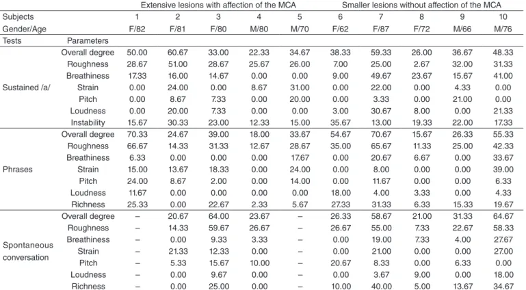
All subjects in the sample had some degree of breathi-ness and roughbreathi-ness in their voice, instability in the emission of sustained vowels, and richness during speech; at least half of the patients showed some degree of vocal strain (Table 2). The laryngeal DDK showed slowness and instability in emis-sions (Table 3). These indings were present regardless of the extent of brain injury.
DISCUSSION
In analyzing the results of the auditory-perceptual evalu-ation, we see generally high values of parameters in all sub-jects in the sample, especially with the sustained /a/ test and independent phrases test, regardless of the type of stroke.
Figure 1. Computed tomography showing an extensive stroke of the middle cerebral artery, affecting more than one cerebral lobe
Figure 2. Computed tomography showing a stroke smaller than 3 cm, affecting the insula
Chart 1. Parameters analyzed in the laryngeal diadochokinesia
Parameters Unit Notes
DDKmT Mean of the DDK rate /s Number of vocalizations per second, which represents the DDK speed DDKdpP Standard deviation of the period of DDK ms
DDKcvp Coefficient of variation of the period of DDK % Measures the degree of variation of the rate in the period, indicating the ability to keep a rate of constant vocalizations
DDKJitP Disturbances of the period of DDK % Measures the degree of cycle to cycle variation in the period, indicating the ability to keep a rate of constant vocalizations DDKcvI Coefficient of variation of the intensity peak of
DDK
% Measures the degree of variation of each vocalization, indicating the ability to keep the intensity of vocalizations
One study(8) showed that acoustic measurements indicating the
presence of noise were more pronounced in patients affected by stroke than those in healthy patients, suggesting greater tendency for noisy voices, with a greater degree of vocal devi-ation in these subjects. Regarding the visual analog scale in voice assessment, the literature refers to the indicated value of 34 voice deviation points as the threshold for considering that the individual failed the voice screening(9). Although the cutoff
values for each of the parameters tested in the CAPE-V were not indicated, it was possible to notice a uniform roughness in the voice of the subjects in the sample, in all tests, as well as
the choice of the items instability and richness in most cases, which is noteworthy.
In the sustained /a/ test, all subjects evaluated presented some degree of instability in the voice, which is characterized by oscillation of any frequency and intensity, regardless of pace. Urban et al.(4), when describing the vocal quality of the
partici-pants in their study, reported vocal instability.
Regarding richness, only one subject was considered to lack this parameter in the evaluation by the three judges; the remainder of the sample presented values ranging from 6 to 31 mm in the sentences test and 5–40 mm in spontaneous
Table 2. Average values of the parameters of the Consensus Auditory-Perceptual Evaluation of Voice protocol evaluated by the judges for each test and for each subject
Extensive lesions with affection of the MCA Smaller lesions without affection of the MCA Subjects
Gender/Age
1 F/82
2 F/81
3 F/80
4 M/80
5 M/70
6 F/62
7 F/87
8 F/72
9 M/66
10 M/76 Tests Parameters
Sustained /a/
Overall degree 50.00 60.67 33.00 22.33 34.67 38.33 59.33 26.00 36.67 48.33 Roughness 28.67 51.00 28.67 25.67 26.00 7.00 25.00 2.67 32.00 31.33 Breathiness 17.33 16.00 14.67 0.00 0.00 9.00 49.67 23.67 15.67 41.00 Strain 0.00 24.00 0.00 8.67 31.00 0.00 22.00 0.00 4.33 0.00 Pitch 0.00 8.67 7.33 0.00 20.00 0.00 3.33 0.00 21.00 0.00 Loudness 0.00 20.00 7.33 0.00 0.00 3.00 30.67 8.00 0.00 21.33 Instability 15.67 30.33 23.00 12.33 15.00 35.67 13.00 19.33 22.00 17.33
Phrases
Overall degree 70.33 24.67 39.00 18.00 33.67 54.67 70.67 15.67 26.33 55.33 Roughness 66.67 14.33 31.33 12.67 28.67 35.00 65.67 11.33 25.00 42.33 Breathiness 6.33 0.00 0.00 0.00 17.67 0.00 20.67 6.67 0.00 33.67 Strain 15.00 13.67 18.33 0.00 24.00 0.00 8.00 0.00 0.00 39.00 Pitch 24.00 8.67 2.00 0.00 14.00 0.00 11.67 0.00 0.00 6.33 Loudness 11.67 0.00 0.00 0.00 0.00 18.00 4.00 3.33 0.00 4.33 Richness 25.33 0.00 22.67 2.33 5.67 27.33 31.33 6.33 15.33 19.67
Spontaneous conversation
Overall degree – 20.67 64.00 23.67 – 26.33 58.67 21.00 31.33 64.67 Roughness – 14.33 59.67 26.67 – 26.67 55.00 7.33 22.67 58.33 Breathiness – 0.00 9.33 3.33 – 0.00 19.00 7.33 4.00 27.67 Strain – 21.33 12.33 0.00 – 0.00 21.00 0.00 0.00 27.00 Pitch – 5.33 15.67 10.00 – 20.67 8.33 0.00 6.33 0.00 Loudness – 0.00 9.67 0.00 – 0.00 3.67 9.00 0.00 18.00 Richness – 0.00 25.00 0.00 – 10.00 40.00 5.00 13.67 34.67
Caption: MCD = middle cerebral artery; F = female; M = male; (–) refers to the nonevaluation of the parameter, due to difficulty or nonexecution of the task requested by the participant
Table 1. Characterization of the sample according to gender, age, time of brain affection in the evaluation and characteristics of the lesion observed on computed tomography
Case Gender Age (years) Time of stroke Brain hemisphere Lobe Extension Lesion of the middle
cerebral artery Other
1 F 82 3.8 years L FITP > Yes V
2 F 81 11 months R FTP > Yes V
3 F 80 3 years R FITP > Yes –
4 M 80 1.7 years R FT > Yes –
5 M 70 2.6 years R FP > Yes –
6 F 62 8 months L Insular < No –
7 F 87 3 years R F < No V
8 F 72 6 months R F > No MCM
9 M 66 3 years R O > No CBG
10 M 76 2 years L I < No –
conversation. Some authors reported that richness in voice relects slowness, imprecise articulation, and hypernasality, characteristics that may be present in neurological disorders such as stroke(4,10), due to loss of muscle tone(10,11). Interestingly,
subjects who showed absence of this parameter presented extensive strokes of the middle cerebral artery in the right cerebral hemisphere, but we can relate this good performance with other studies(4,5) that showed the evolution in the speech
of these patients after the acute phase of the onset.
The literature suggests the DDK task to assess motor coordination and speed of movement of the of the phonoar-ticulatory organs in patients with neurological disorders(4,11).
Some authors(12) reported that normal individuals
pos-sess appropriate adjustments in laryngeal mechanisms that enable rapid glottal opening and closure during production of the consonants and vowels of syllables. This adjustment can be modiied in cases with neurological disorders.
Dividing individuals from the sample into two groups, extensive strokes in the middle cerebral artery and smaller strokes in varied locations, through the average, it is pos-sible to observe that subjects with extensive strokes in the middle cerebral artery tend to have higher DDK speed, both of /a/ and of /i/, also a smaller standard deviation of DDK in both tests, a higher coeficient of variation of the DDK period in the DDK of /a/ and equated to the other group in the DDK of /i/ lower values of disruption for the DDK period (JitP) in both tests and higher values relate to the coeficient of variation of the peak intensity of the DDK (cvI), also in the two tests.
Magalhães(13) evaluated the DDK of elderly individuals
aged 70–79 years. Although we know that it is not pos-sible to correlate the data of the elderly individuals in this study because there is no statistical analysis of the results, and considering that these parameters are poorly studied, it is observed that the rate of the DDK is similar in healthy elderly patients and those who had stroke. However, even if a statistical comparison is not possible, it is possible to describe that, regarding instability parameters (cvP, JitP,
and cvI), women affected by stroke showed higher values than those in the study cited, both for the DDK of /a/ and of /i/. Men affected by stroke showed higher values than healthy elderly only for the DpP and cvP parameters of the DDK of /i/; in the rest of the tests, the values in both populations are quite similar. Therefore, the women in this study showed difficulty in maintaining constant vocaliza-tions, both in period and in cycle to cycle, and a difficulty in maintaining the intensity of vocalizations when compared to the elderly population between 70 and 79 years without neurological changes. When analyzing case by case, we can say that only subjects 2 and 4 had a good performance on the laryngeal DDK task, close to healthy elderly(13) in all
parameters; the remaining subjects showed slower DDK and greater instability in period, cycle to cycle, and relate to intensity.
Data in the literature indicate that the laryngeal DDK is slower in the case of neurological changes(6). Few authors
have addressed the assessment of oral DDK in post-stroke patients(4,11) and only one of them evaluated the laryngeal
DDK in this population(11), having found a DDK rate
vary-ing between 0.75 and 4.75 emissions per second.
When we look jointly to all vocal assessments carried out, we can see that, regardless of the location and extent of brain injury, the subjects who achieved good performance tended to show that in all of the evaluations, as especially shown by cases 2, 4, 6, and 9. Subjects who had a worse performance also tended to have dificulty in all tests, which was observed mainly in subjects 1, 7, and 10.
There are no reports that compare the types of voices to the area of brain injury. However, both the perceptual indings on vocal quality and the perceptual aspects of oral DDK are described in the literature relate to the types of dysarthria, and these can be deined according to the area of injury in the CNS.
Spastic dysarthrias result from upper motor neuron injury and are extensively described in stroke cases. There is also a separate description, made by some authors, of unilateral
Table 3. Laryngeal diadochokinesia values provided by the Motor Speech Profile – Advanced software, by KayPENTAX, for each subject Extensive lesion with affection of the MCA Smaller lesion without affection of the MCA
Subjects Gender/Age
1 F/82
2 F/81
3 F/80
4 M/80
5 M/70
6 F/62
7 F/87
8 F/72
9 M/66
10 M/76 Parameters Unit
A
mT (/s) 3.00 3.67 4.25 2.30 2.19 2.20 3.02 2.30 3.33 2.37 dpP (ms) 29.07 169.0 23.36 21.02 98.82 65.39 72.90 21.02 49.54 28.51 cvP (%) 8.73 61.95 9.94 4.83 21.69 14.42 22.00 4.83 16.49 6.77 JitP (%) 1.77 11.30 3.50 1.67 16.65 10.54 9.04 1.67 6.94 3.65 cvI (%) 2.57 5.05 1.69 3.68 1.59 1.78 3.59 3.68 3.05 2.23
I
mT (/s) 2.80 1.57 3.30 2.49 2.17 2.37 2.78 2.49 3.13 2.62 dpP (ms) 24.36 15.26 32.46 77.94 63.42 149.0 1.82 77.94 89.21 63.94 cvP (%) 6.83 2.39 10.72 19.37 2 5 6 19.37 27.88 16.75 JitP (%) 2.24 1.04 4.68 6.87 F/62 F/87 F/72 6.87 5.92 8.28
upper motor neuron dysarthria, in which 90% of the causes are unilateral strokes in this brain region. Flaccid dysar-thrias, resulting from injuries between the brainstem and the neuromuscular junctions, lead to muscle weakness, paresis, or paralysis that may affect orofacial muscles or laryngeal structures, which makes voice and speech weak. Hypokinetic dysarthrias in stroke cases are commonly associated with bilateral ischemia in the thalamic region. These cases can be termed as vascular parkinsonism. Hyperkinetic dysar-thrias, not very common in stroke cases (about 1% of this type of dysarthria are due to some vascular injury), appear when there is brainstem stroke, which leads to movement disorders such as chorea, dystonia, and essential tremor. Mixed dysarthrias, also common in this population, arise from various types of injuries, as there is a combination of one or more types of dysarthria(14).
Some patients in this sample, with slight vocal changes and absence of changes in other speech components, did not fall into the category of diagnosis of dysarthria. No cases of ataxic dysarthria were observed, because no participant showed cerebellar lesion. Moreover, no lesions in the brain-stem were observed in the tests, which rules out cases of hyperkinesis.
Other patients in this sample it more easily in the descrip-tion of spastic dysarthria or unilateral upper motor neuron dysarthria, whose phonatory characteristics are lowered pitch, roughness, strained voice, frequency breaks, short sentences, and reduced speed in tests such as the DDK. Another possi-ble description is mixed dysarthria, wherein speech charac-teristics from more than one type of dysarthria occur, com-mon in patients with multiple strokes. Owing to the vocal characteristics of this sample, such as weakness or richness in speech and articulatory imprecision, some cases could be described as spastic dysarthria and laccid characteristics.
It is important to consider that this study shows that lesions in similar areas showed distinct vocal expressions; therefore, the most basic classiication of dysarthrias(2) does
not always cover all aspects to be described. Due to brain injuries resulting from stroke being quite variable, which may affect the brain in different locations and sizes, clas-sifying neurological dysphonia in these cases is complex. According to Aronson(15), the vocal manifestations presented
by patients in this study fall in relatively constant neurologi-cal disorders of voice.
Factors such as neuronal plasticity, early intervention, overall health data, and the aging process are directly related to rehabilitation and patient performance. Therefore, one may suggest that vocal changes in brain injury will not always be speciic for each type of injury, because the recovery of the functions observed in the stable phase of onset and common vocal deterioration in aging are often associated. In addi-tion, decline in cognitive ability and apathy observed in the elderly with stroke can also affect speech and, consequently, the vocal aspects of this patient.
This study reported the characteristics of the vocal behav-ior of some individuals after a stroke in a stable phase of the onset, some with extensive lesion and the involvement of
the middle cerebral artery and others with smaller lesions in varied locations. Although case reports present limitations due to the small number of participants and the inability to create multiple groups with more speciic brain lesions, as there is no similar report of such vocal expressions in the literature, the presentation of cases contributed to the char-acterization of the voice of these and individuals, and to the information that the voice quality and laryngeal motor con-trol do not always have speciic characteristics according to the affection of the brain.
Studies involving a large number of post-stroke patients and individuals of the same age without neurological dis-eases, using instruments for auditory-perceptual evaluation, laryngeal motor control, and other procedures, should be undertaken to provide more data for a better understanding of vocal manifestations in this population.
FINAL COMMENTS
Vocal characteristics found in all subjects were roughness, breathiness, sometimes strain, instability (during the emission of sustained vowels), and richness (during speech). The pres-ence of changes in laryngeal motor control was observed, as evidenced by the slowness and instability in emissions of laryn-geal DDK. These features occurred both in patients with exten-sive stroke lesion of the middle cerebral artery and in patients with minor strokes in varying locations in the brain.
*JFG participated in the data collection and drafting of the article; AGB participated as a co-advisor to the study, supervising all collection procedures, actively participating in the conception and design of the study and drafting of the article; GBF actively participated in the conception and design of the study and drafting of the article; AYF participated as an advisor to the study, also aiding in the conception and design of the study, drafting of the article and interpretation of imaging tests.
REFERENCES
1. Mansur LL, Radanovic M. Neurolinguística: princípios para clínica. São Paulo: Edições Inteligentes; 2004.
2. Darley FL, Aronson AE, Brown JR. Differential diagnostic patterns of dysarthria. J Speech Hear Res. 1969;12(2):246-69.
3. Urban PP, Wicht S, Vukurevic G, Fitzek C, Fitzek S, Stoeter P, et al. Dysarthria in acute ischemic stroke: lesion topography, clinicoradiologic correlation, and etiology. Neurology. 2001;56(8):1021-7.
4. Urban PP, Rolke R, Wicht S, Keilmann A, Stoeter P, Hopf HC, et al. Left-hemispheric dominance for articulation: a prospective study on acute ischaemic dysarthria at different localizations. Brain. 2006; 129(Pt 3):767-77.
5. Canbaz DH, Celebisoy M, Ozdemirkiran T, Tokucoglu F. Dysarthria in acute ischemic stroke: localization and prognosis. J Neurol Sci Turk. 2010;27(1):20-7.
6. Depret MMP. Análise da diadococinesia articulatória e laríngea em indivíduos com e sem transtornos neurológicos [Dissertação]. São Paulo: Universidade Federal de São Paulo; 2005.
7. Ortiz KZ, Carrillo L. Comparação entre as análises auditiva e acústica nas disartrias. Rev Soc Bras Fonoaudiol. 2008;13(4):325-31.
9. Yamasaki R, Leão SHS, Madazio G, Padovani M, Azevedo R. Análise perceptivo-auditiva de vozes normais e alteradas: escala analógica visual. In: 15º Congresso Brasileiro de Fonoaudiologia; 2007. Gramado. 10. Altman KW, Schaefer SD, Yu G-P, Hertegard S, Lundy DS, Blumin JH,
et al. The voice and laryngeal dysfunction in stroke: a report from the Neurolaryngology Subcommittee of the American Academy of Otolaryngology-Head and Neck Surgery. Otolaryngol Otolaryngology-Head Neck Surg. 2007;136(6):873-81. 11. Pereira AC, Brasolotto AG, Berretin-Felix G, Padovani CR.
Diadococinesia oral e laríngea em pacientes pós-acidente vascular encefálico. Pró-fono. 2004;16(3):283-92.
12. Leeper HA, Jones E. Frequency and intensity effects upon temporal and aerodynamic aspects of vocal fold diadochokinesis. Percept Mot Skills. 1991;73(3 Pt 1):880-2.
13. Magalhães FF. Diadococinesia oral e laríngea em indivíduos a partir de cinqüenta anos de idade [Dissertação]. Bauru: Universidade de São Paulo; 2008.
14 Duffy JR. Motor speech disorders: substrates, differential diagnosis, and management. 2nd edition. Rochester: Elsevier Mosby; 2005.