Inhibition of K+ Channel Activity and Vasoconstrictor
Response to Angiotensin II in Rat Renal Microvessels
Fan Fan
1☯, Cheng-Wen Sun
2☯, Kristopher G. Maier
3, Jan M. Williams
1, Malikarjuna R. Pabbidi
1, Sean P.
Didion
1, John R. Falck
4, Jialong Zhuo
1, Richard J. Roman
1*1 Department of Pharmacology and Toxicology, University of Mississippi Medical Center, Jackson, Mississippi, United States of America, 2 Department of Pharmaceutical Sciences, North Dakota State University, Fargo, North Dakota, United States of America, 3 Division of Vascular Surgery and Endovascular Services, SUNY Upstate Medical University, Syracuse, New York, United States of America, 4 Department of Molecular Genetics, University of Texas Southwestern Medical Center, Dallas, Texas, United States of America
Abstract
The present study examined whether 20-hydroxyeicosatetraenoic acid (HETE) contributes to the vasoconstrictor effect of angiotensin II (ANG II) in renal microvessels by preventing activation of the large conductance Ca2+
-activated K+ channel (K
Ca) in vascular smooth muscle (VSM) cells. ANG II increased the production of 20-HETE in rat
renal microvessels. This response was attenuated by the 20-HETE synthesis inhibitors, 17-ODYA and HET0016, a phospholipase A2 inhibitor AACOF3, and the AT1 receptor blocker, Losartan, but not by the AT2 receptor blocker,
PD123319. ANG II (10-11 to 10-6 M) dose-dependently decreased the diameter of renal microvessels by 41 ± 5%. This
effect was blocked by 17-ODYA. ANG II (10-7 M) did not alter K
Ca channel activity recorded from cell-attached
patches on renal VSM cells under control conditions. However, it did reduce the NPo of the KCa channel by 93.4 ±
3.1% after the channels were activated by increasing intracellular calcium levels with ionomycin. The inhibitory effect of ANG II on KCa channel activity in the presence of ionomycin was attenuated by 17-ODYA, AACOF3, and the
phospholipase C (PLC) inhibitor U-73122. ANG II induced a peak followed by a steady-state increase in intracellular calcium concentration in renal VSM cells. 17-ODYA (10-5 M) had no effect on the peak response, but it blocked the
steady-state increase. These results indicate that ANG II stimulates the formation of 20-HETE in rat renal microvessels via the AT1 receptor activation and that 20-HETE contributes to the vasoconstrictor response to ANG II
by blocking activation of KCa channel and facilitating calcium entry.
Citation: Fan F, Sun C-W, Maier KG, Williams JM, Pabbidi MR, et al. (2013) 20-Hydroxyeicosatetraenoic Acid Contributes to the Inhibition of K+ Channel Activity and Vasoconstrictor Response to Angiotensin II in Rat Renal Microvessels. PLoS ONE 8(12): e82482. doi:10.1371/journal.pone.0082482
Editor: Jean-Claude Dussaule, INSERM, France
Received July 30, 2013; Accepted November 2, 2013; Published December 4, 2013
Copyright: © 2013 Fan et al. This is an open-access article distributed under the terms of the Creative Commons Attribution License, which permits unrestricted use, distribution, and reproduction in any medium, provided the original author and source are credited.
Funding: This work was funded in part by grants HL36279, HL29587 (RJR), RO1DK067299 (JZ) and HL089884 and HL107632 (SPD) from the National Institutes of Health. The funders had no role in study design, data collection and analysis, decision to publish, or preparation of the manuscript. No additional external funding received for this study.
Competing interests: The authors have declared that no competing interests exist. * E-mail: rroman@umc.edu
☯ These authors contributed equally to this work.
Introduction
Angiotensin II (ANG II) plays a crucial role in the regulation of body fluid volume homeostasis and the long term control of arterial pressure by altering sodium excretion and vascular tone. ANG II is a potent constrictor of renal microvessels that regulates renal blood flow and glomerular filtration rate [1-3]. However, the underlying mechanism is not completely understood. Previous studies have demonstrated that ANG II activates phospholipase A2 (PLA2) and phospholipase C (PLC) in aortic VSM cells to increase the release of arachidonic acid (AA) and the production of prostaglandin E2, prostacyclin, EETs
and 12-, 19- and 20-hydroxyeicosatetraenoic acid (HETE) [4-6]. Several of these metabolites modulate the vasoconstrictor response to ANG II [1,4,7]. For example, the renal vasoconstrictor response to ANG II is potentiated by blockade of cyclooxygenase and the ANG II-induced increase in intracellular calcium concentration ([Ca2+]
effects of ANG II on the formation of 20-HETE, vascular tone, KCa channel activity and intracellular calcium concentration in renal microvessels in the presence and absence of inhibitors of the synthesis of 20-HETE.
Materials and Methods
Animals
Experiments were performed on 178 male, 12-14 week-old SD rats purchased from Charles River Laboratories (Wilmington, MA). The rats were housed in the animal care facilities at the Medical College of Wisconsin and the University of Mississippi Medical Center that are both approved by the American Association for the Accreditation of Laboratory Animal Care. The rats had free access to food and water through the study and all protocols involving animals received prior approval by the Institutional Animal Care and Use Committees (IACUC) of the Medical College of Wisconsin and the University of Mississippi Medical Center.
Measurement of 20-HETE production in renal microvessels
Rat renal microvessels were isolated using an Evans blue sieving procedure similar to that previously described in the cerebral circulation [9]. The rats were anesthetized with isoflurane and a cannula was placed in the lower aorta below the renal arteries. The aorta above the renal arteries was tied off and the kidneys were flushed with 10 ml of iced-cold low calcium Tyrode’s solution containing (in mM): 145 NaCl, 5 KCl, 4.2 NaHCO3, 1 MgCl2, 0.05 CaCl2, 10 HEPES, and 10 glucose. Then, 5 ml of the Tyrode’s solution containing 3% albumin stained with 1% Evans blue was injected to fill the renal microcirculation. The kidney was rapidly removed and hemisected, and the inner medulla and outer medulla were excised. Pieces of the renal cortex were forced through a 150-μm stainless steel sieve with the barrel of a 30 ml glass syringe to mechanically separate tubules and glomeruli from the vascular trees. The tissue retained on the screen was repeatedly rinsed with ice-cold physiological salt solution (PSS) containing (in mM): 119 NaCl, 4.7 KCl, 1.2 MgSO4, 1.6 CaCl2, 1.2 NaH2PO4, 18 NaHCO3, 0.03 EDTA, 10 glucose, and 5 HEPES. The retained vascular tissue on the top of the screen was collected, resuspended in ice-cold PSS solution, and any adherent tubules were removed from the vessels by microdissection using a stereomicroscope.
The freshly isolated renal microvessels were incubated in 1 ml of PSS containing: a) vehicle, b) ANG II alone (10-7 M), c) ANG II plus 17-ODYA (10-5 M), d) ANG II plus HET0016 (10-8 M), e) ANG II plus Losartan (10-6 M), f) ANG II plus AACOF
3 (2 X 10⁻⁵ M), and g) ANG II plus PD123319 (10-7 M) in the presence of 2 µM indomethacin, 1 mM NADPH and 40 μM of AA at 37° C for 30 min. Stock solution of ANG II (Sigma A9525) was dissolved in distilled water and 0.05% acetic acid at a concentration of 10-4 M. Stock solutions of HET0016 (Enzo Scientific) and indomethacin (Sigma I7378), 17-ODYA (Enzo BML, EI-1030), U73122 Sigma U6756), AACOF3 and AA were prepared in ethanol at concentrations of 10-2 M to 10-3 M. Losartan Potassium (Sigma 61188) was dissolved in assay
buffer at a concentration of 10-3 M. All of the drugs were used in 1:500 to 1:1000 dilutions in PSS or assay buffer to the final concentrations used in the various experiments.
We have previously reported that the formation of 20-HETE by the CYP4A enzymes is linearly related to PO2 over the range of 20-140 torr in the incubation media[10]. Thus, the renal microvessels were shaken under an atmosphere of 100% oxygen, which we have previously reported is necessary to maintain a PO2 of approximately 100 torr [11] in the incubation media [12,13]. The reactions were stopped by acidification to pH 3.5 with formic acid and the vessels were homogenized with a ground glass homogenizer on ice until no tissue was visible. A 100 µl aliquot was taken for measurement of protein concentration and the remainder of the sample was extracted twice with 3 ml of ethyl acetate after the addition of 2 ng of an internal standard d6-20-HETE. After centrifugation, the organic phase was dried under nitrogen and stored at -80° C until further analysis.
The metabolites of AA were separated using a Dionex (Sunnyvale, CA) HPLC system and an ABsciex 4000 Q trap tandem mass spectrometer with electrospray ionization (ABsciex, Foster, City, CA.). Separation of the metabolites was achieved using a reverse phase column (Beta basic C18, 150 X2.1 mm, 3 μm; Thermo Hypersil-Keystone, Bellefonte, PA) and the following mobile phase conditions at a flow rate of 300 µl/minute. The column was first equilibrated with 66.7% of a mobile phase A containing water/acetonitrile/methanol/acetic acid 90/8.5/1.4/0.1 (v/v/v/v), and 33.3% mobile phase B, acetonitrile/methanol 85/15 for 5 minutes following injection of the sample. The percentage of mobile phase B was ramped to 53.5% over a 10 min period and then held there for 5 minutes followed by a linear increase to 94.4% mobile phase B over a 7 min period and then held there for another 5 min. Column temperature was maintained at 35° C.
The products were ionized using the negative ion mode and analyzed using multiple reaction monitoring (MRM) with the following instrument settings: Electrospray voltage -4500 volts, curtain gas 30, gas 1-50, temperature 600, gas 2-50, and unit resolution. The following transitions were monitored for each metabolite of AA; m/z 337-207 (14,15-DIHETE), m/z 337-167 (11,12-DIHETE), m/z 337-127 (8,9-DIHETE), m/z 319-231 (19-HETE), m/z 319-245 (20-(19-HETE), m/z 319-261 (18-(19-HETE), m/z 337-145 (5,6-DIHETE), m/z 319-233 (16-HETE), m/z 319-175 (15-HETE), m/z 319-149 (11-HETE), m/z 319-179 (12-HETE), m/z 319-155 (8HETE), m/z 319-203 (5-HETE), m/z 319-175, (14,15-EET), m/z 319- 167 (11,12-EET), m/z 319-127 (8,9-EET), m/z 319-191 (5,6-(8,9-EET), and m/z 325-281/307 (d6 20-HETE) for the internal standard. Standard curves were generated over a range of 0.02 to 20 ng for each metabolite.
Expression of ANG II receptors in renal microvessels
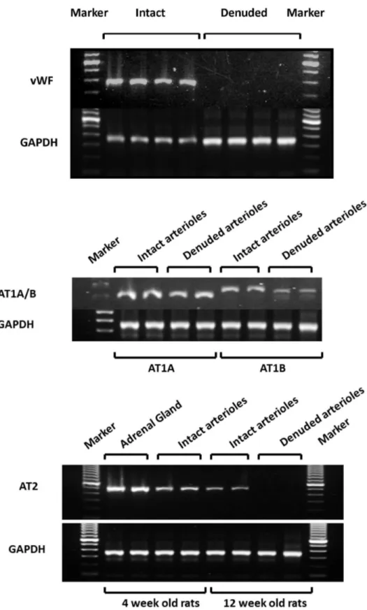
reaction using the iScript cDNA Synthesis Kit (Bio-Rad, Hercules, CA). The reactions were incubated at 25° C for 5 min, 42° C for 30 min followed by inactivation at 85° C for 5 min. The 25 μl PCR reactions contained 25 ng of the forward and reverse primers, 20 mM Tris-HCl buffer (pH 8.4), 50 mM KCl, 1.5 mM MgCl2, 200 μM of each dNTP, 0.5U Taq DNA Polymerase (QIAGEN, Valencia, CA). The reaction mixtures were initially denatured at 94° C for 5 min and then cycled 35 times at 94° C (denaturation) for 30 sec, 64° C (annealing) for 30 sec, and 72° C (elongation) for 30 sec followed by extension for 7 min at 72° C. The RT-PCR products were separated on 1 % agarose gel in a 1X Tris-borate-EDTA (TBE) buffer containing ethidium bromide (Sigma, St. Louis, MO) and the band intensity analyzed using a ChemiDoc MP Imaging System (Bio-Rad, Hercules, CA). The primer sequences for rat AT1AR (NM_030985) corresponded to 5’-CGT CAT CCA TGA CTG TAA AAT TTC-3’ (sense, 1071-1094) and 5’-GGG CAT TAC ATT GCC AGT GTG-3’ (antisense, 1376-1356). The final PCR product size is 306 bp. The primer sequences for rat AT1BR (NM_031009) corresponded to 5’-CAT TAT CCG TGA CTG TGA AAT TG-3’ (sense, 972-994) and 5’-GCT GCT TAG CCC AAA TGG TCC-3’ (antisense: 1316-1296) [14]. The final PCR product size is 345 bp. The primer sequences for rat AT2R (NM_012494) corresponded to 5’-GCT GTG GCT GAC TTA CTC CT-3’ (sense, 259-278) and 5’-GGT CAC GGG TAA TTC TGT TC-3’ (antisense, 757-738) [15], the final PCR product size is 499 bp. The primer sequences for rat GAPDH (NM_017008) corresponded to 5’- CCC CTT CAT TGA CCT CAA CTA C-3’ (sense, 174-195) and 5’-ATG CAT TGC TGA CAA TCT TGA G-3’ (antisense, 520-499), the final PCR product size is 347 bp. The primer sequences for rat von Willebrand factor (vWf) (NM_053889) corresponded to 5’- CTC CCA GCA CTA ACT GCA CCA GC-3’ (sense, 843-865) and 5’-CAA GAA CAG TCA GAG CTC TGC AC-3’ (antisense, bp 1278-1256), the final PCR product size is 436 bp.
Western blotting to verify the purity of isolated renal microvessel preparations
The renal microvessels were powdered in liquid nitrogen and then homogenized in an ice cold RIPA buffer (R0278, Sigma-Aldrich, St. Louis, MO) using a ground glass homogenizer followed by the FastPrep-24 homogenizer (MP Biomedicals, Santa Ana, CA). The samples (50 µg) were separated by electrophoresis on 10% SDS-polyacrylamide gel, transferred to nitrocellulose membranes using Trans-Blot Turbo Transfer System (Bio-Rad, Hercules, CA) and the membranes were blocked at room temperature for one hour in a buffer containing 10% nonfat dry milk. The membranes were incubated overnight at 4° C with a 1:500 dilution of anti-alkaline phosphatase primary antibody (sc-137213, Santa Cruz Biotechnology, Santa Cruz, CA) followed by a 1:5000 dilution of a horseradish peroxidase coupled anti-mouse secondary antibody (sc-2005, Santa Cruz Biotechnology, Santa Cruz, CA) for 1 h. The membrane was re-probed with 1: 8000 dilution of anti-beta actin (ab6276, Abcam, Cambridge, MA) and 1: 20000 of anti-mouse antibody. The blots were exposed to SuperSignal West Dura Extended Duration Substrate (34076, Thermo Scientific, Pittsburgh, PA) and the relative intensities of the bands at 80
KD for alkaline phosphatase and 42 KD for beta actin were imaged using a ChemiDoc photodocumentation system (Bio-Rad, Hercules, CA) .
Vascular reactivity studies
Rats were anesthetized with pentobarbital (50 mg/kg body weight, i.p.) and the abdominal aorta was cannulated below the left renal artery. The kidneys were flushed via the aorta with 10 ml of ice-cold low calcium Tyrode’s solution. The left kidney was removed and renal microvessels (50 to 100 μm) were isolated by microdissection, mounted on glass micropipettes and intraluminal pressure was maintained at 80 mmHg. The vessels were de-endothelialized by perfusion with anti-factor VIII-related antigen antibody (1:1000, Sigma-Aldrich, St. Louis, MO) and complement (20 mg/ml, Sigma-Aldrich, St. Louis, MO) for 5 min as previously described to remove the influence of endothelial dependent vasodilatory factors (NO, prostaglandins and EETs) on the vascular response to ANG II [16]. After a 30-minute equilibration period, the effects of various agonists and inhibitors on the inner diameter of the vessel were determined using a video system composed of a stereomicroscope (Carl Zeiss Inc. Thornwood, NY), a television camera (KP-130 AV, Hitachi, Woodbury, NY), a videocassettes recorder (A6 to 7330, Panasonic) and a video caliper (VIA-100, Boeckeler Instrument Co.). The vessels were perfused with PSS that was equilibrated with a 95% O2, 5% CO2 gas mixture to maintain pH at 7.4. Indomethacin (5 x 10⁻⁶ M) and baicalein (5 x 10⁻⁶ M) were added to the bath to block the endogenous metabolism of AA via the cyclooxygenase and lipoxygenase pathways as previously described [17,18]. The vasoconstrictor response to ANG II was evaluated before and after the addition of vehicle or 17-ODYA (10-5 M) to the bath.
Patch clamp studies
Isolation of renal VSM cells. The kidneys were flushed with ice-cold low calcium Tyrode’s solution and renal microvessels were microdissected. The vessels were then sequentially incubated in 1 ml of a low calcium Tyrode’s solution containing 1.5 mg/ml, papain (14 U/mg), 1 mg/ml DTT for 15 minutes at 37° C, followed by incubation in low calcium Tyrode’s solution containing 90 U/ml elastase, 10000 U/ml soybean trypsin inhibitor, and 196 U/ml collagenase for about 18 minutes or until free cells were found in the media. The supernatant was collected and the cells were centrifuged at 500g for 5 minutes, resuspended in fresh low calcium Tyrode’s solution, and stored at 4° C. Patch-clamp experiments were performed within 4 hours after cell isolation.
Figure 1. The expression of ANG II receptor subtypes in renal microvessels. The upper panel indicates the endothelium specific marker-von Willebrand factor (vWf) is expressed in intact vessels but not in vessels in which the endothelium was removed. Message for AT1A and AT1B receptors was detected by RT-PCR in both intact vessels and vessels with the endothelium removed indicating that they are expressed in VSM cells. Expression of the AT2 receptor was only detected in renal microvessels with an intact endothelium (lower panel). GAPDH was amplified in all of the samples.
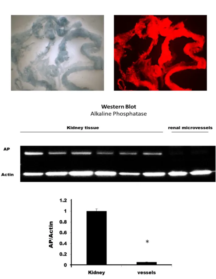
Figure 2. Typical appearance of the isolated renal microvessels. The upper left panel presents appearance of the vessels under white light illumination. The upper right panel presents the appearance of the vessels with excitation of 550 nm, emission of 610 nm. The vessels that were stained with Evans blue in the isolation procedure exhibit red fluoresce while the adherent tubules do not fluoresce. The lower panel compares alkaline phosphatase activity which is highly expressed in the proximal tubules in renal homogenates versus that seen in the renal microvessel preparation. * indicates a significant difference from control.
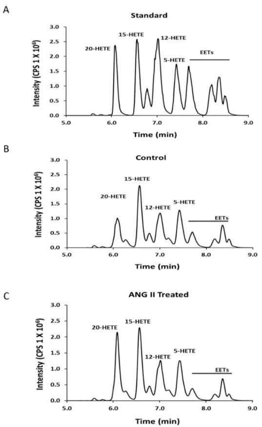
representative chromatogram showing the metabolites of AA produced by renal microvessels before and after addition of ANG II are presented in Figure 3B and 3C. These results indicate that renal microvessels produce 5-, 12-, 15-, and 20-HETE along with various EETs under control conditions. ANG II increased the formation of 20-HETE in the vessels but it had little effect on the formation of other metabolites of AA.
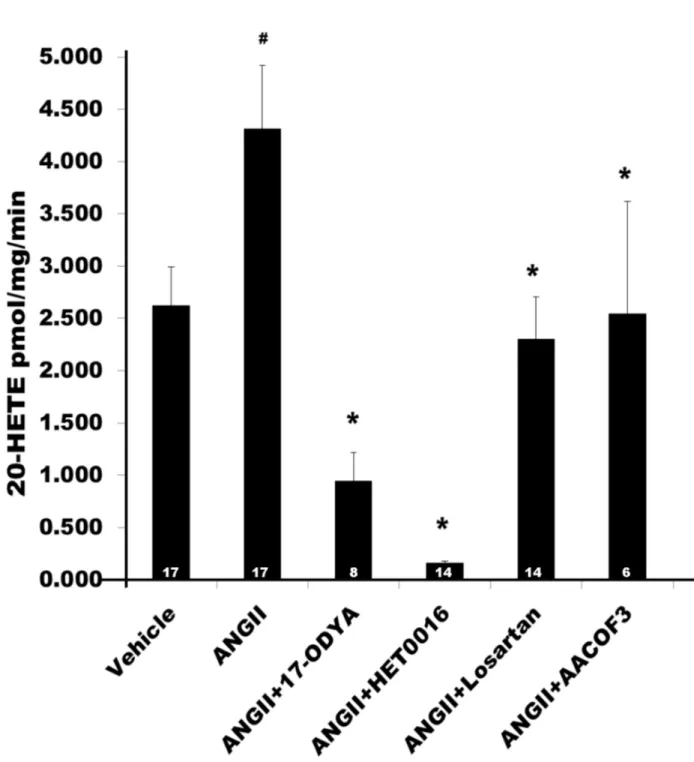
A summary of the effects of ANG II (10-7 M) on the production of 20-HETE in renal microvessels is presented in Figure 4. Treatment of renal microvessels with ANG II significantly increased the production of 20-HETE from 2.6 ± 0.4 to 4.3 ± 0.6 pmol/mg/min (n = 17, P < 0.05). It had no significant effect on the formation of EETs and other HETEs (Table 1). 17-ODYA is an inhibitor of the formation of both EETs and 20-HETE [1,26,27], while HET0016 is a more specific inhibitor of the formation of 20-HETE [28]. The stimulatory effect of ANG II on the production of 20-HETE was completely blocked by 17-ODYA (10-5 M), HET0016 (10-8 M), the PLA2 inhibitor, AACOF3 (2X10⁻⁵ M) and the AT1 receptor antagonist, Losartan (10-6 M). In contrast, blockade of the AT
2 receptor with PD123319 (10-7 M, n=8) had no effect on ANG II-stimulated 20-HETE production (data not shown). These results indicate that treatment of renal microvessels with ANG II selectively increases the formation of 20-HETE in renal microvessels and the stimulatory effect of ANG II is mediated by activation of the AT1 receptor and a PLA2- dependent pathway.
Role of 20-HETE in the vasoconstrictor response to ANG II in rat renal microvessels
The contribution of 20-HETE to the vasoconstrictor response to ANG II was determined by comparing the response of renal interlobular arterioles to ANG II under control conditions and after blocking the formation of 20-HETE with 17-ODYA (10-5 M). In order to eliminate the influence of vasodilator mediators from the endothelium, the vessels were denuded by perfusion with anti-factor VIII antibody and complement as previously described [16] and the efficiency of the removal of the endothelium was confirmed by measuring the expression of message for the endothelium specific marker-von-Willebrand factor (vWf) [21,22]. The effectiveness of removal of the endothelium was also confirmed by measuring the response of the vessels to acetylcholine (Ach, 10-6 M). The control diameter of the vessels were 130 ± 7 μm. Addition of phenylephrine (1 μM) reduced the inner diameter by 50 % to 66 ± 5 μm. and Ach (10-6 M) had no significant effect on the diameter of these denuded renal arterioles (67 ± 5 μm, n=6).
The effects of 17-ODYA on the vasoconstrictor response to ANG II in the denuded renal microvessels are summarized in Figure 5. ANG II decreased the inner diameter of the denuded renal interlobular arterioles in a concentration dependent manner by 41 ± 5 %. After addition of the 20-HETE inhibitor 17-ODYA (10-5 M), the EC50 for ANG II was not significantly altered but the maximal vasoconstrictor response was reduced by 50%.
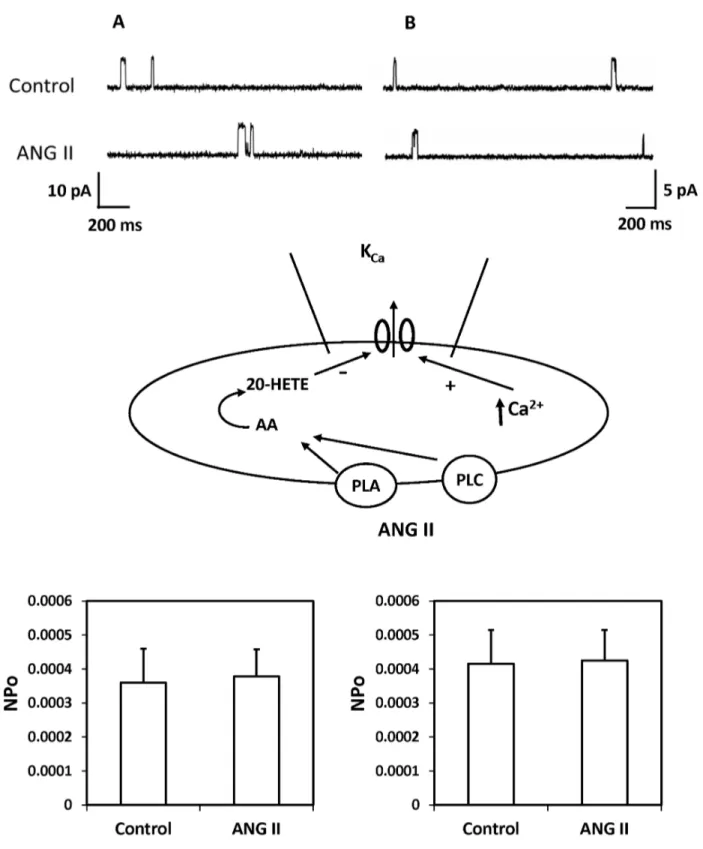
Effect of ANG II on KCa channel activity in renal VSM
cells
A representative recording depicting the effects of ANG II on single-channel K+ currents recorded using cell-attached patches on freshly isolated renal arteriolar VSM cells is presented in Figure 6. The single KCa channel currents were recorded with the cells bathed in a high K+ solution to null membrane potential at pipette potential of –40 mV in panel A or in normal PSS with 140 mM KCl in the pipette at the resting membrane potential (pipette potential = 0 mV) in panel B. ANG II (10-7 M) did not alter the NPo of K
Ca channels under either of these conditions. ANG II also had no effect on KCa single channel amplitude which averaged 9.6 ± 0.1 before and 9.5 ± 0.2 pA after addition of ANG II to the bath.
Effect of ANG II on [Ca2+]
i of renal VSM cells The effects of ANG II on [Ca2+]
i was examined in renal VSM cells bathed in normal PSS in the presence and absence of the calcium ionophore, ionomycin (10-6 M). The results of these experiments are presented in Figure 7. Baseline [Ca2+]
i of VSM cells averaged 71 ± 3 nM. ANG II (10-7 M) increased [Ca2+]
i to a peak value of 425 ± 21 nM that returned to an elevated level of 194 ± 7 nM. 100-200 msec after administration of ANG II. In another group of cells, addition of ionomycin produced a large sustained increase of [Ca2+]
i from 90 ± 9 nM to 728 ± 53 nM. In the presence of ionomycin, ANG II had no effect on [Ca2+]
i.
Effect of ANG II on KCa channel activity response in
renal VSM cells in the presence of ionomycin
The effects of ANG II on KCa channel activity in renal arteriolar VSM cells bathed in normal PSS recorded in the cell attached mode at resting membrane potential (0 mv pipette potential) is presented in Figure 8. The baseline activity of this channel was very low (NPo, 0.0004 ± 0.0001). Raising [Ca2+] i with ionomycin (10-6 M) increased channel activity 10-fold. But, with the addition of ANG II (10-7 M) to the bath in the presence of ionomycin reduced KCa channel activity by 93 ± 3%.
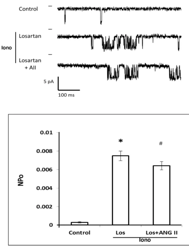
We next determined which ANG II receptor contributes to the inhibitory action of ANG II on KCa channel activity. The results of these experiments are presented in Figure 9. The addition of ionomycin produced a 10-fold increase in KCa channel activity. In the presence of the AT1 receptor antagonist, Losartan, ANG II had no effect on KCa channel activity.
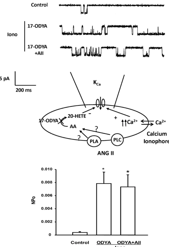
The effect of ANG II on KCa channel activity was also studied before and after blockade of 20-HETE formation with 17-ODYA. The representative traces are shown in the upper panel of Figure 10. Addition of 17-ODYA (10-5 M) and ionomycin (10-6 M) to the bath solution significantly increased the NPo of the KCa channel by 20-fold. Subsequent addition of ANG II had no significant effect on KCa channel activity after the synthesis of 20-HETE in renal VSM was blocked by 17-ODYA.
Figure 3. Production of 20-HETE in renal microvessels measured by LC/MS/MS. Panel A presents a typical chromatograph indicating the retention times of a mixture of 20 ng aliquots of various CYP450 metabolites of AA. Panel B presents a typical chromatograph of the metabolites of AA produced by control incubation of renal microvessels (0.68 mg protein). Panel C presents a typical chromatograph of the metabolites of AA produced when an aliquot of these same vessels were incubated with ANG II (0.66 mg protein).
Figure 4. Effect of ANG II on the production of 20-HETE in renal microvessels. Comparison of the production of 20-HETE in renal microvessels treated with vehicle, ANG II and ANG II plus inhibitors of the synthesis of 20-HETE, 17-ODYA (10-5 M) and HET0016 (10-6 M), a phospholipase A
2 inhibitor, AACOF3 (2 X 10⁻⁵ M), a phospholipase C inhibitor, of U-73122 (10-5 M), and the AT1 receptor blocker, Losartan (10-6 M). Numbers in the bars indicate the number of vessel preparations studied. # indicates a significant difference from the corresponding value in vessels treated with vehicle. * indicates a significant difference from the corresponding value in vessels treated with ANG II (10-7 M).
activity, single KCa channel currents were recorded from VSM cells before and after addition of ionomycin (10-6 M) and the selective PLC inhibitor, U-73122 (10-5 M) to the bath and then after addition of ANG II (10-7 M). Addition of ionomycin and U-73122 significantly increased the activity of KCa channel by 6-fold. In the presence of the ionomycin and U-73122, ANG II reduced the NPo of the KCa channel by 62% (Figure 11, left panel).
Effects of PLA2 on the inhibitory effect of ANG II on KCa
channel activity
We also examined the role of PLA2 on ANG II-induced inhibitory effect on KCa channel currents in VSM cells. The KCa channel currents were recorded before and after addition of ionomycin (10-6 M) and the selective PLA
2 inhibitor, AACOF3 (2 X 10⁻⁵ M) to the bath followed by ANG II (10-7 M). The results are presented in the right panel of Figure 11. Addition of AACOF3 and ionomycin to the bath increased KCa channel activity 4-fold. Under these conditions, addition of ANG II did not significantly change the NPo of the KCa channel.
Effect of 17-ODYA on the effects of ANG II on [Ca2+] i The effect of blockade of the synthesis of 20-HETE with 17-ODYA on the effects of ANG II on [Ca2+]
i in freshly isolated renal arteriolar VSM cells is presented in Figure 12. ANG II (10-7 M) induced a rapid increase in [Ca2+]
i from 58 ± 7 to 452 ± 21 nM that was followed by a recovery to a steady-state level of 195 ± 6 nM. After administration of 17-ODYA (10-5 M), the peak response to ANG II was similar to that seen in control cells (from 65 ± 6 to 459 ± 31 nM), however, the steady-state response to ANG II was markedly reduced to (76 ± 5 nM).
Table 1. Summary of the production of various metabolites of arachidonic acid by renal microvessels under control conditions and after addition of ANG II (10-7 M).
Vehicle pmol/mg/min ANG II pmol/mg/min
5-HETE 1.43 ± 0.50 1.85 ± 0.33
12-HETE 2.30 ± 0.54 2.48 ± 0.60
15-HETE 1.48 ± 0.42 2.22 ± 0.38
14,15-DiHETE 1.07 ± 0.24 1.27 ± 0.19
11,12-DiHETE 2.26 ± 0.48 3.06 ± 0.47
8,9-DiHETE 1.20 ± 0.22 1.56 ± 0.25
5,6-DiHETE 0.02 ± 0.01 0.25 ± 0.13
14,15-EET 0.25 ± 0.10 0.47 ± 0.09
11,12-EET 0.23 ± 0.09 0.59 ± 0.19
8,9-EET 0.42 ± 0.22 0.71 ± 0.17
5,6-EET 0.30 ± 0.12 0.42 ± 0.09
The production of various metabolites of arachidonic acid by renal microvessels (N = 12-22 preparations) are presented. There was no significant difference in the production of any of the metabolites in the vehicle and ANG II treated vessels. doi: 10.1371/journal.pone.0082482.t001
Discussion
Previous studies have indicated that ANG II increases the release of AA in VSM cells by activation of PLA2 and/or PLC [4-6]. AA is a substrate for the formation of 20-HETE in renal arterioles and 20-HETE has been reported to constrict renal and cerebral arteries through depolarization of VSM cells by blocking the large conductance KCa channel [7,30,31]. However, the role of 20-HETE in modulating its vasoconstrictor actions of ANG II by affecting KCa channel activity remains to be determined. Therefore, the present study examined the role of 20-HETE in mediating the inhibitory effects of ANG II on KCa channel activity in rat renal VSM cells.
The effects of ANG II on the production of 20-HETE in renal microvessels were first studied utilizing LC/MS. The results indicate that ANG II selectively increases 20-HETE production in renal microvessels but it has little effect on the production of EETs and other metabolites of AA. This effect was abolished by the AT1 receptor blocker Losartan, whereas administration of the AT2 receptor antagonist had no effect on the ability of ANG II to stimulate the production of 20-HETE. In the presence of calcium ionophore to fix intracellular calcium concentration at a high level, ANG II reduced KCa channel activity in freshly isolated rat renal VSM cells. The inhibitory effect of ANG II was blocked by administration of 17-ODYA which is a specific inhibitor of the production of 20-HETE in renal VSM cells. These results are consistent with previous reports that 20-HETE has a direct effect to inhibit the KCa channel activity in renal VSM cells even though it increases intracellular calcium levels which normally would activate these channels [7,32].
The present results indicating that ANG II stimulates production of 20-HETE in renal microvessels is also consistent with previous findings in our lab [17,31] and others [4,5]. In this regard, Croft et al (2000) reported that the effect of ANG II on the endogenous production of 20-HETE in renal microvessels was mediated by the AT2 receptor and could be blocked by PD-123319 at a concentration of 10-4 M, but not by AT
1 receptor antagonist Losartan (10-4 M). In contrast, the results of the present study indicate that a much lower dose of losartan (10-6 M) was effective in blocking the effects of ANG II on the production of 20-HETE, whereas PD-123319 (10-7 M) had no effect. The difference in the results may be due to the different concentrations of the AT1 and AT2 receptor blockers used in the two studies. High concentration of PD-123319 (10-4 M) have been reported to block both AT1 and AT2 receptors [33]. Another difference is that we studied the ability of ANG II to stimulate the production of 20-HETE following addition of a saturating concentration of the substrate AA to the bath to eliminate the potential influence of release of preformed 20-HETE, whereas the studies by Croft et al [4] were done in the absence of substrate and focused on the formation and release of 20-HETE from endogenous phospholipid pools.
20-HETE by > 95%. These results indicate that ANG II increases the formation of 20-HETE in renal vascular smooth muscle by stimulating synthesis from AA rather than releasing preformed 20-HETE from phospholipid pools.
We also examined the effect of inhibitors of PLC and PLA2 on the ability of ANG II to increase the formation of 20-HETE in renal microvessels. Both inhibitors of PLC and PLA2 reduced 20-HETE levels in response to ANG II, however the PLA2 inhibitor had a greater effect than the PLC inhibitor. Since the vessels were incubated in the presence of a saturating concentration of the substrate AA, inhibition of the release of
AA from membrane phospholipid pools does not account for these findings. Rather, we suggest that 20-HETE, once formed, is likely rapidly reincorporated into phospholipid pools in renal VSM and ANG II and sustained activation of PLC and PLA2 promotes the release of 20-HETE from these pools essentially opposing the reuptake process. According to this view, the PLA2 and PLC inhibitors likely reduced free 20-HETE levels in the vessels by promoting reuptake into membrane phospholipid pools. .
The functional significance of the effects of 20-HETE on the inhibitory action of ANG II on the KCa channel to the Figure 5. Effects of 20-HETE on vasoconstrictor responses to ANG II in renal microvessels. Concentration response curves for the vasoconstrictor response to ANG II (10-11 to 10-6 M) in pressurized renal microvessels with the endothelium removed (N=5) are presented before and after addition of 17-ODYA (10-5 M). 17-ODYA an inhibitor of the formation of 20-HETE had no significant effect on the EC50 but it reduced the maximal response to ANG II by 50%. # indicates a significant difference from the value at the lowest concentration of ANG II concentration (10-11 M). * indicates a significant difference from the corresponding value prior to treatment of 17-ODYA (10-5 M).
Figure 6. Effect of ANG II on the KCa channel activity in renal VSM cells. The upper panel presents a representative current recording of the effects of ANG II on of the KCa channel activity recorded using a cell-attached patches of VSM cells freshly isolated from renal microvessels at a pipette potential -40 mV in high K+ solution with 140 mM KCl in the pipette to null membrane potential (panel A) or in normal PSS with 140 mM KCL in the pipette at the resting membrane potential (pipette potential = 0 mV, panel B). The middle panel summarizes the cell-attached patch clamp recording mode. The lower panel presents a summary of the effects of ANG II on the activity of KCa channel recorded in depolarized cells (panel A) in a high K+ solution and in cells bathed in PSS at a normal depolarized membrane potential (panel B). Recordings were obtained from 6-8 cells under each condition.
Figure 7. Effect of ANG II on intracellular calcium response in renal VSM cells. The upper panel presents a representative tracing of a [Ca2+]
i response of a single renal microvessel VSM cell to ANG II (10-7 M) bathed in PSS before and after addition of ionomycin (10-6 M). ANG II elicited a rise in [Ca2+]
i in renal VSM cells under control conditions. Addition of ionomycin raised baseline [Ca2+]
i and under these conditions ANG II had no additional effect on [ Ca2+]i. The lower left panel summarizes the effects of on peak and plateau [Ca2+]
i responses to ANG II (10-7 M) in the renal VSM cells. The right panel presents the [Ca2+]i response to ANG II after addition of ionomycin. * indicates a significant rise in [Ca2+]
i over baseline. Mean values + SE recorded from 45-70 cells (5-10 cells per experiment) isolated from 9 different renal VSM cell isolation are presented.
Figure 8. Effect of ANG II on KCa channel activity in renal VSM cells with high [Ca2+]i. The upper panel presents a representative current recording of the effects of ANG II on KCa channel activity recorded using cell-attached patches on renal arteriolar VSM cells. The middle panel summarizes the patch clamp recording mode. KCa channel currents were recorded with 140 mM KCl in the pipette at the resting membrane potential (pipette potential = 0 mV) and the cells were bathed in normal physiological salt solution in the presence of ionomycin. The lower panel summarizes the effects of ionomycin and subsequent addition of ANG II on the KCa channel activity. * indicates the significant difference from the control value. # indicates a significant difference from the corresponding value recorded in the presence of ionomycin.
Figure 9. Effect of Losartan on the inhibitory action of ANG II on KCa channel activity in renal VSM cells. The upper panel presents a representative current traces recording of the effects of Losartan and ANG II on the KCa channel activity recorded in the presence of ionomycin. KCa channel currents were recorded with 140 mM KCl in the pipette at the resting membrane potential (pipette potential = 0 mV) and the cells were bathed in normal physiological salt solution in the presence of ionomycin. The lower panel summarizes the effects of Losartan and ANG II on KCa channel activity recorded in the cell attached mode in the presence of ionomycin in the bath. * indicates a significant difference from the control value recorded from the same cells. # indicates a significant difference from the corresponding value recorded in the presence of ionomycin.
Figure 10. Effect of 17-ODYA on the inhibitory action of ANG II on KCa channel activity in renal VSM cells. Upper panel presents a representative tracing KCa channel activity in the cell-attached mode before and after addition of 17-ODYA (10-7 M) and 17-ODYA plus ANG II to the bath. KCa channel currents were recorded with 140 mM KCl in the pipette at the resting membrane potential (pipette potential = 0 mV) and the cells were bathed in normal physiological salt solution in the presence of ionomycin. The middle panel summarizes the patch clamp recording mode. Lower panel summarizes the effects of 17-ODYA and 17-ODYA and ANG II on the open probability of the KCa channel in renal VSM cells (n = 4 cells from 4 rats). * indicates a significant difference versus the corresponding control value.
Figure 11. Effect of blockade of PLC and PLA2 on the inhibitory action of ANG II on KCa channel activity in renal VSM cells. The upper panel presents representative tracings of single KCa channel currents recorded at the resting membrane potential (pipette potential = 0 mV) in the cell-attached mode before and after addition of PLC inhibitor U-73122 (10-5 M) and U-73122 plus ANG II (10-7 M) to the bath (left). The effects of the PLA
2 inhibitor, AACOF3 (2 x 10⁻⁵ M) and AACOF3 plus ANGII (10-7 M) are presented in the right panel. KCa channel currents were recorded with 140 mM KCl in the pipette at the resting membrane potential (pipette potential = 0 mV) and the cells were bathed in normal physiological salt solution in the presence of ionomycin. The middle panel summarizes the patch clamp recording mode. KCa channel currents were recorded as described in Figure 8. The lower left panel summarizes the effect of U-73122 and ANG II on the open probability of the KCa channel in renal VSM cells while the lower right panel depicts the effects of AACOF3 on the response to ANGII. * indicates a significant difference versus control and # indicates a significant difference from the corresponding value recorded after PLC activity was inhibited with U-73122 or PLA2 activity was inhibited with AACOF3.
Figure 12. Effects of ANG II on [Ca2+]i in renal VSM cells before and after 17-ODYA. The upper panel presents representative tracings of the effect of ANG II on [Ca2+]
i in renal VSM before and after blockade of the synthesis of 20-HETE with 17-ODYA (10-5 M). The lower panel presents the peak and steady state [Ca2+]
i responses to ANG II measured before and after addition of 17-ODYA to the bath. Mean values + SE recorded from 25-30 cells (5-8 cells per experiment) isolated from 6 different renal VSM isolations are presented. * indicates a significant difference between ANG II and ANG II plus 17-ODYA treated vessels.
vasoconstrictor response to ANG II was studied in pressurized renal interlobular arterioles. The endothelium was removed to reduce the modulation of the vasoconstrictor response by release of NO, prostaglandins and EETs secondary to stimulation of the AT2 receptor on the endothelium [34-36]. ANG II dose-dependently decreased the diameter of renal interlobular arteries by 40%, however, in the presence of the 17-ODYA, the maximum vasoconstrictor response to ANG II was reduced by 50%. These results indicating that 20-HETE potentiates the vasoconstrictor response to ANG II in renal interlobular arteries are consistent with previous findings of Alonso-Galicia et al (2002), who reported that administration of a 20-HETE inhibitor attenuated the acute pressor response to ANG II in rats in vivo by about 50% and chronic blockade attenuated the development of ANG II induced hypertension [1].
Patch clamp studies were performed to explore the mechanism by which 20-HETE potentiates the renal vasoconstrictor response to ANG II. The results of these experiments confirmed previous findings of Inscho et al and Fellner and Arendshorst that ANG II produces a large transient increase [Ca2+]
i in renal VSM cells [37,38]. However, ANG II had no significant effect on the activity of the KCa channel in renal arteriolar VSM recorded in the cell attached mode using a pipette potential of -40 mv with the cells bathed in a high K+ solution to null the membrane potential or in VSM cells bathed in PSS solution at a physiological resting membrane potential (pipette potential = 0 mV). This finding is surprising since ANG II increased [Ca2+]
i which should increase KCa channel activity in renal VSM [39] unless some other factor intervened to oppose this effect. This finding raised the possibility that ANG II may block the activation of KCa channel in VSM that normally would be expected to attenuate the vasoconstrictor response to ANG II by hyperpolarizing the cell and blocking subsequent calcium entry via voltage gated calcium channels.
To address this hypothesis, renal VSM cells were treated with the calcium ionomycin to clamp [Ca]i at high levels to activate the KCa channels prior to administration of ANG II. Ionomycin raised [Ca2+]
i and markedly increased KCa channel activity. Subsequent administration of ANG II reduced the
activity of the KCa channel by >90%. Blockade of the formation of 20-HETE with 17-ODYA, blockade of the AT1 receptor with Losartan or administration of inhibitors of PLC and PLA2 opposed the inhibitory effects of ANG II on the KCa channel. In other experiments, we found that pretreatment of renal VSM cells with 17-ODYA had no effect on the peak [Ca2+]
i response to ANG II but it reduced the sustained elevation in [Ca2+]
i. This result is consistent with previous reports that the sustained [Ca2+]
i response to ANG II is dependent of membrane depolarization and calcium entry via voltage-sensitive calcium channels [7,32].
In summary, the results of the present study indicates that ANG II constricts renal VSM cells by binding to AT1 receptors expressed on the VSM cells which activates of PLA2 to release AA from membrane phospholipid pools. AA is then converted into 20-HETE by CYP450 enzymes of the 4A family in VSM cells. 20-HETE then acts to inhibit the KCa channels, which in turn hyperpolarizes the VSM cells and opposes calcium entry through voltage gated calcium channels. The exact mechanism by which 20-HETE inhibits opening of KCa channels remains to be determined. However, previous studies have suggested that that it most likely involves activation of PKC [19,40,41], tyrosine kinase [19,42] and/or the Rho kinase [43] pathways.
Acknowledgements
We thank Dr. Rodney Baker and Mrs. Chris Purser in HPLC/ Mass Spectrometry Analytical Core in the department of Pharmacology and Toxicology at the University of Mississippi Medical Center for their assistance in analyzing the AA metabolites.
Author Contributions
Conceived and designed the experiments: RJR JRF. Performed the experiments: FF CWS KGM JMW MRP. Analyzed the data: FF CWS JMW MRP KGM RJR. Contributed reagents/materials/analysis tools: RJR. Wrote the manuscript: FF CWS KGM RJR JZ SPD.
References
1. Alonso-Galicia M, Maier KG, Greene AS, Cowley AW Jr., Roman RJ (2002) Role of 20-hydroxyeicosatetraenoic acid in the renal and vasoconstrictor actions of angiotensin II. Am J Physiol Regul Integr Comp Physiol 283: R60-R68. PubMed: 12069931.
2. Kohagura K, Arima S, Endo Y, Chiba Y, Ito O et al. (2001) Involvement of cytochrome P450 metabolites in the vascular action of angiotensin II on the afferent arterioles. Hypertens Res 24: 551-557. doi:10.1291/ hypres.24.551. PubMed: 11675950.
3. Imig JD, Zou AP, Ortiz de Montellano PR, Sui Z, Roman RJ (1994) Cytochrome P-450 inhibitors alter afferent arteriolar responses to elevations in pressure. Am J Physiol 266: H1879-H1885. PubMed: 8203587.
4. Croft KD, McGiff JC, Sanchez-Mendoza A, Carroll MA (2000) Angiotensin II releases 20-HETE from rat renal microvessels. Am J Physiol Renal Physiol 279: F544-F551. PubMed: 10966934.
5. Chu ZM, Croft KD, Kingsbury DA, Falck JR, Reddy KM et al. (2000) Cytochrome P450 metabolites of arachidonic acid may be important mediators in angiotensin II-induced vasoconstriction in the rat mesentery in vivo. Clin Sci (Lond) 98: 277-282. doi:10.1042/ CS19990217. PubMed: 10677385.
6. Parmentier JH, Muthalif MM, Nishimoto AT, Malik KU (2001) 20-Hydroxyeicosatetraenoic acid mediates angiotensin ii-induced phospholipase d activation in vascular smooth muscle cells. Hypertension 37: 623-629. doi:10.1161/01.HYP.37.2.623. PubMed: 11230346.
7. Roman RJ (2002) P-450 metabolites of arachidonic acid in the control of cardiovascular function. Physiol Rev 82: 131-185. PubMed: 11773611.
8. Imig JD, Deichmann PC (1997) Afferent arteriolar responses to ANG II involve activation of PLA2 and modulation by lipoxygenase and P-450 pathways. Am J Physiol 273: F274-F282. PubMed: 9277588. 9. Dunn KM, Renic M, Flasch AK, Harder DR, Falck J et al. (2008)
Elevated production of 20-HETE in the cerebral vasculature contributes to severity of ischemic stroke and oxidative stress in spontaneously hypertensive rats. Am J Physiol Heart Circ Physiol 295: H2455-H2465. doi:10.1152/ajpheart.00512.2008. PubMed: 18952718.
10. Harder DR, Narayanan J, Birks EK, Liard JF, Imig JD et al. (1996) Identification of a putative microvascular oxygen sensor. Circ Res 79: 54-61. doi:10.1161/01.RES.79.1.54. PubMed: 8925569.
distribution in the living tissue. Kidney Int 51: 372-380. doi:10.1038/ki. 1997.49. PubMed: 9027709.
12. Ito O, Roman RJ (1999) Regulation of P-450 4A activity in the glomerulus of the rat. Am J Physiol 276: R1749-R1757. PubMed: 10362756.
13. Imig JD, Zou AP, Stec DE, Harder DR, Falck JR et al. (1996) Formation and actions of 20-hydroxyeicosatetraenoic acid in rat renal arterioles. Am J Physiol 270: R217-R227. PubMed: 8769805.
14. Guo DF, Inagami T (1994) The genomic organization of the rat angiotensin II receptor AT1B. Biochim Biophys Acta 1218: 91-94. doi: 10.1016/0167-4781(94)90105-8. PubMed: 8193170.
15. Koike G, Horiuchi M, Yamada T, Szpirer C, Jacob HJ et al. (1994) Human type 2 angiotensin II receptor gene: cloned, mapped to the X chromosome, and its mRNA is expressed in the human lung. Biochem Biophys Res Commun 203: 1842-1850. doi:10.1006/bbrc.1994.2402. PubMed: 7945336.
16. Juncos LA, Ito S, Carretero OA, Garvin JL (1994) Removal of endothelium-dependent relaxation by antibody and complement in afferent arterioles. Hypertension 23: I54-I59. doi:10.1161/01.HYP. 23.1_Suppl.I54. PubMed: 8282376.
17. Ma YH, Gebremedhin D, Schwartzman ML, Falck JR, Clark JE et al. (1993) 20-Hydroxyeicosatetraenoic acid is an endogenous vasoconstrictor of canine renal arcuate arteries. Circ Res 72: 126-136. doi:10.1161/01.RES.72.1.126. PubMed: 8417836.
18. Alonso-Galicia M, Hudetz AG, Shen H, Harder DR, Roman RJ (1999) Contribution of 20-HETE to vasodilator actions of nitric oxide in the cerebral microcirculation. Stroke 30: 2727-2734; discussion: 10.1161/01.STR.30.12.2727. PubMed: 10583004.
19. Sun CW, Falck JR, Harder DR, Roman RJ (1999) Role of tyrosine kinase and PKC in the vasoconstrictor response to 20-HETE in renal arterioles. Hypertension 33: 414-418. doi:10.1161/01.HYP.33.1.414. PubMed: 9931139.
20. Alonso-Galicia M, Falck JR, Reddy KM, Roman RJ (1999) 20-HETE agonists and antagonists in the renal circulation. Am J Physiol 277: F790-F796. PubMed: 10564244.
21. Zanetta L, Marcus SG, Vasile J, Dobryansky M, Cohen H et al. (2000) Expression of Von Willebrand factor, an endothelial cell marker, is up-regulated by angiogenesis factors: a potential method for objective assessment of tumor angiogenesis. Int J Cancer 85: 281-288. doi: 10.1002/(SICI)1097-0215(20000115)85:2. PubMed: 10629090. 22. Horvath B, Hegedus D, Szapary L, Marton Z, Alexy T et al. (2004)
Measurement of von Willebrand factor as the marker of endothelial dysfunction in vascular diseases. Exp Clin Cardiol 9: 31-34. PubMed: 19641694.
23. Akishita M, Ito M, Lehtonen JY, Daviet L, Dzau VJ et al. (1999) Expression of the AT2 receptor developmentally programs extracellular signal-regulated kinase activity and influences fetal vascular growth. J Clin Invest 103: 63-71. doi:10.1172/JCI5182. PubMed: 9884335. 24. Kim S, Iwao H (2000) Molecular and cellular mechanisms of
angiotensin II-mediated cardiovascular and renal diseases. Pharmacol Rev 52: 11-34. PubMed: 10699153.
25. Mifune M, Sasamura H, Shimizu-Hirota R, Miyazaki H, Saruta T (2000) Angiotensin II type 2 receptors stimulate collagen synthesis in cultured vascular smooth muscle cells. Hypertension 36: 845-850. doi: 10.1161/01.HYP.36.5.845. PubMed: 11082154.
26. Zou AP, Ma YH, Sui ZH, Ortiz de Montellano PR, Clark JE et al. (1994) Effects of 17-octadecynoic acid, a suicide-substrate inhibitor of cytochrome P450 fatty acid omega-hydroxylase, on renal function in rats. J Pharmacol Exp Ther 268: 474-481. PubMed: 8301590. 27. Ortiz de Montellano PR, Reich NO (1984) Specific inactivation of
hepatic fatty acid hydroxylases by acetylenic fatty acids. J Biol Chem 259: 4136-4141. PubMed: 6706995.
28. Miyata N, Taniguchi K, Seki T, Ishimoto T, Sato-Watanabe M et al. (2001) HET0016, a potent and selective inhibitor of 20-HETE synthesizing enzyme. Br J Pharmacol 133: 325-329. doi:10.1038/sj.bjp. 0704101. PubMed: 11375247.
29. Sadoshima J (1998) Versatility of the angiotensin II type 1 receptor. Circ Res 82: 1352-1355. doi:10.1161/01.RES.82.12.1352. PubMed: 9648733.
30. Roman RJ, Alonso-Galicia M (1999) P-450 Eicosanoids: A Novel Signaling Pathway Regulating. Renal Function - News Physiol Sci 14: 238-242.
31. Zou AP, Fleming JT, Falck JR, Jacobs ER, Gebremedhin D et al. (1996) 20-HETE is an endogenous inhibitor of the large-conductance Ca(2+)-activated K+ channel in renal arterioles. Am J Physiol 270: R228-R237. PubMed: 8769806.
32. Kaley G (2000) Regulation of vascular tone: role of 20-HETE in the modulation of myogenic reactivity. Circ Res 87: 4-5. doi: 10.1161/01.RES.87.1.4. PubMed: 10884363.
33. Boulay G, Servant G, Luong TT, Escher E, Guillemette G (1992) Modulation of angiotensin II binding affinity by allosteric interaction of polyvinyl sulfate with an intracellular domain of the DuP-753-sensitive angiotensin II receptor of bovine adrenal glomerulosa. Mol Pharmacol 41: 809-815. PubMed: 1569928.
34. Imig JD (2013) Epoxyeicosatrienoic acids, 20-hydroxyeicosatetraenoic acid, and renal microvascular function. Prostaglandins Other Lipid Mediat, 104-105: 2–7. PubMed: 23333581.
35. Roman RJ, Maier KG, Sun CW, Harder DR, Alonso-Galicia M (2000) Renal and cardiovascular actions of 20-hydroxyeicosatetraenoic acid and epoxyeicosatrienoic acids. Clin Exp Pharmacol Physiol 27: 855-865. doi:10.1046/j.1440-1681.2000.03349.x. PubMed: 11071299. 36. Harder DR, Lange AR, Gebremedhin D, Birks EK, Roman RJ (1997)
Cytochrome P450 metabolites of arachidonic acid as intracellular signaling molecules in vascular tissue. J Vasc Res 34: 237-243. doi: 10.1159/000159228. PubMed: 9226306.
37. Fellner SK, Arendshorst WJ (2005) Angiotensin II Ca2+ signaling in rat afferent arterioles: stimulation of cyclic ADP ribose and IP3 pathways. Am J Physiol Renal Physiol 288: F785-F791. PubMed: 15598842. 38. Inscho EW, Imig JD, Cook AK (1997) Afferent and efferent arteriolar
vasoconstriction to angiotensin II and norepinephrine involves release of Ca2+ from intracellular stores. Hypertension 29: 222-227. doi: 10.1161/01.HYP.29.1.222. PubMed: 9039106.
39. Jackson WF (2000) Ion channels and vascular tone. Hypertension 35: 173-178. doi:10.1161/01.HYP.35.1.173. PubMed: 10642294. 40. Nowicki S, Chen SL, Aizman O, Cheng XJ, Li D et al. (1997)
20-Hydroxyeicosa-tetraenoic acid (20 HETE) activates protein kinase C. Role in regulation of rat renal Na+,K+-ATPase. J Clin Invest 99: 1224-1230. doi:10.1172/JCI119279. PubMed: 9077530.
41. Amlal H, LeGoff C, Vernimmen C, Soleimani M, Paillard M et al. (1998) ANG II controls Na(+)-K+(NH4+)-2Cl- cotransport via 20-HETE and PKC in medullary thick ascending limb. Am J Physiol 274: C1047-C1056. PubMed: 9575802.
42. Cheng J, Wu CC, Gotlinger KH, Zhang F, Falck JR et al. (2010) 20-hydroxy-5,8,11,14-eicosatetraenoic acid mediates endothelial dysfunction via IkappaB kinase-dependent endothelial nitric-oxide synthase uncoupling. J Pharmacol Exp Ther 332: 57-65. doi:10.1124/ jpet.109.159863. PubMed: 19841472.
![Figure 7. Effect of ANG II on intracellular calcium response in renal VSM cells. The upper panel presents a representative tracing of a [Ca 2+ ] i response of a single renal microvessel VSM cell to ANG II (10 -7 M) bathed in PSS before and after additi](https://thumb-eu.123doks.com/thumbv2/123dok_br/18194590.332727/13.918.105.814.116.928/figure-effect-intracellular-response-presents-representative-response-microvessel.webp)
![Figure 8. Effect of ANG II on K Ca channel activity in renal VSM cells with high [Ca 2+ ] i](https://thumb-eu.123doks.com/thumbv2/123dok_br/18194590.332727/14.918.177.749.113.943/figure-effect-ang-channel-activity-renal-vsm-cells.webp)