RESEARCH ARTICLE
Activation of
Salmonella
Typhi-Specific
Regulatory T Cells in Typhoid Disease in a
Wild-Type
S
. Typhi Challenge Model
Monica A. McArthur1, Stephanie Fresnay1, Laurence S. Magder2, Thomas C. Darton3, Claire Jones3, Claire S. Waddington3, Christoph J. Blohmke3, Gordon Dougan4, Brian Angus5, Myron M. Levine1, Andrew J. Pollard3, Marcelo B. Sztein1*
1Center for Vaccine Development, University of Maryland School of Medicine, Baltimore, Maryland, United States of America,2Department of Epidemiology and Public Health, University of Maryland School of Medicine, Baltimore, Maryland, United States of America,3Oxford Vaccine Group, Department of
Paediatrics, University of Oxford and the National Institute for Health Research Oxford Biomedical Research Centre, Oxford, United Kingdom,4Microbial Pathogenesis Group, Wellcome Trust Sanger Institute, Hinxton, United Kingdom,5Nuffield Department of Medicine, University of Oxford, United Kingdom
Abstract
SalmonellaTyphi (S. Typhi), the causative agent of typhoid fever, causes significant morbid-ity and mortalmorbid-ity worldwide. Currently available vaccines are moderately efficacious, and identification of immunological responses associated with protection or disease will facilitate the development of improved vaccines. We investigatedS. Typhi-specific modulation of activation and homing potential of circulating regulatory T cells (Treg) by flow and mass cy-tometry using specimens obtained from a human challenge study. Peripheral blood mono-nuclear cells were obtained from volunteers pre- and at multiple time-points post-challenge with wild-typeS. Typhi. We identified differing patterns ofS. Typhi-specific modulation of the homing potential of circulating Tregbetween volunteers diagnosed with typhoid (TD) and those who were not (No TD). TD volunteers demonstrated up-regulation of the gut homing molecule integrinα4ß7 pre-challenge, followed by a significant down-regulation post-chal-lenge consistent with Treghoming to the gut. Additionally,S. Typhi-specific Tregfrom TD volunteers exhibited up-regulation of activation molecules post-challenge (e.g., HLA-DR, LFA-1). We further demonstrate that depletion of Tregresults in increasedS. Typhi-specific cytokine production by CD8+ TEMin vitro. These results suggest that the tissue distribution of activated Treg, their characteristics and activation status may play a pivotal role in typhoid fever, possibly through suppression ofS. Typhi-specific effector T cell responses. These studies provide important novel insights into the regulation of immune responses that are likely to be critical in protection against typhoid and other enteric infectious diseases.
OPEN ACCESS
Citation:McArthur MA, Fresnay S, Magder LS, Darton TC, Jones C, Waddington CS, et al. (2015) Activation ofSalmonellaTyphi-Specific Regulatory T Cells in Typhoid Disease in a Wild-TypeS. Typhi Challenge Model. PLoS Pathog 11(5): e1004914. doi:10.1371/journal.ppat.1004914
Editor:Denise M. Monack, Stanford University School of Medicine, UNITED STATES
Received:February 4, 2015
Accepted:April 27, 2015
Published:May 22, 2015
Copyright:© 2015 McArthur et al. This is an open access article distributed under the terms of the
Creative Commons Attribution License, which permits unrestricted use, distribution, and reproduction in any medium, provided the original author and source are credited.
Data Availability Statement:All relevant data are within the paper and its Supporting Information files.
Author Summary
In this manuscript, we describe, for the first time, a potential role for regulatory T cells (Treg) as an important factor in determining disease outcome in humans following
expo-sure to wild-typeS. Typhi. We studied in considerable depth the modulation of Treg
activa-tion characteristics and their homing potential in the development of typhoid disease following a wild-typeS. Typhi challenge in a unique human infection model. We show
thatS. Typhi-specific up-regulation of the gut homing molecule integrinα4β7 pre-chal-lenge is associated with subsequent development of typhoid disease. We further demon-strate that increasedS. Typhi-specific expression of molecules associated with Treg
activation as well as distinct kinetics of the expression of key activation molecules involved in Tregfunction are present in volunteers diagnosed with typhoid disease. We also provide
the first evidence that Tregcan functionally suppressS. Typhi-specific CD8+ T cellsin
vitro. These intriguing results suggest that Tregare likely to play a role in the development
of typhoid fever and potentially other enteric infections.
Introduction
Salmonella entericaserovar Typhi (S. Typhi), the causative agent of typhoid fever, is a major
public health threat throughout the developing world. An estimated 26.9 million cases resulting in approximately 217,000 deaths occur annually [1,2]. Furthermore, antibiotic resistance in-creasingly limits treatment options in many areas [3,4]. Current typhoid vaccines licensed in the US provide modest protection and are only moderately immunogenic [5,6]. In order to ef-fectively develop new vaccine candidates that will provide robust, long-lasting protection, an improved understanding of the immune correlates of protection is desirable. The recent re-es-tablishment of the human challenge model with wild-typeS. Typhi provides a unique
opportu-nity to investigate in detail the immune responses following exposure to this pathogen [7]. While multiple studies have investigated cell-mediated immune (CMI) responses againstS.
Typhi immunization and infection [8,9,10,11,12,13,14,15], to date there are no published stud-ies of the potential role of regulatory T cell (Treg) responses against this organism. Tregare a
specialized subset of CD4+ T cells that are responsible for regulating other immune cells [16,17,18]. They are characterized by expression of interleukin (IL)-2 receptorα(CD25) and the transcription factor Forkhead box protein (Fox)P3 [18]. Tregmay be derivedin vivoin the
thymus (tTreg) or the periphery (pTreg) as well as followingin vitroactivation (iTreg) [19]. At
present, there is considerable controversy regarding the expression of specific molecules (e.g., Helios for tTreg) that enable the distinction among these subsets [19]. For simplicity, since we
did not measure expression of the considerable number of molecules required to potentially differentiate among these subsets, in the current studies we refer to circulating Tregas those
which were obtainedex vivofrom the peripheral blood and are likely to represent a
combina-tion of tTregand pTreg. Activated Tregmay traffic to the sites of specific immune responses and
exert their regulatory functions via cytotoxic T-lymphocyte-associated protein 4 (CTLA-4; CD152) competition for co-stimulatory molecules (CD80 and CD86) on antigen presenting cells, consumption of IL-2, and production of suppressive cytokines [17]. Alterations in hom-ing molecules/chemokine receptors expressed by Tregaffect their ability to traffic to the site of
specific immune responses [16,20,21,22,23]. In addition to their roles in autoimmunity and cancer biology, Treghave been shown to play a role in suppression of immune responses against
multiple pathogens, potentially contributing to disease [24,25].
Research [CETR], and a Passano Foundation Clinical Investigator Award (to MAM). SF was funded in part by NIH Fellowship Training Program in Vaccinology T32-AI07524. Fellowship Training Program in Vaccinology. The content is solely the responsibility of the authors and does not necessarily represent the official views of the National Institute of Allergy and Infectious Diseases, the National Institutes of Health, the National Health Service, the National Institute for Health Research (NIHR) or the UK Department of Health. The funders had no role in study design, data collection and analysis, decision to publish, or preparation of the manuscript.
In the present studies we have evaluated the characteristics and kinetics of Treghoming
po-tential and activation, as well as the functional capacity of Tregto suppressS. Typhi-specific T
cell responses following wild-type challenge of healthy adult volunteers. Of importance, we identified distinct homing potential and activation patterns associated with typhoid diagnosis indicating that Tregmay play an important role in the development of typhoid fever. In fact, it
is likely that immune homeostasis between suppressive and inflammatory responses is critical to the prevention of disease. These studies describe for the first time, the role ofS.
Typhi-specif-ic modulation of Treghoming potential and activation characteristics in typhoid disease, a role
which may be broadly applicable to other enteric infections.
Results
Similar levels of circulating Treg
and
ex vivo
Treg
proliferation were
observed in volunteers diagnosed with typhoid (TD) and those who were
not (No TD)
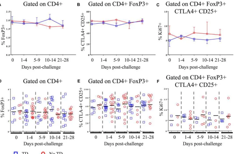
Peripheral blood mononuclear cells (PBMC) from healthy adult volunteers were obtained prior to and at multiple time-points following challenge with ~2 x 10⁴colony forming units (cfu) of wild-typeS. Typhi (S1 Fig) [7]. Volunteers who developed a fever38°C sustained for 12 hours and/or blood culture-confirmedS. Typhi bacteremia were diagnosed with typhoid
as described [7]. In the present study, randomly selected volunteers meeting criteria for typhoid diagnosis (TD, n = 6) and volunteers who did not meet criteria (No TD, n = 6) were assessed for Tregphenotype, activation status, and homing potential. In the randomly selected TD
vol-unteers, the time of typhoid diagnosis ranged from 6–9 days post-challenge. Flow cytometry was used to detect the percentages of circulating CD4+ FoxP3+ Tregas well as the more
strin-gently defined CD4+ FoxP3+ CTLA-4+ CD25+ Tregsubset in unstimulated PBMC. The gating
strategy is shown inS2 Fig. Percentages of FoxP3+ cells ranged from<1% to 3.5% of total
CD4+ T cells, a finding consistent with previous reports [26,27]. CTLA-4+ CD25+ Tregranged
from 23–88% of CD4+ FoxP3+ Treg. There was considerable variation among volunteers in
both groups and we found no significant difference between TD and No TD volunteers in the pre- or post-challenge percentages of circulating Tregas defined by either strategy (Fig 1A, 1B,
1D and 1E). Furthermore, no statistically significant differences in the percentage of circulating Tregover time were noted in either group (Fig 1A, 1B, 1D and 1E). Furthermore, in a subset of
volunteers, we measured Ki67 expressionex vivoas a surrogate of proliferation. While
circulat-ing Tregexpressed Ki67 indicating that a small proportion of them were proliferatingin vivo,
there was no difference in the magnitude or kinetics of Ki67 expression between TD and No TD volunteers (Fig 1C and 1F).
Differential expression of homing molecules in circulating T
regfrom TD
and No TD volunteers
Although no differences were identified between TD and No TD volunteers in the total per-centage of circulating Treg, we hypothesized that differences in theS. Typhi-specific modulation
of the homing potential of Tregmight be associated with typhoid diagnosis. To evaluate this
possibility, PBMC from volunteers challenged with wild-typeS. Typhi (as described above)
were stimulated withS. Typhi-infected autologous Epstein Barr Virus (EBV)-transformed B
lymphoblastoid cell lines (B-LCL) or non-infected B-LCL (negative control). Flow cytometry was utilized to detect expression of the gut homing molecule integrinα4β7, as well as CXCR3 (homing to sites of inflammation) and CCR6 (homing to sites of TH17 inflammation). Relative
(net)S. Typhi-specific modulation of the expression of homing molecules was determined by
subtracting the values obtained following stimulation with non-infected B-LCL from stimula-tion withS. Typhi-infected B-LCL.
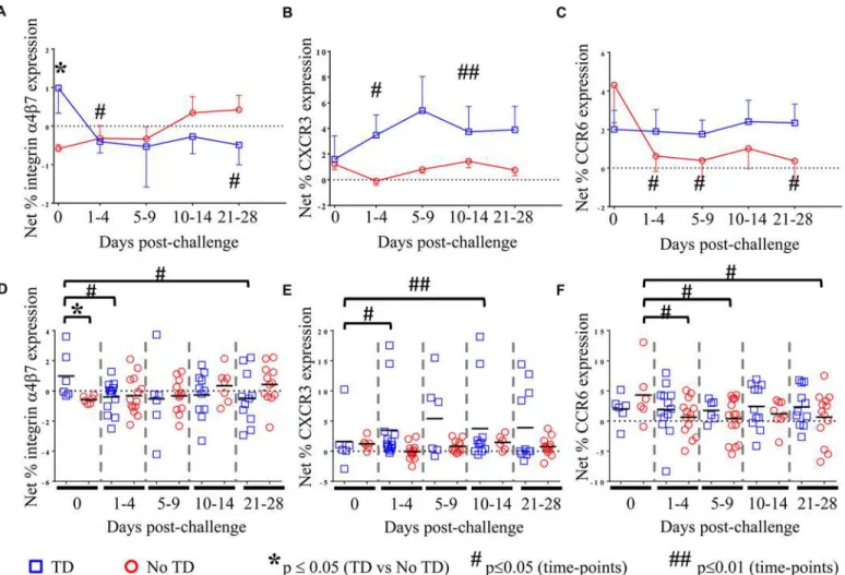
As an enteric pathogen, the gut is the first site of immune encounter withS. Typhi, and
therefore, we conjectured that the ability ofS. Typhi to up-regulate the expression of integrin
α4β7 (a gut homing molecule) on circulating Tregmight be associated with suppression of
pro-tective host responses contributing to disease. We observed that pre-challenge levels ofS.
Typhi-specific modulation of integrinα4β7 expression were indeed higher on circulating Treg
isolated from volunteers who were subsequently diagnosed with typhoid (TD; p = 0.054—
mixed effects regression model (Fig 2A and 2D)). Of note,S. Typhi-specific expression of
integrinα4β7 was significantly down-regulated in TD volunteers in the 1–4 day time-frame after challenge (p = 0.047—mixed effects regression model), remaining at relatively stable levels thereafter (Fig 2A and 2D). Interestingly, there was an opposite trend in No TD volunteers
Fig 1. Percentages of circulating and proliferating Tregin TD and No TD volunteers. A)Percentage of CD4+ T cells positive for FoxP3 expression and
B)percentage of CD4+ FoxP3+ T cells positive for CTLA-4 and CD25 in PBMC obtained pre-challenge and at multiple time-points after challenge (TD n = 6; No TD n = 6).C) Percentage of CD4+ FoxP3+ CTLA-4+ CD25+ T cells expressing Ki67 (a marker of proliferation)ex vivo(TD n = 2, No TD n = 3).
Values are shown as the mean +/- SEM. Scatter plots showingD)the percentage of CD4+ T cells positive for FoxP3 expression andE)the percentage of CD4+ FoxP3+ T cells positive for CTLA-4 and CD25 in PBMC obtained pre-challenge and at multiple time-points after challenge (TD n = 6; No TD n = 6).F) the percentage of CD4+ FoxP3+ CTLA-4+ CD25+ T cells expressing Ki67 (a marker of proliferation)ex vivo(TD n = 2, No TD n = 3). Means are indicated with
a black horizontal line. P-values were determined using a mixed effects regression model. TD (blue squares); No TD (red circles). Values from multiple time-points were grouped together in time segments (1–4, 5–9, 10–14, and 21–28 days post-challenge) to account for variability in the samples available from
each volunteer. Some volunteers had samples from multiple time-points in a time-segment resulting in more data points than the corresponding number of volunteers.
with up-regulation ofS. Typhi-specific integrinα4β7 expression in the 10–14 and 21–28 days
post-challenge time frames; however, these changes did not reach statistical significance. We further investigated whether differences in CXCR3 expression are able to distinguish TD and No TD volunteers asS. Typhi-specific Treghoming to sites of active inflammation
could also suppress a protective immune response. Both TD and No TD volunteers exhibited
S. Typhi-specific modulation of CXCR3 expression prior to challenge; however, there was no
difference observed between the groups (Fig 2B and 2E). Following challenge, CXCR3 expres-sion was significantly up-regulated onS. Typhi-specific Tregin TD volunteers (p = 0.04—
mixed effects regression model) with 2 volunteers showing particularly high levels ofS.
Typhi-specific up-regulation of CXCR3 expression (Fig 2E). Expression of CXCR3 upon exposure to
S. Typhi-infected targets remained relatively constant over time in No TD volunteers.
Additionally, because TC17 responses have been identified following Ty21a immunization
[8], we hypothesized that expression of CCR6 may also lead to homing of Tregto sites ofS.
Typhi-induced inflammation. WhileS. Typhi-specific modulation of the expression of CCR6
Fig 2.S. Typhi-specific homing potential of circulating Treg.NetS. Typhi-specific expression ofA) integrin 47, (TD n = 6, No TD n = 6)B)CXCR3, (TD n = 6, No TD n = 6)C)CCR6 (TD n = 6, No TD n = 6) Values are shown as the mean +/- SEM. Scatter plots showing the net expression ofD)integrinα4β7,E)
CXCR3, andF)CCR6 onS. Typhi-specific Treg. Means are indicated with a black horizontal line. Time points with statistically significant differences between TD and No TD volunteers (*) or among time-points within each group (#) are identified. P-values were determined using a mixed effects regression model. TD (blue squares); No TD (red circles). Values from multiple time-points were grouped together in time segments (1–4, 5–9, 10–14, and 21–28 days
post-challenge) to account for variability in the numbers of samples available from each volunteer. Some volunteers had samples from multiple time-points in a time-segment resulting in more data points than the corresponding number of volunteers.
doi:10.1371/journal.ppat.1004914.g002
by circulating Tregwas detected, we did not find significant differences between TD and No TD
volunteers prior to challenge (Fig 2C and 2F).S. Typhi-specific up-regulation of CCR6
expres-sion by circulating Tregremained relatively constant in TD volunteers with no significant
dif-ferences in the mean pre-challenge expression compared to later time-points. In contrast, a significant decrease inS. Typhi-specific expression of CCR6 by circulating Tregwas evidenced
after challenge in No TD volunteers (p = 0.02–0.03—mixed effects regression model) (Fig 2C and 2F). Of interest, no differences were detected inS. Typhi-specific modulation of CCR6
ex-pression between TD and No TD volunteers following challenge.
Activation of peripheral
S
. Typhi-specific Treg
after challenge is
associated with typhoid diagnosis
We hypothesized that increases inS. Typhi-specific expression of additional activation
mole-cules on circulating Tregmight be associated with suppression of protective responses resulting
in typhoid diagnosis. To test this hypothesis, modulation ofS. Typhi-specific expression levels
of activation molecules, including HLA-DR, CD11a (lymphocyte function-associated antigen-1; LFA-1), T cell immunoglobulin mucin (Tim)-3, CD304 (neuropilin-antigen-1; NRP-1), CD279 (pro-grammed cell death-1; PD-1), CD27, and CD39 (ectonucleoside triphosphate diphosphohy-drolase 1; ENTPD1) on Tregwere measured using flow cytometry. As for the measurement of
homing potential, PBMC from volunteers challenged with wild-typeS. Typhi were stimulated
withS. Typhi-infected autologous B-LCL or non-infected B-LCL. Relative (net)S.
Typhi-spe-cific modulation of the expression of the activation molecules listed above was determined by subtracting the values obtained following stimulation with non-infected B-LCL from stimula-tion withS. Typhi-infected B-LCL.
There was considerable variability among volunteers in theS. Typhi-specific modulation of
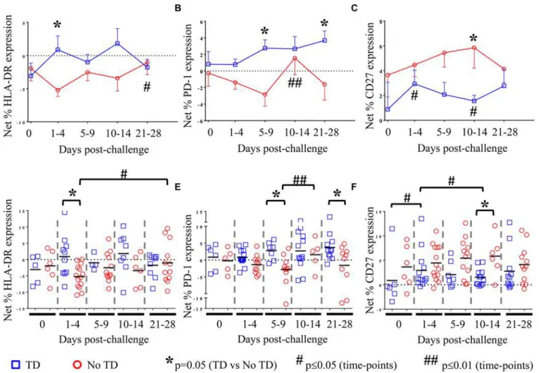
the expression of all activation molecules. We observed thatS. Typhi-specific up-regulation of
PD-1, CD27, LFA-1, NRP-1, and Tim-3 was present before challenge in many volunteers (Fig 3B, 3C, 3E and 3F,Fig 4A and 4BandS3B Fig). However, we identified no significant differ-ences in theS. Typhi-specific expression of activation molecules prior to challenge in TD
vol-unteers compared with No TD volvol-unteers (Fig 3A and 3F, andS3 Fig). At early time-points (days 1–4) following challenge, however, we observed a notable increase inS. Typhi-specific
ex-pression of HLA-DR resulting in significantly higher exex-pression in TD than in No TD volun-teers (p = 0.015—mixed effects regression model) (Fig 3A and 3D). In contrast,S.
Typhi-specific HLA-DR expression on circulating Tregin No TD volunteers decreased slightly after
challenge (days 1–4), returning to baseline levels by 21–28 days post-challenge (Fig 3A and 3D).
We also identified marked up-regulation of the expression of PD-1 byS. Typhi-infected
tar-gets in circulating Tregisolated from TD compared to No TD volunteers post-challenge (Fig
3B and 3E).S. Typhi-specific up-regulation of PD-1 expression increased gradually in TD
vol-unteers following challenge with the highest levels identified on days 21–28 post-challenge (Fig 3B and 3E). Distinctly, however, among those volunteers who did not develop disease we noted a general down-regulation ofS. Typhi-specific PD-1 expression in circulating Treg1–9
days post-challenge with a significant increase between the days 5–9 and days 10–14 post-chal-lenge time groups (Fig 3B and 3E).S. Typhi-specific PD-1 expression returned to baseline
lev-els by days 21–28 post-challenge (Fig 3B and 3E). These opposite trends resulted in
significantly higher up-regulation ofS. Typhi-specific expression of PD-1 in circulating Tregin
Interestingly, we observed increased up-regulation of the expression of CD27 onS.
Typhi-specific Tregin No TD compared to TD volunteers (Fig 3C and 3F). While the trend was
pres-ent at most time-points, this difference was statistically significant only in the day 10–14 time frame (after typhoid diagnosis and initiation of antibiotics), (p = 0.031—mixed effects regres-sion model). In a subset of volunteers,S. Typhi-specific CD39 expression was also measured.
Although there were only a small number of samples tested, we identified up-regulation ofS.
Typhi-specific CD39 expression on circulating Tregfollowing challenge in TD volunteers. This
increase peaked at days 10–14 post-challenge and was significantly higher than pre-challenge (p = 0.02—mixed effects regression model) (S3A Fig). WhileS. Typhi-specific Tim-3
expres-sion was present on circulating Tregthere was no difference noted between TD and No TD
vol-unteers or in either group over time (S3B Fig).
Fig 3.S. Typhi-specific activation of circulating Treg.NetS. Typhi-specific expression ofA) HLA-DR, (TD n = 5, No TD n = 4)B) PD-1, (TD n = 6, No TD n = 6) andC) CD27, (TD n = 6, No TD n = 6) on Treg. Values are shown as the mean +/- SEM. Scatter plots showing the net expression ofD)HLA-DR, (TD n = 5, No TD n = 4)E) PD-1, (TD n = 6, No TD n = 6) andF) CD27, (TD n = 6, No TD n = 6) onS. Typhi-specific Treg. Means are indicated with a black horizontal line. Time points with statistically significant differences between TD and No TD volunteers (*) or among time-points within each group (#) are identified. P-values were determined using a mixed effects regression model. TD (blue squares); No TD (red circles). Values from multiple time-points were grouped together in time segments (1–4, 5–9, 10–14, and 21–28 days post-challenge) to account for variability in the numbers of samples available from
each volunteer. Some volunteers had samples from multiple time-points in a time-segment resulting in more data points than the corresponding number of volunteers.
doi:10.1371/journal.ppat.1004914.g003
Differential kinetics of activation of circulating
S
. Typhi-specific Treg
between TD and No TD volunteers
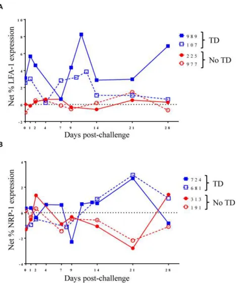
To further explore changes inS. Typhi-specific modulation of the expression of activation
mol-ecules over time, we examined kinetic curves of individual volunteers. While significant differ-ences were not detected in the mean expression of LFA-1 between TD and No TD volunteers, the kinetic patterns were remarkably different in volunteers diagnosed, or not, with typhoid following challenge. Despite considerable variation among volunteers, a pattern of increased expression of LFA-1 around the time of disease was identified in a majority of TD volunteers (4/5) while expression remained relatively constant for most No TD volunteers (Fig 4A). We also identified differences in the kinetic patterns ofS. Typhi-specific NRP-1 expression. Unlike
LFA-1, we observedS. Typhi-specific up-regulation of NRP-1 expression in TD volunteers
after diagnosis and initiation of antibiotics (days 14–21 post-challenge) (Fig 4B).
Fig 4. Kinetics of S. Typhi-specific modulation of LFA-1 and NRP-1 expression on circulating Treg
following challenge.Kinetic curves from representative volunteers showing netS. Typhi-specific modulation
of the expression ofA)LFA-1 andB)NRP-1 from day 0 (pre-challenge) until day 28 post-challenge.
Circulating T
regsuppress
S
. Typhi-specific T
effresponses
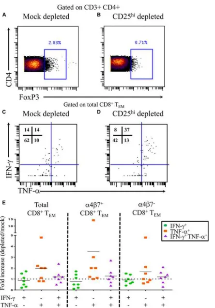
To further assess the functionality of Tregin the setting of typhoid disease, we performed CD25
depletion assays. PBMC from 4 TD volunteers were either mock depleted (pan anti-mouse IgG) or CD25 depleted (anti-human CD25) using magnetic bead separation. Time-points were selected based on knownS. Typhi-specific cytokine responses. A total of 7 independent
volun-teer-time points were used for depletion studies. Depletion resulted in a 55–74% reduction in FoxP3+ CD4+ T cells (Fig 5A and 5B). Following stimulation withS. Typhi-infected B-LCL,
CD8+ T effector memory (TEM) were evaluated forS. Typhi-specific cytokine production in
the presence (mock-depleted) or absence (CD25-depleted) of Tregusing mass cytometry. We
found higher percentages of IFN-γand TNF-αsingle cytokine producing and multi-functional
Fig 5.S. Typhi-specific cytokine production following Tregdepletion.Percentage of CD4+ FoxP3+ T cells followingA)mock depletion orB)CD25 depletion in a representative volunteer. Production of IFN-γand/ or TNF-αbyS. Typhi-specific CD8+ TEMfollowingC)mock orD)CD25 depletion.E)Data are presented as fold increases in IFN-γand/or TNF-αproduction byS. Typhi-specific total CD8+ TEMin depleted vs mock-depleted cultures, as well as by CD8+ TEMexpressing, or not, integrinα4β7.
doi:10.1371/journal.ppat.1004914.g005
(IFN-γ+ TNF-α+)S. Typhi-specific CD8+ TEMwhen Tregwere depleted (Fig 5C and 5E).
In-terestingly, increases in cytokine production were observed inS. Typhi-specific CD8+ TEM
with or without gut homing potential (integrinα4β7+ and integrinα4β7-, respectively) (Fig 5E).
Discussion
Effective immune responses must balance the need for pathogen-specific inflammatory re-sponses to fight infection with the need to protect the host from the consequences of excessive inflammation. Homeostasis between regulatory and effector T cells is a major component of this balance. Tregsuppress Teffby multiple mechanisms including contact dependent
mecha-nisms, such as CTLA-4, as well as contact-independent mechanisms such as IL-10 production. We aimed to investigate the characteristics, kinetics, and functionality of Tregresponses in anS.
Typhi human controlled infection model. Homing of Tregto sites of specific inflammation has
been previously shown [16,20,21,22,23]. Integrinα4β7 is an important molecule associated with homing of lymphocytes to the gut, the site of initial encounter withS. Typhi [28]. Here we
identified, for the first time, significantly higher pre-challenge gut homing potential of circulat-ing Treg(up-regulation ofS. Typhi-specific integrinα4β7 expression) in volunteers who were
subsequently diagnosed with typhoid disease compared to those who were not. Following chal-lenge, however, there was a significant decrease inS. Typhi-specific integrinα4β7 expression
on circulating Treg, suggesting that these Tregleft the peripheral blood, presumably as a result
of homing to the gut microenvironment. It is currently unclear whyS. Typhi-specific Treg
ex-pressing differential levels of integrinα4β7 were observed among volunteers before challenge. Participants were recruited in a non-endemic area and are, therefore, unlikely to have previous-ly encounteredS. Typhi. However, theS. Typhi genome has a high degree of homology with
other Enterobacteriaceae. Thus, differences in baseline Tregresponses could be the result of
pre-vious encounters with other enteric Gram negative bacilli, including those present in the nor-mal gut microbiota. We have previously reported that oral immunization of volunteers with attenuated oralS. Typhi vaccines elicitsS. Typhi-specific TEMwhich expressed, or not, the gut
homing molecule integrinα4β7 [11,29]. It has been shown that T cells activated in the gut pref-erentially express high levels of integrinα4β7 compared to T cells primed in peripheral lymph nodes [30]. Therefore, it is possible that Treginitially primed in the gut would express higher
levels of integrinα4β7 upon re-stimulation resulting in recirculation to the site of initial anti-gen encounter. It is thus reasonable to speculate that higher levels of Treghoming to the gut
may suppress local Teffresponses resulting in ineffectual control of the infection ultimately
leading to typhoid diagnosis. This hypothesis is further supported by our findings showing the capacity of Tregto suppressS. Typhi–specific responses by integrinα4β7+ TEMelicited in
vol-unteers following exposure to wild-typeS. Typhi. Of note, we have also observed in these
vol-unteers, that TregsuppressS. Typhi–specific responses by integrinα4β7- TEM, suggesting that
specific Tregmight also exert their regulatory activity at systemic sites.
In addition to early homing to the gut, we identifiedS. Typhi-specific up-regulation of the
expression of both CCR6 and CXCR3. CXCR3 expression on Tregis associated with homing to
sites of TH1/TC1 inflammation [21]. It is known that immunization withS. Typhi vaccines, as
well as natural infection withS. Typhi, induce predominantly TH1/TC1 type responses
[8,9,10,11,12,13,14,15,29]. While not significant, there was a trend toward higher levels ofS.
Typhi-specific up-regulation of CXCR3 expression on circulating Tregin TD volunteers
[20,22]. We have previously identifiedS. Typhi-specific production of IL-17A by CD8+ TEM
following Ty21a immunization [8]. While no significant differences were noted inS.
Typhi-specific up-regulation of CCR6 expression on circulating Tregin TD versus No TD volunteers
prior to or following challenge, No TD volunteers exhibited a significantS. Typhi-specific
de-crease in the levels of expression of CCR6 on Tregfollowing challenge, then remained at
rela-tively constant levels through day 28 post challenge. These results suggest that suppression of
S. Typhi-specific TH17/Tc17 responses plays a role in protection from typhoid disease. TH17/
Tc17 responses are known to produce inflammation including recruitment of neutrophils [31] and increased TH17 infiltration has been identified in gut inflammatory conditions, such as in
Crohn’s disease [32]. It is possible that excessive inflammation could result in increased gut permeability and subsequent dissemination ofS. Typhi. It is important to note however, that
TH17 cells also play an important role in gut mucosal integrity [33]. Therefore, it is likely that
the balance of Tregand TH17/Tc17 effector responses may be critical in determining disease
outcome.
Taken together, these results highlight the likely importance of Treglocalization in the
devel-opment of typhoid fever. Interestingly, integrinα4β7 is the only molecule measured which showed significant pre-challenge differences inS. Typhi-specific expression on circulating Treg
between TD and No TD volunteers, highlighting the potential importance of the local re-sponses early in infection.
In addition to homing to appropriate sites, the activation status of Tregis likely to affect
their potential to suppressS. Typhi-specific inflammatory responses. Expression of the
activa-tion molecule HLA-DR has been associated with increased contact-dependent activity of human Treg[34]. Furthermore, it has been shown that HLA-DR+ Treg, while more active, are
also more susceptible to apoptosis [35]. We identified significant increases in HLA-DR expres-sion on circulatingS. Typhi-specific Tregin TD compared to No TD volunteers in the early
time-points (day 1–4) post-challenge suggesting that increasedS. Typhi-specific activation of
circulating Tregmay play a role in the development of typhoid fever. Furthermore, we also
identified significant differences in the intracellular PD-1 content inS. Typhi-specific
circulat-ing Tregbetween TD and No TD volunteers. While only a small percentage of natural Treg
express PD-1 on the surface, higher levels of PD-1 transcript have been associated with sup-pressive function, suggesting that PD-1 expression is also involved in the development of ty-phoid fever [36]. Similarly, we observed up-regulation ofS. Typhi–specific surface expression
of CD39 in TD volunteers, albeit at later time points. Tregexpression of CD39 has been
associ-ated with suppression of TH17/TC17 responses [37] which, as previously mentioned, have been
identified followingS. Typhi immunization [8]. It is therefore possible that CD39 expressing
Tregin TD volunteers exert their activity, at least in part, by modulating TH17/TC17 responses.
In contrast, we did not identify differences inS. Typhi-specific Tim-3 expression between TD
and No TD volunteers. The fact that Tim-3 expression on Treghas been associated with
in-creased suppressive Tregactivity [38], but no differences were observed between TD and No
TD, suggests that the mechanism(s) of Tregactivation may vary depending on the model
stud-ied. The observations that increased levels of Tregactivation appear to play a role in the
devel-opment of typhoid fever are supported by the results of depletion studies that show increased
S. Typhi-specific cytokine production by TEMfollowing depletion of Treg.
CD27 has been proposed as a marker for Tregsuppressive activity as well as a marker for
CD4+ T memory phenotype [39,40]. We identified higher levels ofS. Typhi-specific CD27
ex-pression in No TD volunteers, particularly at days 10–14 post-challenge. This is in striking contrast to other markers of activation which were all increased in TD volunteers. Both CD27+ and CD27- populations displaying suppressive characteristics have been identified in expanded human Treg[41]. Interestingly, CD27+ Tregwere predominantly CD62L+ compared to
CD27- Tregsuggesting that CD27+ Tregmay be localizing to peripheral lymph nodes [41].
Therefore, the tissue distribution of activated Treg, their characteristics and levels of activation
may constitute important determining factors in protection from typhoid fever by contributing to an appropriate balance between suppressive and inflammatory responses.
In addition toS. Typhi-specific increase in HLA-DR expression and up-regulation of PD-1
and CD39 in Treg, we also identified differences in the kinetic patterns of other molecules
asso-ciated with Tregactivation including LFA-1 and NRP-1. LFA-1 plays an important role in the
formation of Tregaggregates that block access of responder T cells to dendritic cells [18]. In
contrast, the precise function of NRP-1 up-regulation on human Tregremains to be elucidated
and in some studies NRP-1 expression was not identified on human Treg[42]. However, other
studies have shown NRP-1 up-regulation to be associated with Tregactivation in humans [43].
Furthermore, in mice, NRP-1 has been suggested as a marker for tTreg; however, this has not
been definitively shown in humans [19,42]. Here we identifiedS. Typhi-specific up-regulation
of NRP-1 in TD volunteers and report differences in the observed kinetics ofS. Typhi-specific
NRP-1 expression between TD and No TD volunteers. The difference in kinetics ofS.
Typhi-specific expression of both LFA-1 and NRP-1 molecules in TD compared to No TD volunteers may indicate that multiple mechanisms of increased Tregactivation play a role in Tregresponses
followingS. Typhi challenge. Furthermore, these findings suggest that not only the precise
bal-ance, but also the timing of Tregresponses with inflammatory responses might ultimately
deter-mine disease outcome.
While there are no animal models for typhoid fever that fully recapitulate human disease, there have been studies in mice using infection withS. Typhimurium which reveal a potential
role for Tregin suppressing specific Teffresponses [44]. In this mouse model, increased Treg
suppressive capacity, including upregulated CTLA-4 (CD152) expression, is associated with higherS. Typhimurium bacterial burden. Furthermore, Tregablation results in enhanced Teff
activation leading to reduced pathogen burden. These results support our findings that in-creased Tregactivation is associated with typhoid disease and that Tregare capable of
suppress-ingS. Typhi-specific Teffin humans.
In summary, we have shown thatS. Typhi-specific up-regulation of the gut homing
mole-cule integrinα4β7 prior to challenge is associated with typhoid diagnosis. Moreover, despite differences in the kinetics of the responses among various Tregactivation molecules, with the
notable exception of CD27, there was a clear trend for circulating Tregfrom TD volunteers to
display increased levels ofS. Typhi-specific activation. Of great importance, we have also
dem-onstrated that Tregare functionally capable of suppressingS. Typhi-specific CD8+ TEM
cyto-kine responses. While the small sample size is a limitation, these studies provide an important first description of Tregresponses followingS. Typhi exposure in humans. Further investigation
into how these responses may relate to protection following immunization with attenuated strains ofS. Typhi will provide much needed information to inform and accelerate the
develop-ment of novel vaccines for typhoid and other enteric fevers, as well as other enteric infections. For example, strategies to identify vaccines that activate Teffwithout the concomitant activation
of suppressive Tregresponses, or that elicit an optimal balance between Teffand Tregresponses
may result in improved protective efficacy.
Materials and Methods
challenged with 1–5x10⁴CFU of wt-S. Typhi (Quailes strain) suspended in sodium bicarbonate
at Oxford University in compliance with the National Research Ethic Service (NRES), Oxford Research Ethics Committee A [7]. Close monitoring was performed throughout the study, and at the time of typhoid fever diagnosis (TD, as determined by blood culture-confirmedS. Typhi
bacteremia or development of a fever38°C for12 hours), volunteers were treated with a 2-week course of antibiotics (Ciprofloxacin, 500mg twice daily). Those volunteers who did not developed typhoid fever (No TD) received a 2-week course of antibiotics at day 14 post-chal-lenge. PBMC collected from 12 randomly selected volunteers (TD n = 6, No TD n = 6) partici-pating in the challenge trial were used in this study. Selection was made based on the number of available PBMC with those volunteers having more PBMC utilized for the studies. PBMC were isolated prior to challenge and at 9–11 time-points following challenge (S1 Fig). Isolation was performed by Lymphoprep gradient centrifugation (Axis-Shield, Oslo, Norway) and PBMC were cryopreserved in liquid nitrogen following standard techniques within four hours of initial blood draw. Viability of cryopreserved PBMC was assessed after thawing of cells and an overnight rest at 37°C with 5% CO2(as described inex vivostimulation).
Target/stimulator cells
B-LCL were generated from autologous PBMC for each volunteer as previously described [45]. Briefly, B-LCL were established using supernatant from the B95.8 cell line (ATCC CRL1612; American Type Culture Collection) as the source of EBV. PBMC from each volunteer were incu-bated with EBV containing supernatant and cyclosporine (0.5μg/mL; Sigma, St. Louis, MO) at 37°C with 5% CO2for 2–3 weeks. B-LCL were maintained in culture or cryopreserved until use.
Infection of target/stimulator cells
Target cells were infected by incubation with wild-typeS. Typhi strain ISP1820 in RPMI 1640
media (Gibco, Carlsbad, CA) without antibiotics for 3 hours at 37°C with 5% CO2as previously
described [45]. On the day following infection the cells were gamma irradiated (6000 rad). To confirm that targets were infected withS. Typhi, cells were stained with anti-Salmonella
com-mon structural Ag (CSA-1)-FITC (Kierkegaard & Perry, Gaithersburg, MD) and analyzed by flow cytometry on an LSRII flow cytometer (BD Biosciences, San Jose, CA) [8]. The percentage of cells infected withS. Typhi was recorded for each experiment and the infected targets were
only used if infection rates were>30% of viable cells.
Ex vivo stimulation
PBMC were thawed and rested overnight at 37°C. Cells were then resuspended in RPMI 1640 media (Gibco) supplemented with 100 U/mL penicillin (Sigma), 100μg/mL streptomycin (Sigma), 50μg/mL gentamicin (Gibco), 2 mM L-glutamine (Gibco), 2.5 mM sodium pyruvate (Gibco), 10 mM HEPES buffer (Gibco), and 10% fetal bovine serum (Gemini Bioproducts, West Sacramento, CA) at a concentration of 1x10⁶cells/mL in sterile 5 mL round bottom tubes (BD Falcon, Franklin Lakes, NJ). PBMC were stimulated withS. Typhi-infected B-LCL or B-LCL
alone (negative control). After 2 hours, Golgi Stop (containing monensin) and Golgi Plug (con-taining brefeldin A) from BD were added at concentrations of 0.5μl/mL and cultures continued overnight at 37°C in 5% CO2. Media alone was used as an additional negative control.
Conventional flow cytometric analyses
Following stimulation as described above, cells were plated in 96-well V-bottom plates for staining. Cells were washed once with staining buffer (phosphate buffered saline with 0.5%
BSA and 0.1% sodium azide) and stained for live/dead discrimination using Invitrogen LIVE/ DEAD fixable yellow dead cell stain kit (Invitrogen, Carlsbad, CA). Fc receptor blocking was performed with human immunoglobulin (Sigma; 3μg/mL) followed by surface staining, per-formed as previously described.[8] Briefly, cells were surface stained with panels that included the following fluorochrome-conjugated monoclonal antibodies against: CD14-BV570 (M5E2, Biolegend, San Diego, CA), CD19-BV570 (HIB19, Biolegend), CD3-BV650 (OKT3, Biole-gend), CD4-APC-H7 (RPA-T4, BD), CD25-PECy7 (M-A251, BD), CCR6/CD196-PE (11A9, BD), HLA-DR-Qdot 800 (Life technologies, Grand Island, NY), integrinα4β7-Alexa 647 (clone ACT-1, conjugated in-house), CXCR3/CD183-Alexa 700 (1C6/CXCR3, BD), LFA-1/ CD11a-Alexa 488 (HI111, Biolegend), NRP-1/CD304-APC (12C2, Biolegned), CD27-BV605 (4S.B3, Biolegend), CD39-BV421 (A1, Biolegend), and Tim-3-Alexa 700 (344823, R&D, Min-neapolis, MN) at 4°C for 30 minutes. The cells were then fixed and permeabilized using FoxP3 IC fixation and permeabilization buffers from eBiosciences according to manufacturer’s rec-ommendations. Intracellular staining with FoxP-PerCP-Cy5.5 (236A/E7, BD), CTLA-4/ CD152-PECy5 (BNI3, BD), PD-1/CD279-BV421 (EH12.1, BD) and Ki67-BV605 (Ki67, Biole-gend) was performed for 20 minutes at room temperature. After staining, cells were fixed in 1% paraformaldehyde and stored at 4°C until analyzed. Flow cytometry was performed using a customized LSRII flow cytometer (BD). Flow cytometry data were analyzed using WinList ver-sion 7 (Verity Software House, Topsham, ME) software package. Graphs were generated using GraphPad Prism version 6 (Graphpad Software, San Diego, CA).
CD25-depletion studies
In a subset of volunteers, CD25 cells were depleted- or mock-depleted using anti-CD25 or pan anti-mouse IgG Dynabeads, respectively (Invitrogen) as previously described [25]. Briefly, thawed PBMC were rested overnight as described above. Following the overnight rest, PBMC were divided into two aliquots consisting of 2.1–3 x 10⁶cells and either mock-depleted or de-pleted of CD25 cells using magnetic bead separation. Dede-pleted (mock and CD25) PBMC were stimulated withS. Typhi-infected B-LCL or non-infected B-LCL (negative control) as
described above.
Mass cytometry
Following CD25- or mock-depletion and stimulation withS. Typhi-infected B-LCL, cells were
stained for mass cytometry with a panel of 22 metal-conjugated mAb to detect both Tregand
responder T cells. A table of the mAb used is shown in supplementary materials (S1 Table). Vi-ability staining was performed with cisplatinum (Sigma; 25μM) for 60 seconds. Following cis-platinum, samples were Fc-blocked with human immunoglobulin (Sigma; 3μg/mL) followed by surface staining, performed as previously described. Fixation and permeabilization were performed with FoxP3 IC fixation and permeabilization buffers (eBiosciences) followed by in-tracellular staining. Samples were stained with an Ir191/193DNA intercalator for cell detection by mass cytometry within 48 hours of sample acquisition and re-suspended in EQ4 normaliza-tion beads (Fluidigm, Sunnyvale, CA). Acquisinormaliza-tion was performed using a CyTOF mass cytom-eter (Fluidigm, formerly DVS Sciences). Data were analyzed with Fluidigm Cytobank.
Statistical analyses
independence between multiple measures from the same volunteer at the same time period and across time periods, we used mixed effects models, including a random effect for subject, fit by restricted maximum likelihood. Through simulation experiments we confirmed that this approach provided valid statistical inference for data sets of this size.
Ethics statement
The human challenge study was performed in compliance with the National Research Ethic Service (NRES), and approved by the Oxford Research Ethics Committee A. All volunteers provided written informed consent.
Supporting Information
S1 Fig. Outline of study design.Volunteers were challenged with wild-typeS. Typhi on day 0.
PBMC were obtained prior to challenge and at up to 10 time-points after challenge. (TIF)
S2 Fig. Gating strategy for identification of Treghoming potential and activation. Lympho-cytes were gated on forward versus side scatter, followed by exclusion of doublets. Yellow Via-bility Dye live/dead staining was used to exclude dead cells and anti-CD14 and CD19 were used to exclude monocytes and B cells, respectively with positive selection of CD3+ T cells. CD4+ FoxP3+ cells were identified followed by gating on CTLA-4 and CD25. Tregwere defined
as CD4+ FoxP3+ CTLA4+ CD25+. Representative histograms depicting the expression of homing molecules integrinα4β7, CXCR3, and CCR6, as well as the expression of activation molecules HLA-DR, PD-1, CD27, LFA-1, NRP-1, CD39, and Tim-3.
(TIF)
S3 Fig.S. Typhi-specific activation of circulating Treg.NetS. Typhi-specific modulation of
the expression ofA) CD39, (TD n = 3, No TD n = 3) andB) Tim-3, (TD n = 5, No TD n = 6) on Treg. Values are shown as the mean +/- SEM. Statistically significant differences in the
mod-ulation of the expression of CD39 between pre- and days 10–14 post-challenge in TD volun-teers (#) are indicated. TD (blue squares); No TD (red circles). Values from multiple time-points were grouped together in time segments (1–4, 5–9, 10–14, and 21–28 days post-chal-lenge) to account for variability in the samples available from each volunteer. Some volunteers had samples from multiple time-points in a time-segment resulting in more data points than the corresponding number of volunteers.
(TIF)
S1 Table. Monoclonal antibodies used for mass cytometry staining. (XLSX)
Acknowledgments
We thank Ms. Catherine Storrer and Ms. Regina Harley for excellent technical assistance.
Author Contributions
Conceived and designed the experiments: MAM SF MBS. Performed the experiments: MAM SF. Analyzed the data: MAM SF LSM MBS. Contributed reagents/materials/analysis tools: TCD CSW CJ CJB BA. Wrote the paper: MAM SF LSM TCD CJ CSW CJB GD BA MML AJP MBS. Set up challenge model and generated clinical data: TCD CSW GD MML AJP.
References
1. Crump JA, Luby SP, Mintz ED (2004) The global burden of typhoid fever. Bull World Health Organ 82: 346–353. PMID:15298225
2. Buckle GC, Walker CL, Black RE (2012) Typhoid fever and paratyphoid fever: Systematic review to es-timate global morbidity and mortality for 2010. J Glob Health 2: 010401. doi:10.7189/jogh.02.010401 PMID:23198130
3. Bhutta ZA (1996) Impact of age and drug resistance on mortality in typhoid fever. Arch Dis Child 75: 214–217. PMID:8976660
4. Rowe B, Ward LR, Threlfall EJ (1997) Multidrug-resistant Salmonella typhi: a worldwide epidemic. Clin Infect Dis 24 Suppl 1: S106–109. PMID:8994789
5. Levine MM, Ferreccio C, Abrego P, Martin OS, Ortiz E, et al. (1999) Duration of efficacy of Ty21a, atten-uated Salmonella typhi live oral vaccine. Vaccine 17 Suppl 2: S22–27. PMID:10506405
6. Sur D, Ochiai RL, Bhattacharya SK, Ganguly NK, Ali M, et al. (2009) A cluster-randomized effective-ness trial of Vi typhoid vaccine in India. N Engl J Med 361: 335–344. doi:10.1056/NEJMoa0807521
PMID:19625715
7. Waddington CS, Darton TC, Jones C, Haworth K, Peters A, et al. (2014) An outpatient, ambulant-de-sign, controlled human infection model using escalating doses of salmonella typhi challenge delivered in sodium bicarbonate solution. Clin Infect Dis 58: 1230–1240. doi:10.1093/cid/ciu078PMID:
24519873
8. McArthur MA, Sztein MB (2012) Heterogeneity of multifunctional IL-17A producing S. Typhi-specific CD8+ T cells in volunteers following Ty21a typhoid immunization. PLoS One 7: e38408. doi:10.1371/ journal.pone.0038408PMID:22679502
9. Salerno-Goncalves R, Fernandez-Vina M, Lewinsohn DM, Sztein MB (2004) Identification of a human HLA-E-restricted CD8+ T cell subset in volunteers immunized with Salmonella enterica serovar Typhi strain Ty21a typhoid vaccine. J Immunol 173: 5852–5862. PMID:15494539
10. Salerno-Goncalves R, Pasetti MF, Sztein MB (2002) Characterization of CD8(+) effector T cell re-sponses in volunteers immunized with Salmonella enterica serovar Typhi strain Ty21a typhoid vaccine. J Immunol 169: 2196–2203. PMID:12165550
11. Salerno-Goncalves R, Wahid R, Sztein MB (2005) Immunization of volunteers with Salmonella enterica serovar Typhi strain Ty21a elicits the oligoclonal expansion of CD8+ T cells with predominant Vbeta repertoires. Infect Immun 73: 3521–3530. PMID:15908381
12. Salerno-Goncalves R, Wahid R, Sztein MB (2010) Ex Vivo kinetics of early and long-term multifunction-al human leukocyte antigen E-specific CD8+ cells in volunteers immunized with the Ty21a typhoid vac-cine. Clin Vaccine Immunol 17: 1305–1314. doi:10.1128/CVI.00234-10PMID:20660136
13. Salerno-Goncalves R, Wyant TL, Pasetti MF, Fernandez-Vina M, Tacket CO, et al. (2003) Concomitant induction of CD4+ and CD8+ T cell responses in volunteers immunized with Salmonella enterica sero-var typhi strain CVD 908-htrA. J Immunol 170: 2734–2741. PMID:12594304
14. Sztein MB (2007) Cell-mediated immunity and antibody responses elicited by attenuated Salmonella enterica Serovar Typhi strains used as live oral vaccines in humans. Clin Infect Dis 45 Suppl 1: S15–
19. PMID:17582562
15. Sztein MB, Salerno-Goncalves R, McArthur MA (2014) Complex adaptive immunity to enteric fevers in humans: lessons learned and the path forward. Front Immunol 5: 516. doi:10.3389/fimmu.2014.00516 PMID:25386175
16. Wing K, Sakaguchi S (2010) Regulatory T cells exert checks and balances on self tolerance and auto-immunity. Nat Immunol 11: 7–13. doi:10.1038/ni.1818PMID:20016504
17. Sakaguchi S, Wing K, Miyara M (2007) Regulatory T cells—a brief history and perspective. Eur J
Immu-nol 37 Suppl 1: S116–123. PMID:17972355
18. Sakaguchi S, Wing K, Onishi Y, Prieto-Martin P, Yamaguchi T (2009) Regulatory T cells: how do they suppress immune responses? Int Immunol 21: 1105–1111. doi:10.1093/intimm/dxp095PMID:
19737784
19. Dhamne C, Chung Y, Alousi AM, Cooper LJ, Tran DQ (2013) Peripheral and thymic foxp3(+) regulatory T cells in search of origin, distinction, and function. Front Immunol 4: 253. doi:10.3389/fimmu.2013. 00253PMID:23986762
21. Koch MA, Tucker-Heard G, Perdue NR, Killebrew JR, Urdahl KB, et al. (2009) The transcription factor T-bet controls regulatory T cell homeostasis and function during type 1 inflammation. Nat Immunol 10: 595–602. doi:10.1038/ni.1731PMID:19412181
22. Yamazaki T, Yang XO, Chung Y, Fukunaga A, Nurieva R, et al. (2008) CCR6 regulates the migration of inflammatory and regulatory T cells. J Immunol 181: 8391–8401. PMID:19050256
23. Engelhardt BG, Sengsayadeth SM, Jagasia M, Savani BN, Kassim AA, et al. (2012) Tissue-specific regulatory T cells: biomarker for acute graft-vs-host disease and survival. Exp Hematol 40: 974–982
e971. doi:10.1016/j.exphem.2012.08.002PMID:22885125
24. Li L, Lao SH, Wu CY (2007) Increased frequency of CD4(+)CD25(high) Treg cells inhibit BCG-specific induction of IFN-gamma by CD4(+) T cells from TB patients. Tuberculosis (Edinb) 87: 526–534. PMID:
17851131
25. Lyke KE, Dabo A, Arama C, Daou M, Diarra I, et al. (2012) Reduced T regulatory cell response during acute Plasmodium falciparum infection in Malian children co-infected with Schistosoma haematobium. PLoS One 7: e31647. doi:10.1371/journal.pone.0031647PMID:22348117
26. Baecher-Allan C, Brown JA, Freeman GJ, Hafler DA (2001) CD4+CD25high regulatory cells in human peripheral blood. J Immunol 167: 1245–1253. PMID:11466340
27. Presicce P, Moreno-Fernandez ME, Lages CS, Orsborn KI, Chougnet CA (2010) Association of two clones allows for optimal detection of human FOXP3. Cytometry A 77: 571–579. doi:10.1002/cyto.a.
20875PMID:20162533
28. Mora JR, Iwata M, Eksteen B, Song SY, Junt T, et al. (2006) Generation of gut-homing IgA-secreting B cells by intestinal dendritic cells. Science 314: 1157–1160. PMID:17110582
29. Wahid R, Salerno-Goncalves R, Tacket CO, Levine MM, Sztein MB (2008) Generation of specific effec-tor and memory T cells with gut- and secondary lymphoid tissue- homing potential by oral attenuated CVD 909 typhoid vaccine in humans. Mucosal Immunol 1: 389–398. doi:10.1038/mi.2008.30PMID:
19079203
30. Stagg AJ, Kamm MA, Knight SC (2002) Intestinal dendritic cells increase T cell expression of alpha4-beta7 integrin. Eur J Immunol 32: 1445–1454. PMID:11981833
31. Witowski J, Ksiazek K, Jorres A (2004) Interleukin-17: a mediator of inflammatory responses. Cell Mol Life Sci 61: 567–579. PMID:15004696
32. Kleinschek MA, Boniface K, Sadekova S, Grein J, Murphy EE, et al. (2009) Circulating and gut-resident human Th17 cells express CD161 and promote intestinal inflammation. J Exp Med 206: 525–534. doi:
10.1084/jem.20081712PMID:19273624
33. Raffatellu M, Santos RL, Verhoeven DE, George MD, Wilson RP, et al. (2008) Simian immunodeficien-cy virus-induced mucosal interleukin-17 deficienimmunodeficien-cy promotes Salmonella dissemination from the gut. Nat Med 14: 421–428. doi:10.1038/nm1743PMID:18376406
34. Baecher-Allan C, Wolf E, Hafler DA (2006) MHC class II expression identifies functionally distinct human regulatory T cells. J Immunol 176: 4622–4631. PMID:16585553
35. Ashley CW, Baecher-Allan C (2009) Cutting Edge: Responder T cells regulate human DR+ effector reg-ulatory T cell activity via granzyme B. J Immunol 183: 4843–4847. doi:10.4049/jimmunol.0900845
PMID:19801510
36. Tang Q, Henriksen KJ, Bi M, Finger EB, Szot G, et al. (2004) In vitro-expanded antigen-specific regula-tory T cells suppress autoimmune diabetes. J Exp Med 199: 1455–1465. PMID:15184499
37. Fletcher JM, Lonergan R, Costelloe L, Kinsella K, Moran B, et al. (2009) CD39+Foxp3+ regulatory T Cells suppress pathogenic Th17 cells and are impaired in multiple sclerosis. J Immunol 183: 7602–
7610. doi:10.4049/jimmunol.0901881PMID:19917691
38. Gupta S, Thornley TB, Gao W, Larocca R, Turka LA, et al. (2012) Allograft rejection is restrained by short-lived TIM-3+PD-1+Foxp3+ Tregs. J Clin Invest 122: 2395–2404. doi:10.1172/JCI45138PMID:
22684103
39. Duggleby RC, Shaw TN, Jarvis LB, Kaur G, Gaston JS (2007) CD27 expression discriminates between regulatory and non-regulatory cells after expansion of human peripheral blood CD4+ CD25+ cells. Im-munology 121: 129–139. PMID:17425604
40. Ruprecht CR, Gattorno M, Ferlito F, Gregorio A, Martini A, et al. (2005) Coexpression of CD25 and CD27 identifies FoxP3+ regulatory T cells in inflamed synovia. J Exp Med 201: 1793–1803. PMID:
15939793
41. Koenen HJ, Fasse E, Joosten I (2005) CD27/CFSE-based ex vivo selection of highly suppressive allo-antigen-specific human regulatory T cells. J Immunol 174: 7573–7583. PMID:15944257
42. Yadav M, Stephan S, Bluestone JA (2013) Peripherally induced tregs—role in immune homeostasis
and autoimmunity. Front Immunol 4: 232. doi:10.3389/fimmu.2013.00232PMID:23966994
43. Chaudhary B, Khaled YS, Ammori BJ, Elkord E (2014) Neuropilin 1: function and therapeutic potential in cancer. Cancer Immunol Immunother 63: 81–99. doi:10.1007/s00262-013-1500-0PMID:24263240 44. Johanns TM, Ertelt JM, Rowe JH, Way SS (2010) Regulatory T cell suppressive potency dictates the
balance between bacterial proliferation and clearance during persistent Salmonella infection. PLoS Pathog 6: e1001043. doi:10.1371/journal.ppat.1001043PMID:20714351