Oral Challenge with Wild-Type
Typhi Induces Distinct Changes in B Cell
Subsets in Individuals Who Develop Typhoid
Disease
Franklin R. Toapanta1,2*, Paula J. Bernal1,3, Stephanie Fresnay1,3, Laurence S. Magder4, Thomas C. Darton5, Claire Jones5, Claire S. Waddington5, Christoph J. Blohmke5, Brian Angus6, Myron M. Levine1,2,3, Andrew J. Pollard5, Marcelo B. Sztein1,2,3*
1Center for Vaccine Development, University of Maryland School of Medicine, Baltimore, Maryland, United States of America,2Department of Medicine, University of Maryland School of Medicine, Baltimore, Maryland, United States of America,3Department of Pediatrics, University of Maryland School of Medicine, Baltimore, Maryland, United States of America,4Department of Epidemiology and Public Health, University of Maryland School of Medicine, Baltimore, Maryland, United States of America,5Oxford Vaccine Group, Department of Paediatrics, University of Oxford and the NIHR Oxford Biomedical Research Centre, Oxford, United Kingdom,6Nuffield Department of Medicine, University of Oxford, Oxford, United Kingdom
*[email protected](FRT);[email protected](MBS)
Abstract
A novel human oral challenge model with wild-typeSalmonellaTyphi (S. Typhi) was recently established by the Oxford Vaccine Group. In this model, 104CFU ofSalmonella resulted in 65% of participants developing typhoid fever (referred here as typhoid diagnosis -TD-) 6–9 days post-challenge. TD was diagnosed in participants meeting clinical (oral tem-perature38°C for12h) and/or microbiological (S. Typhi bacteremia) endpoints. Changes in B cell subpopulations followingS. Typhi challenge remain undefined. To address this issue, a subset of volunteers (6 TD and 4 who did not develop TD -NoTD-) was evaluated. Notable changes included reduction in the frequency of B cells (cells/ml) of TD volunteers during disease days and increase in plasmablasts (PB) during the recovery phase (>day 14). Additionally, a portion of PB of TD volunteers showed a significant increase in activa-tion (CD40, CD21) and gut homing (integrinα4β7) molecules. Furthermore, all BMsubsets of TD volunteers showed changes induced byS. Typhi infections such as a decrease in
CD21 in switched memory (Sm) CD27+ and Sm CD27- cells as well as upregulation of CD40 in unswitched memory (Um) and Naïve cells. Furthermore, changes in the signaling profile of some BMsubsets were identified afterS. Typhi-LPS stimulation around time of
dis-ease. Notably, naïve cells of TD (compared to NoTD) volunteers showed a higher percent-age of cells phosphorylating Akt suggesting enhanced survival of these cells. Interestingly, most these changes were temporally associated with disease onset. This is the first study to describe differences in B cell subsets directly related to clinical outcome following oral chal-lenge with wild-typeS. Typhi in humans.
a11111
OPEN ACCESS
Citation:Toapanta FR, Bernal PJ, Fresnay S, Magder LS, Darton TC, Jones C, et al. (2016) Oral Challenge with Wild-TypeSalmonellaTyphi Induces Distinct Changes in B Cell Subsets in Individuals Who Develop Typhoid Disease. PLoS Negl Trop Dis 10(6): e0004766. doi:10.1371/journal.pntd.0004766
Editor:Edward T. Ryan, Massachusetts General Hospital, UNITED STATES
Received:January 28, 2016
Accepted:May 17, 2016
Published:June 14, 2016
Copyright:© 2016 Toapanta et al. This is an open access article distributed under the terms of the Creative Commons Attribution License, which permits unrestricted use, distribution, and reproduction in any medium, provided the original author and source are credited.
Data Availability Statement:All relevant data are within the paper and its Supporting Information files.
Author Summary
Typhoid fever continues to be a public health problem and novel more effective vaccines are needed. To gain new insights into the host-pathogen interactions, which could aid in novel
vaccine design, an improved human oral challenge model with wild-typeSalmonellaTyphi
(S. Typhi) was recently developed. In this model, 65% of the challenged subjects developed typhoid fever (referred here as typhoid diagnosis -TD-). B cells, whose main function is
anti-body production, are crucial components of the adaptive immune system. The effects thatS.
Typhi infection has on B cells have not been explored before. Here we demonstrate thatS.
Typhi induces several changes in various B cell subsets of participants that developed typhoid fever. Notable changes included upregulation of activation markers by a B cell sub-set that actively produces antibodies (plasmablasts). These cells also upregulated markers
that guide them to the gut, the main site ofS. Typhi infection. Various other changes were
identified in other B cell subsets (e.g., Sm CD27+, SmCD27- and naïve) including upregula-tion of activaupregula-tion molecules (e.g., CD40) and downregulaupregula-tion of co-stimulators (e.g., CD21) that might indicate that each subset plays a different role during typhoid disease. Impor-tantly, these changes were identified mainly in volunteers diagnosed with typhoid disease.
Introduction
Salmonella entericaserovar Typhi (S. Typhi) is a human-restricted pathogen and the agent responsible for typhoid fever, a disease that continues to be a major global public health problem
[1–3]. Due in part to the absence of a suitable animal model, several aspects of the human
response toS. Typhi infection remain to be explored [4,5]. A successful human oral infection
model ofS. Typhi, which allowed studying various aspects of the host-pathogen interaction as
well as test vaccines and alternative treatment options, was developed forty years at the
Univer-sity of Maryland [4,6–10]. A new controlled human infection model ofS. Typhi was recently
developed at the Centre for Clinical Vaccinology and Tropical Medicine, University of Oxford
(Oxford Vaccine Group). In this new model, participants were challenged with up to 104CFU of
S. Typhi (Quailes strain) in a sodium bicarbonate buffered solution. This dose resulted in 65% of
participants being diagnosed with typhoid fever (referred here as typhoid diagnosis -TD-) [11].
Immunity toS. Typhi is not well understood, though it is believed to be complex involving
local and systemic antibody and cell mediated immunity (CMI) components. Until very recently, the principal role of B cells was considered to be antibody production and antigen pre-sentation; however, recent reports have demonstrated that the B cell compartment is quite
complex and involves multiple subsets [12–16]. Interestingly, various B memory (BM) subsets
have been associated with certain diseases and novel functions [12,17–19]. For example, using
one of the most widely accepted classification schemes (IgD/CD27), four BMsubsets are
defined [12]: (i) naïve [IgD+CD27-], (ii) unswitched memory (Um) [CD27+IgD+], (iii)
switched memory CD27+ (Sm CD27+) [CD27+IgD-] and (iv) Sm CD27- [CD27-IgD-]. Among these subsets, the frequency of Sm CD27- cells has been shown to increase in patients
with respiratory syncytial virus (RSV) infections [12]. Additionally, Um cells seem to play a
crucial role in response to encapsulated pathogens (e.g.,S.pneumoniaorN.gonorrhea) [18,20,
21]. In the case ofS. Typhi, there is evidence of the importance of the B cell compartment in
protection from disease. For example, the purified Vi polysaccharide administered as a paren-teral vaccine, which is a T-independent antigen that activates only B cells, is efficacious in the prevention of typhoid fever; therefore, demonstrating that serum Vi antibodies can mediate protection. Additionally, another typhoid vaccine, Ty21a, elicits serum IgG antibodies against NIHR Oxford Biomedical Research Centre, to CSW
and TCD,http://oxfordbrc.nihr.ac.uk/; The European Union 7th Framework Programme, Marie Curie Fellowship, #MCF_IIF.Blohmke2012; NIH—University
of Maryland Fellowship Training Program in Vaccinology, #T32-AI07524 to SF.http://www.nih.gov; and UMB-CCHI Pilot project, a component of NIH-NIAID #U19 AI082655 to FRT. The funders had no role in study design, data collection and analysis, decision to publish, or preparation of the manuscript.
lipopolysaccharide (LPS) O-antigen, which correlates with the level of protection conferred by
some, but not other, formulations and immunization schedules [4,22]. Further evidence
comes from studies showing that B cells are able to cross-presentSalmonellaantigens and
acti-vate CD8+ T cells, a process that depends on CD4 T cell help [23]. In the rodent model of
typhoid (S. Typhimurium) more evidence of the role of B cells has been reported using B
cell-deficient mice (Igh-6-/-or Igμ-/-) [24–26]. For example, B cell-deficient mice that were
vacci-nated with a live-attenuated strain ofSalmonellaand subsequently challenged with a virulent
strain (SL1344) were unable to resist infection [27]. Of note, adoptive transfer of immune
serum to vaccinated B cell-deficient mice (Igμ-/-) the day previous to challenge (virulent
Salmo-nellaSL1344) successfully reconstituted their immunity [25]. Moreover, in B cells, Toll-like receptor (TLR) stimulation appears to drive appropriate development of humoral responses, as
demonstrated in mice with B cells deficient in MyD88. In these animals,Salmonellainfections
resulted in impaired IgG2b, IgG2c, IgA and IgM responses compared to mice with functional
MyD88 [28]. These animals also showed impairment in the development of IFN-γeffector
cells mainly due to deficient cytokine production by B cells [29], suggesting a role for B cells in
T cell differentiation, which depended on TLR stimulation. Importantly, in human B cells, TLR stimulation (e.g., TLR-2, TLR-5, TLR-7 and TLR-9, but not TLR-4 since human B cells do not
express this receptor) has also been suggested as a requirement for effective activation [30].
Other studies are providing insights into the interactions betweenSalmonellaand B cells [31].
For example, B cell infection byS. Typhimurium was reported and this process depended on
antigen-specific BCRs (on the B cell side) and a functional Type-III secretion system (T3SS)
(on the bacterium side) [32–34]. Additionally,S. Typhimurium is able to modulate ongoing
immune responses by facilitating the development of regulatory B cells (immune-suppressive)
[35,36]. Finally,S. Typhimurium can induce B cells survival, a process that dependents on
inhibition of the inflammasone and that requires the bacteria T3SS SPI-1 [37]. Induction of B
cell survival benefitsSalmonellabecause the bacteria use the cells as a survival and
dissemina-tion niche [33]. Finally, while the existence of human BMcells toS. Typhi was suspected for
many years, only recently has our group provided the first direct evidence for the presence ofS.
Typhi-specific BMcells (IgA and IgG anti-LPS and -Vi) in volunteers immunized with vaccines
forS. Typhi [38,39]. Despite these advances, our knowledge regarding human B cell responses in typhoid fever is still limited. For example, it is unknown whether a specific B cell subset has a predominant function in typhoid disease as described for other pathogens and the changes induced in these cells following immunization and/or infection. Furthermore, whether similar Salmonella-B cell interaction as described above forS. Typhimurium are operational in
humans infected withS. Typhi remain to be explored. Evaluation of these phenomena in
humans has been impaired since specimens from individuals infected with wild-type (wt)S.
Typhi are difficult to obtain in field settings. The development of a new human infection model of typhoid fever has provided a unique opportunity to explore important questions about the role of circulating B cells and their various memory subsets in this disease. In the
cur-rent study we report changes in frequency, activation and migration of various BMsubsets in
participants with typhoid diagnosis (TD) and those who did not developed disease (NoTD)
fol-lowing wild-type challenge withS. Typhi. Furthermore, we explore changes in activation ofS.
Typhi-LPS-specific BMcells and contrast the differences between TD and NoTD volunteers.
Methods
Human volunteers, clinical trial description and ethics statement
Vaccinology and Tropical Medicine) aimed at developing a new human model ofS. Typhi
infection. The clinical results of this study have already been published [11]. In short, healthy
adult (18–60 years-old) individuals without previous history of typhoid vaccination or
resi-dence (>6 months) in endemic areas were included in the study. Previous to oral challenge, the
volunteers fasted for 90 minutes before ingesting 120 mL/2.1 g NaHCO3(aq). The bacteria
inocula (S. Typhi -Quailes strain- 104CFU) were prepared in 30 mL/0.53 g NaHCO3(aq)
which was administered 2 minutes after the volunteers ingested the 120 mL/2.1 g NaHCO3(aq). Following oral challenge, the participants were evaluated daily for at least 14 days. During this time, solicited and unsolicited symptoms experienced by the participants as well as oral tem-perature readings (2 times per day) were recorded. Typhoid fever diagnosis included reaching
clinical (temperature38°C sustained for12 hours) and/or microbiological (blood culture
confirmedS. Typhi bacteremia) endpoints. Antibiotic treatment (ciprofloxacin, 500 mg twice
daily, 14 days) was indicated when (i) typhoid was diagnosed, (ii) unmanageable symptoms were present or (iii) due to clinical necessity. Additionally, all volunteers who did not develop typhoid fever received antibiotic treatment at day 14. Additional follow-up visits were com-pleted at days 21 and 28 days post-challenge. In the current study a subset of individuals (6 TD and 4 NoTD) were evaluated for changes in B cells. These volunteers were selected based on specimen availability at critical time points to evaluate B cell responses.
All volunteers enrolled in the study provided a written informed consent and the procedures were approved by the Oxfordshire Research Ethics Committee A (10/H0604/53). This trial was registered on the UK Clinical Research Network (identifier UKCRN ID 9297). Additionally, in order to optimize flow cytometry panels and other assays, PBMC from healthy adult volunteers recruited from the Baltimore-Washington area and the Center for Vaccine Development (CVD) of the University of Maryland (UMB) were used. These volunteers also provided writ-ten informed consent and the procedures approved by the UMB IRB (HCR-HP-00040025).
Isolation of PBMC
PBMC isolation (density gradient centrifugation) and cell cryopreservation from blood
sam-ples of volunteers challenged withS. Typhi (Quailes strain) were performed before (day 0) and
after (various time points) challenge as previously described [40,41]. The time points evaluated
differed slightly between TD and NoTD volunteers. Days 0 (pre-challenge), 1, 2, 4, 7, 9, 14, 21 and 28 were evaluated in all subjects. Additional samples were collected in TD volunteers,
which included the time at which typhoid was diagnosed (6–9 days after challenge [11]) as well
as 48 and 96 hours later.
Staining for flow-cytometry
Cryopreserved PBMC were thawed and allowed to rest overnight (37°C, 5% CO2) as previously
described [41,42]. Plating of cells (1x10⁶), staining for viability, bacteria binding (50:1—
bacte-ria:cells ratio [43]), blocking (human IgG -25μl of a 1 mg/ml solution; mouse IgG -25μl of a
200μg/ml solution) and staining of surface targets with monoclonal antibodies were performed
as described in detail in [40]. Monoclonal antibodies (mAbs) against the following molecules
were used: CD19-ECD (clone J3-119; Beckman Coulter -BC-), CD38-PE-Cy5 (clone LS1298-4-3; BC), CD14-QDot 655 (clone TuK; Invitrogen), CD21-BV711 (clone B-ly4;
Becton-Dickin-son -BD-), integrinα4β7-Alexa647 (clone ACT-1; Millennium, The Takeda Oncology Co),
Phosphoflow assay
Stimulants used. PBMC were stimulated withS. Typhi-LPS-QDot655 micelles of
nano-particle size (approx. 30–60 nm) (LPS-nanoparticles) which were generated as previously
described [40,44–46]. Stimulation of PBMC was with approximately 5μg of LPS (contained
in theS. Typhi-LPS-nanoparticles). Additionally, as positive and negative stimulation
con-trols H2O2(6 mM) (a general phosphatase inhibitor) and 1% BSA (in PBS) were used,
respectively.
Stimulation and staining. Thawing, staining for viability, stimulation (LPS-nanoparticles,
6 mM H2O2and 1% BSA), fixation (80% methanol), blocking (human IgG -25μl of a 1 mg/ml
solution; mouse IgG -25μl of a 200μg/ml solution) and staining of surface and intracellular
tar-gets were performed as described in detail in [40]. mAbs used included: IgD-FITC (goat
poly-clonal; SouthernBiotech), CD27-PE (clone L128; BD), pAKT-S473-Ax647 (clone D9E; Cell Signaling Technologies), CD20-PerCP-Cy5.5 (clone H1; BD), p38MAPK-T180/Y182-Pacific Blue (clone 36/p38 (pT180/pY182); BD), Erk1/2-T202/Y204-PE-CF594 (clone 20A; BD), p38MAPK-T180/Y182- PE-CF594 (clone 36/p38 (pT180/pY182), Btk-Y551-Alexa647 (clone
BtkY551 & ItkY511; BD) and/or NFκB p65-pS529-PE-Cy7 (clone K10-895.12.50 (pS529; BD).
Stained cells were fixed with 1% PFA in PBS until data collection in a LSRII (BD) instrument. Finally, all samples were analyzed using FlowJo (Tree Star, San Francisco, CA) and Cytobank (Palo Alto, CA) software packages.
Statistical methods
The frequency of the cells (per ml of blood) or percentages of the various B cell subsets in TD and NoTD volunteers before challenge were compared using Mann-Whitney tests. The fre-quency of cells (cell/ml) was calculated using the number of lymphocytes (per ml of blood) as determined in the white cell counts (WCC) for each volunteer. WCC were performed at
vari-ous time points after challenge and these data have already been published in [11]. Among
those who acquired disease, the disease onset occurred 6–9 days post-challenge (104CFU);
however, since not all volunteers developed typhoid at the same time, the data was grouped in narrow time frames, defined by the exact onset date, to facilitate analysis and interpretation of the data. These time frames included: Around Time of Disease (AroundTD) and After Time of Disease (AfterTD). AroundTD in TD volunteers encompassed the day in which typhoid was diagnosed (TD+0h) until 96 hours post-diagnosis (TD+96h). Importantly, in TD volunteers two extra blood samples were collected at 48 and 96 hours post-diagnosis (TD+48h and TD
+96h, respectively). In NoTD volunteers, the AroundTD time frame corresponds to days 7–11
(D7-11). The AfterTD time frame in TD volunteers involves all time points>TD+96h.
Mean-while, AfterTD in NoTD volunteers encompassed all time points>day 11 (>D11) after
chal-lenge. We used mixed effects models in order to compare mean values by time period (AroundTD and AfterTD) and group (TD and NoTD), while still accounting for the lack of independence between multiple measures from the same volunteer at the same time period and across time periods. The mixed effects models evaluation included a random effect for sub-ject, fitted by restricted maximum likelihood. We have confirmed that this approach provides valid statistical inference for data sets of this size through various simulation experiments. All
hypotheses in the study were evaluated using two-sided tests and p values<0.05 (two-sided)
Results
Changes in B cells and plasmablasts (PB) induced by
S
. Typhi infection
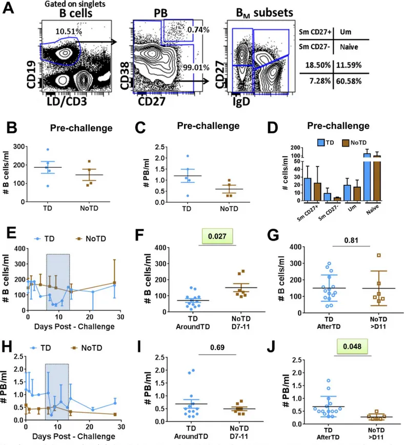
B cells were identified using CD19 (CD19+CD3-) (Fig 1A). Within B cells, plasmablasts (PB)
were identified as cells expressing high levels of CD27 and CD38 (CD27highand CD38high).
Non-PB cells were further categorized into various B memory (BM) subsets using the IgD/
CD27 classification scheme as proposed by Sanz et al [12], which delineates four
subpopula-tions: (i) Sm CD27+ [CD27+IgD-], (ii) Sm CD27- [CD27-IgD-], (iii) Um [CD27+IgD+] and (iv) naïve [CD27-IgD+] cells. No significant differences were observed in the frequency of all B
cells (B cells/ml) (Fig 1B), PB (PB/ml) (Fig 1C) or the various BMsubsets (cells/ml) (Fig 1D)
beforeS. Typhi challenge between participants who were diagnosed with typhoid (TD) and
those who were not (NoTD). We next assessed changes in the number of whole B cells (cell/
ml) after challenge (Fig 1E–1G). A significant reduction in the number of B cells in TD
(com-pared to NoTD) volunteers was observed AroundTD (Fig 1F). The reduction of these cells was
transitory as no differences between TD and NoTD volunteers was observed AfterTD (Fig
1G). Of note, PB did not show statistically significant differences in the frequencies between
TD and NoTD AroundTD (Fig 1H and 1I). However, an increase in frequency of these cells
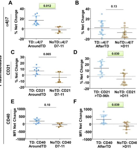
was noted AfterTD (Fig 1J). We then evaluated various molecules on PB including gut homing
(integrinα4β7), activation (CD40 and CD21) and class switching markers (IgA) as well as the
ability of these cells to bindS. Typhi (Fig 2andS1 Fig). These data are reported as net
differ-ences (percentages or MFI) AroundTD or AfterTD relative to day 0. TD volunteers showed a
significant upregulation of integrinα4β7, during the AroundTD time frame, compared to
NoTD volunteers (Figs2A,S1A and S1B). This homing marker returned to pre-challenge
lev-els AfterTD (Fig 2B). An up-regulation of CD21 and CD40 was also noted (Fig 2C–2Fand
S1D–S1F Fig) in TD volunteers and the increase was significant in the AfterTD time frame (Fig 2Dand2F). Moreover, the upregulation of CD21 by PB (AfterTD) correlated with the
increase of anti-flagella IgG antibody titers, but not anti-LPS or anti-Vi, AfterTD (S1I Fig)
[11]. Examples of the differences between pre-challenge and post-challenge in TD volunteers
as well as the time courses of the changes in these markers are included inS1 FigFinally, no
significant differences between TD and NoTD volunteers were noted in the expression of IgA
or the ability of PB to bindS. Typhi (S1 Fig).
Changes in B
Msubsets following wt
S
. Typhi infection
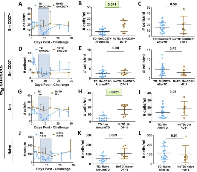
Evaluation of the frequency (cells/ml) of the BMsubsets (IgD/CD27 classification) between TD
and NoTD volunteers before challenge showed no significant differences among these groups (Fig 1D). Changes in the frequency (cells/ml) of the various BMsubsets after challenge (Fig 3) demonstrated that SmCD27+ and Um cells of TD volunteers decreased significantly
AroundTD, compared to NoTD volunteers (Fig 3A, 3B, 3G and 3H). However, the decrease
was transitory, since the frequency of the cells returned to pre-challenge levels by day 21 (Fig
3C and 3I). Changes in frequency were not evident in Sm CD27- or naïve cells (Fig 3D, 3E, 3J and 3K). When the data was analyzed as net percentage changes, only Um cells of TD
volun-teers (compared to NoTD) showed a significant decrease in AroundTD time frame (S2A–S2D
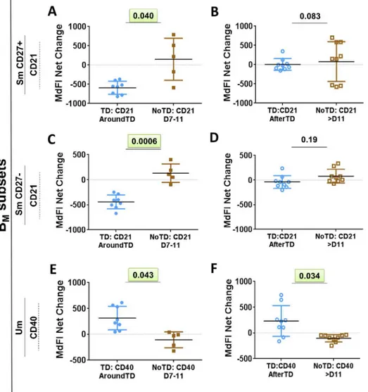
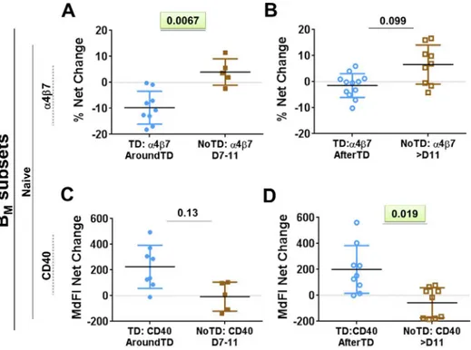
Fig). Next, we evaluated changes in the expression of various molecules (e.g., CD21, CD40,
integrinα4β7 and IgA, as well asS. Typhi binding) within each BMsubset (reported as net
changes relative to day 0) (Figs4,5andS2). Interestingly, despite that only SmCD27+ and
Um cells showed reduction in their frequency AroundTD, all BMsubsets showed significant
changes in some of the evaluated markers and most of them were AroundTD. However, in
Fig 1. Changes in frequency (cells/ml) of B cells induced by wtS. Typhi infection.PanelAdisplays the gating strategy used to identify various B cell subsets. Whole B cells were identified using CD19. Plasmablasts (PB) were identified as B cells (CD19+) expressing high levels of CD27 and CD38. Non-PB cells were classified into various memory B (BM) subsets using the IgD/CD27 classification: (i) Sm CD27+ (CD27+IgD-), (ii) Sm
CD27-(CD27-IgD-), (iii) Um (CD27+IgD+) and (iv) Naïve (CD27-IgD+). The frequency (cells/ml) of whole B cells, PB and BMsubsets in TD (blue symbols) and
Sm CD27+ and Sm CD27- cells AroundTD, compared to NoTD volunteers (Fig 4A–4Dand S2E–S2H Fig). In these volunteers, CD21 returned to pre-challenge levels AfterTD (Fig 4B– 4DandS2E–S2H Fig). In contrast, Um cells showed a significant up-regulation of CD40 frequency of B cells after challenge in TD (solid blue circles) and NoTD (solid brown squares) volunteers. The time frame AroundTD is indicated by the blue rectangle with dotted lines. AroundTD in TD volunteers include the day in which typhoid was diagnosed (TD+0h) and two extra time points, 48h and 96h post-diagnosis (TD+48h and TD+96h, respectively), in which blood samples were collected. Shown inFare the data of B cells from TD (solid blue circles) and NoTD (solid brown squares) volunteers in the AroundTD time frame. Shown inGare the data of B cells from TD (open blue circles) and NoTD (open brown squares) in the AfterTD time frame (days 14, 21 and 28 for TD and NoTD volunteers). Time course, AroundTD and AfterTD data of the frequency of plasmablasts (PB) is shown inH,IandJ, respectively. Shown is the P value from the mixed effects model analysis. Significant differences are highlighted in light green. Mean±SD are presented in all graphs.
doi:10.1371/journal.pntd.0004766.g001
Fig 2. Changes in expression of surface molecules in PB induced by wtS. Typhi infection.Shown in panels A,CandEare the expression of integrinα4β7, CD21 and CD40, respectively, from TD (solid blue circles) and NoTD (solid brown squares) volunteers in the AroundTD time frame. PanelsB,DandFshow the expression of integrinα4β7, CD21 and CD40, respectively, from TD (open blue circles) and NoTD (open brown squares) in the AfterTD time frame. The P value from the mixed effects model analysis is shown. Significant differences are highlighted in light green. Mean±SD are presented in all graphs.
AroundTD and AfterTD (compared to NoTD) (Fig 4E–4FandS2I and S2J Fig). Naïve cells of
TD volunteers showed a significant down-regulation of integrinα4β7 AroundTD (compared
to NoTD) and this marker returned to pre-challenge levels around day 21 (Figs5A, 5B,S2K
and S2L). In naïve cells, up-regulation of CD40 was also present. This marker started to
increase AroundTD (S3A and S3B Fig) in TD volunteers, but statistically significant
differ-ences were demonstrated only AfterTD (Fig 5D). The levels of IgA remained unchanged in Sm
CD27+ and Sm CD27- cells compared to pre-challenge and no differences between TD and
NoTD volunteers were identified in any time frame (S3CandS3D Fig). Similarly, no BM
sub-set showed significant changes in the binding toS. Typhi (S3E–S3H Fig). A table summarizing
all the markers evaluated in the various B subsets is displayed inS4 Fig.
Fig 3. Changes in frequency (cells/ml) of BMsubsets induced by wtS. Typhi infection.BMsubsets (IgD/CD27
classification) were evaluated for changes in frequency (cells/ml) elicited by exposure to wtS. Typhi. PanelsA,D, GandJ display the time courses of the changes in Sm CD27+, Sm CD27-, Um and Naïve cells, respectively, in TD and NoTD volunteers
(blue and brown simbols, respectively). The AroundTD time frame is indicated in panelsA, D, GandJby the blue rectangles with dotted lines. PanelsB,E, HandKshown changes in Sm CD27+, Sm CD27-, Um and Naïve subsets, respectively, from TD (solid blue circles) and NoTD (solid brown squares) volunteers in the AroundTD time frame. PanelsC,F, Iand L, show changes in Sm CD27+, Sm CD276-, Um and Naïve subsets, respectively, from TD (open blue circles) and NoTD (open brown squares) in
the AfterTD time frame. The P value from the mixed effects model analysis is shown. Significant differences are highlighted in light green. Mean±SD are presented in all graphs.
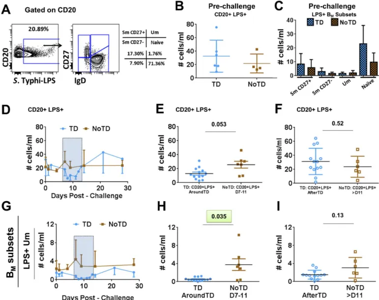
Identification of antigen-specific B cells by
S
. Typhi-LPS micelles
LPS is one of the most abundant antigens on the surface ofS. Typhi and to which anti-O-LPS
antibodies are directed. Additionally, LPS-specific memory B cells have recently been reported
[38,39]. To identify and evaluateS. Typhi-LPS-specific B cells,S. Typhi-LPS micelles of
nano-particle size (LPS-nanonano-particles) were used [40]. These LPS-nanoparticles contain fluorescent
Quantum dot (Qdot655) cores to allow identification of antigen-specific cells. Furthermore, LPS-nanoparticles are used as stimulants to determine if the receptor(s) interacting with this reagent induce activation of signaling pathways. Since human B cells lack TLR4 and CD14, these receptors cannot induce signaling. However, LPS-specific memory B cells have been reported suggesting that clones specific for this molecule are developed upon infection or vacci-nation and therefore, the B cell receptor (BCR), which drives the initial clonal selection, is the most likely candidate to interact with LPS. Thus, the main objective of these experiments was Fig 4. Changes in surface molecules of Sm CD27+, Sm CD27- and Um subsets induced by wtS. Typhi infection.Shown in panelsA,CandEare the expression of CD21 and CD40 from TD (solid blue circles) and NoTD (solid brown squares) volunteers in the AroundTD time frame. PanelsB,DandFshow the expression of CD21 and CD40 from TD (open blue circles) and NoTD (open brown squares) volunteers in the AfterTD time frame. The P value from the mixed effects model analysis is shown. Significant differences are highlighted in light green. Mean±SD are presented in all graphs.
to determine changes in the intracellular signaling pathways of B cells induced byS. Typhi infection and identify whether differences were present between volunteers who developed dis-ease (TD), and those who did not (NoTD). PBMC were stimulated with LPS-nanoparticles
(~5–10 ug; 10 min; 37°C) and then fixed for analysis by phospho-flow.S. Typhi-LPS-specific B
cells were identified and the distribution of these cells within the different BMsubsets was
eval-uated (Fig 6A). No differences in the frequency (cells/ml) ofS. Typhi-LPS-specific B cells or
within the BMsubsets between TD and NoTD volunteers before challenge (day 0) were
identi-fied (Fig 6B and 6C) (S5A Figdisplay gating examples ofS. Typhi-LPS-specific B cells from
TD volunteers on day 0. Additionally,S5B FigandS5C Figshow percentage comparison
between TD and NoTD groups at day 0 and the percentage distribution in the different BM
subsets). Furthermore, after challenge, no differences in the frequency ofS. Typhi-LPS-specific
B cells between TD and NoTD volunteers was identified (Fig 6D–6F). Subsequently, we
exam-ined changes in the frequency of BMsubsets within CD20+ LPS+ B cells. Only Um (CD20
+ LPS+ IgD+ CD27+) cells of TD volunteers (compared to NoTD) showed a significant
decrease in their frequency AroundTD (Fig 6G and 6H). The Um cell frequency was back to
pre-challenge levels AfterTD (Fig 6G–6I). The other BMsubsets did not show significant
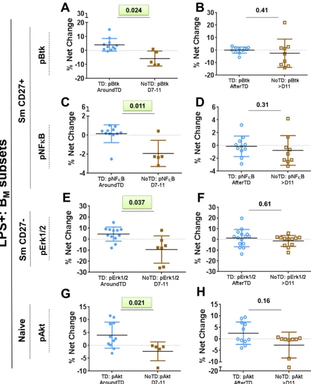
changes (TD vs. NoTD) in their frequencies (S5DandS5F Fig). We next evaluated changes
(net percentage relative to day 0) in the BMsubsets phosphorylating signaling proteins
associ-ated with the BCR after challenge. The results were compared between TD and NoTD
volun-teers (Fig 7). The BCR-associated signaling proteins evaluated included: Spleen tyrosine kinase
(Syk); Bruton’s tyrosine kinase (Btk), Protein kinase B (Akt), p38 mitogen-activated protein
kinase (p38MAPK), extracellular-signal-regulated kinase (Erk) 1/2 and nuclear factor kappa-Fig 5. Changes in surface molecules of Naive cells induced by wtS. Typhi infection.Shown in panelsA andCare the expression of integrinα4β7 and CD40 from TD (solid blue circles) and NoTD (solid brown squares) volunteers in the AroundTD time frame. PanelsBandDshow the expression of integrinα4β7 and CD40 from TD (open blue circles) and NoTD (open brown squares) volunteers in the AfterTD time frame. The P value from the mixed effects model analysis is shown. Significant differences are highlighted in light green. Mean±SD are presented in all graphs.
light-chain-enhancer of activated B cells (NFκB).S6A Figshows examples of the changes in
signaling (pAkt) induced byS. Typhi-LPS micelle stimulation in naïve (CD20+ LPS+ IgD
+ CD27-) cells in 3 volunteers. The examples show the percentage of cells phosphorylating Akt pre-challenge (day 0) and post-challenge (TD+48h or TD+96h). The gates were set up based on cells stimulated with media only (unstimulated control) and the data was reported as per-centage differential (% Net changes). Significant differences in the net perper-centage of cells phos-phorylating signaling proteins were identified in TD volunteers (compared to NoTD
volunteers) and the changes were concentrated in the AroundTD time frame. These changes were evident in Sm CD27+ (CD20+ LPS+ CD27+), Sm CD27- (CD20+ LPS+ IgD-Fig 6. Identification ofS. Typhi-LPS-specific B cells.PanelsAshows the gating strategy used to identifyS. Typhi-specific B cells using
LPS-nanoparticles and the different BMsubsets within theS. Typhi-LPS specific cells in a representative volunteer. Panels B and C show the frequency
(cells/ml) of allS. Typhi-LPS-specific B cells, as well as within the BMsubsets, in TD (blue symbols) and NoTD (brown symbols) volunteers before
wild-type challenge. PanelsD, EandFshow the time course, AroundTD and AfterTD, respectively, of the frequency (cells/ml) ofS. Typhi-LPS-specific B cells in TD (blue symbols) and NoTD (brown symbols) volunteers. Shown in panelsG, HandIare the time course, AroundTD and AfterTD,
respectively, of the frequency (cells/ml) ofS. Typhi-LPS-specific Um (CD20+ LPS+ IgD+ CD27+) cells in TD (blue symbols) and NoTD (brown
symbols) volunteers. The P value from the mixed effects model analysis is shown. Significant differences are highlighted in light green. Mean±SD are presented in all graphs.
Fig 7. Phosphorylation of diverse signaling molecules in BMsubsets following wtS. Typhi challenge.The
percentages ofS. Typhi-LPS-specific B cells phosphorylating specific signaling proteins (e.g., Btk, NFkB, Erk1/2 and Akt) were evaluated in each BMsubset. Phosphorylation was evaluated after stimulation withS. Typhi-LPS-nanoparticles.
PanelsAandBshow the % net changes of Btk phosphorylation (pBtk) in AroundTD and AfterTD, respectively, of Sm CD27+ cells in TD (blue symbols) and NoTD (brown symbols) volunteers. PanelsCandDshow the % net changes of NFκB phosphorylation (pNFκB) in AroundTD and AfterTD, respectively, in Sm CD27+ cells in TD (blue symbols) and NoTD (brown symbols) volunteers. PanelsEandFshow the % net changes of Erk1/2 phosphorylation (pErk1/2) AroundTD and AfterTD, respectively, in Sm CD27- cells in TD (blue symbols) and NoTD (brown symbols) volunteers. PanelsGandHshow the % net changes of Akt phosphorylation (pAkt) AroundTD and AfterTD, respectively, in naïve cells
in TD (blue symbols) and NoTD (brown symbols) volunteers. The P value from the mixed effects model analysis is shown. Significant differences are highlighted in light green. Mean±SD are presented in all graphs.
CD27-) and naïve (CD20+ LPS+ IgD+ CD27-) cells (Fig 7andS6B–S6E Fig). In Sm CD27
+ cells of TD volunteers, the percentage of cell phosphorylating Btk and NFκB was higher than
NoTD volunteers AroundTD (Fig 7A and 7C). Sm CD27- cells (from TD volunteers) showed
a significantly higher percentage of cells phosphorylating Erk1/2 (Fig 7E). Finally, a significant
higher percentage of naïve cells phosphorylating Akt was also noted (Fig 7G).S6B–S6E Fig
shows the time course of the changes induced byS. Typhi infection in B cells signaling proteins
where significant differences were identified. The table shown inS6F Figsummarizes the
results of phospho-proteins evaluated in all the BMsubsets.
Discussion
BecauseS. Typhi is a human restricted pathogen and no animal model is capable of
reproduc-ing all the clinical aspects of this disease, studies in humans are necessary to understand patho-gen-human host immune interactions. These studies ultimately hold the potential to aid in the design of novel vaccines. The specimens used in the current study were collected as part of a
clinical study aimed at developing a new human challenge model to study typhoid disease [11].
B cells are part of the adaptive immune system which, in addition to being responsible for anti-body production, perform other important functions including antigen presentation and cyto-kine production. Recent advances in B cell biology have shown that the B cell compartment is composed of multiple subsets whose functions are not clearly understood, particularly in the context of infectious diseases. In the current study we aimed at studying changes in the
fre-quency, activation, and signaling induced byS. Typhi in peripheral blood B cells of volunteers
challenged with 104CFU of wtS. Typhi (Quailes strain). Importantly, by comparing the results
between TD and NoTD volunteers we aimed at furthering our understanding of the role of this
important component of the human adaptive immune response when first encounteringS.
Typhi, and their potential to impact the development of disease as well as anamnestic responses.
No differences in the frequency (cells/ml) whole B cells (CD19+CD3-) or plasmablasts (PB)
(CD3-CD19+CD27highCD38high) were identified before wild-type challenge, suggesting that it
is not possible to predict which volunteers will develop disease by simply looking at the
fre-quency of these cells in circulation (Fig 1B and 1C). Of note, after challenge the frequency of
whole B cells (Fig 1E–1G) was reduced in volunteers that developed disease (TD), mimicking
the results reported in the parent study (decrease of whole cell counts (WCC) and
lympho-cytes) [11]. We further evaluated the effects of typhoid fever in various B cell subsets.
Interest-ingly, plasmablasts (PB), a B cell subset that produces antibodies early after infection, did not show a reduction in the frequency AroundTD in TD volunteers, but an increase and AfterTD (Fig 1J). This increase in frequency was accompanied by upregulation of CD21 and CD40 (Fig 2D and 2F), which are co-activator and activator markers, respectively. This suggests that these cells are activated and could be the responsible for giving rise to plasma cells and memory
cells [47]. This is further supported by the positive correlation between CD21 upregulation by
PB and the increase in anti-flagella antibody titers (S1I Fig). Of interest, no correlation was
identified with anti-LPS antibody titers.
Integrinα4β7-expressing B and T cells identified in peripheral blood are re-circulating cells
that left Peyer’s patches (PP) and/or mesenteric lymph nodes (MLN) upon being primed by
intestinal DC [48–51]. Upregulation of integrinα4β7 AroundTD by PB (Fig 2A) suggest that a
proportion of these cells are getting ready to migrate to the gut, which is not surprising since it
is the primary site of infection. This upregulation of integrinα4β7 and antibody production by
Similar to observations with whole B cells and PB, no differences were noted in the
fre-quency (cells/ml) of the BMsubsets before challenge between TD and NoTD volunteers (Fig
1D). However, following wild-type challenge, Sm CD27+ and Um cells showed a significant
reduction in their frequency AroundTD in TD volunteers (compared to NoTD) (Fig 3B, 3H
and 3K), which suggest that some B cell subsets are more affected than other by typhoid fever. Further evidence of the changes induced in B cells by typhoid came from the analysis of activa-tion and homing markers. CD40 was significantly upregulated in a significant proporactiva-tion of
the naïve subset (Figs4E–4Fand5D) showing that these cells were activated. Considering that
naïve B cells give rise to all the other subsets, including PB, plasma cells and memory B cells, it was not surprising to observe that these cells were activated. Of note, naïve B cells also showed
downregulation of integrinα4β7 (AroundTD;Fig 5A), which is intriguing since it was
expected that these cells would migrate to the gut. These data might indicate that naïve B cells first migrate to other secondary organ(s) (currently undefined, but different from the gut)
where they differentiate further (e.g., PB) and acquire the expression of integrinα4β7.
Alterna-tively, these data might suggest that different B subsets use different gut homing receptors, for example naïve cells might use CCR9 or CD103 to reach the gut and once these cells commit to
another lineage (e.g., PB) integrinα4β7 is upregulated. Yet another possibility is that naïve cells
have already migrated to the gut using the integrinα4β7 receptor, resulting in lower levels of
circulating integrinα4β7+ B naïve cells. Future studies will be directed to further explore these
possibilities.
Um cells have been described to produce high-affinity IgM in the early phase of infections
and the equivalent of B1 murine cells [53,54]. However, the exact origin and function of these
cells in humans remains controversial. Here, we demonstrate that these cells play a role in
typhoid fever, since despite that their frequency is reduced AroundTD (Fig 3H), some of these
cells show signs of activation (Fig 4E–4F). Their role in typhoid and other enteric infections
remains to be further explored.
CD21 works in conjunction with CD19 (CD19/CD21 complex) to enhance BCR signaling, leading to B cell activation. Downregulation of CD21 by Sm CD27+ and Sm CD27- cells
AroundTD in TD volunteers (Fig 4A and 4C) might represent a physiological process during
the development of a normal B cell response, particularly in its early stages. We observed that
CD21 was upregulated in PB (Fig 2D–2F), which likely represent antigen-specific cells already
producing antibodies and which require activation to continue their developmental process. In contrast, Sm CD27+ and Sm CD27- cells might contain memory B cells with affinity for
anti-gens other thanS. Typhi LPS. In the case of any infectious disease, the immune response can
lead to an environment (e.g., cytokine milieu) that facilitates the activation of innate and anti-gen specific (adaptive) cells. In the case of B cells, downregulation of co-activators in cells that have not encountered an antigen specific for the invading microorganism might represent a mechanism to limit their activation. To our knowledge, CD21 downregulation has been
reported in some autoimmune diseases and these cells are characterized by being anergic [55,
56]. Therefore, these B cells (notS. Typhi-specific) might be refractory to activation.
One of the most abundant molecules on the surface ofS. Typhi and to which antibodies are
directed is LPS. LPS is a well-recognized mouse, but not human, B-cell mitogen. This is due to the fact that unlike mouse B cells, human B cells express neither TLR4 nor CD14. However, in
humans exposed toS. Typhi, anti-LPS-O antibodies are detected and used as an indicator of
successful immunization or challenge. This suggests that B cell clones specific for this antigen
are generated during the development of immune responses toS. Typhi. Considering that
clonal selection for B cells occurs through interaction of the antigen with the BCR, the presence of anti-LPS antibodies indicate that LPS was recognized by B cells that developed into antibody
the identification of changes in signaling profiles elicited by typhoid fever. To this end, we used LPS-nanoparticles (S. Typhi-LPS), which contain a quantum dot (QDot 655) core, as stimu-lants and evaluated the BCR associated signaling pathways induced within the different BM
subsets that interacted with the LPS-nanoparticle [40]. Importantly, LPS is a very“sticky”
mol-ecule and its“sticky”nature is thought to be dependent on ionic interaction [57]. Therefore,
besides the BCR (specific interaction), it is likely that LPS interacts non-specifically with other molecules, especially if they have a net positive charge (LPS has a net negative charge). As
expected, due to the large repetitive nature of LPS and its“stickiness”, a relatively high
percent-age of B cells interacted with the LPS-nanoparticles (high background) (Fig 6A–6C). This is
evidenced in pre-challenge samples (day 0) where LPS interaction of B cells raged from ~10–
25% (S5A Fig), despite the fact that these volunteers did not exhibit anti-S. Typhi antibodies
(anti-LPS, anti-Vi or anti-flagellin) before challenge as previously reported [11]. We expected
that only a small percentage of cells interacting with LPS will actually respond to the stimuli (specific interaction), which in our assay was determined by phosphorylation of signaling pro-teins. To identify the differential in activation of BCR signaling pathways induced by typhoid, each sample was corrected for background using the phosphorylation on each protein induced
in day 0. Following wild-typeS. Typhi challenge, naïve B cells from TD volunteers showed a
significant difference (compared to NoTD) in the net percentage of cells phosphorylating Akt
AroundTD (Fig 7G). Akt is part of the survival pathway and this suggests that naïve B cells of
TD volunteers that interact with the LPS-nanoparticles are more resistant to apoptosis. These
data complemented the observations that naïve B cells were activated (CD40 upregulation–Fig
5D). Interestingly, we also noticed differences in the Sm CD27+ population, in which NFκB
and Btk phosphorylation showed differences between TD and NoTD volunteers AroundTD (Fig 7A and 7C). Sm CD27- B cells from TD volunteers also showed differential in the phos-phorylation of Erk1/2 (compared to NoTD). Whether either one of these pathways leads to the
downregulation of CD21, as identified in the Sm CD27+ and Sm CD27- AroundTD (Fig 4A
and 4C) remains to be explored. Also, whether activation of these signaling proteins indicate that these cells are memory cells, will be evaluated in future studies. We acknowledge, however, that since we evaluated phosphorylation at a single time point (10 minutes stimulation), we might have captured only a fraction of the changes that these cells are undergoing. Future stud-ies will be directed to evaluate in more detail the kinetics of the signaling pathways associated with the BCR activation in TD and NoTD volunteers.
In summary, the changes induced byS. Typhi infection in volunteers that developed disease
(TD) were analyzed in various B cell subpopulations. The cells that showed the most important changes were PB which exhibited significant increases in various key activation and gut hom-ing molecules. Naïve B cells showed activation and enhanced survival, which are likely related to their differentiation into PB as well as plasma cells and possibly long term memory B cells. Interestingly, changes in Sm CD27+ and Sm CD27- cells were also identified including down-regulating CD21 as a possible mechanism to avoid non-specific activation. All these changes
were present only in TD volunteers, suggesting that these were induced by exposure to wtS.
Typhi and are in response to the development of disease. To our knowledge this is the first study to describe these phenomena on human B cells of volunteers that developed typhoid
dis-ease after wild-type challenge. The changes that otherS. Typhi antigens (e.g., flagella, Vi)
induce in the different BMsubsets will be addressed in future studies.
Supporting Information
S1 Fig. Changes in PB induced by wtS. Typhi infection.PanelsAandBshow examples of
(percentage) of this molecule in PBs. TD and NoTD volunteers are indicated by the blue and
brown symbols, respectively. PanelsCandDshow examples of the changes in the expression of
CD21 and the time course of the net change (percentage) of this molecule in PBs. PanelsEandF
show examples of the changes in the expression of CD40 and the time course of the net change
(MFI) of this molecule in PBs. GraphsA,CandEshow the data before challenge and peak
increases after-challenge. In PanelEthe bar color bar indicates the net MFI change over
pre-chal-lenge. Also sown are the raw MFI values. PanelsGandHshow the complete time courses of IgA
expression andS. Typhi binding in PB in TD (blue symbols) and NoTD (brown symbols)
volun-teers. AroundTD is indicated by the blue rectangles with dotted lines in panelsB, D, F, G,and
H. PanelsB, D, F, G,andHdisplay Mean ± SD. PanelIdisplays the Spearman correlation results
between PB CD21+ cells (% net change over day 0; AfterTD time frame) and the anti-flagellin IgG antibody titer (fold-increase over day 0; After TD time frame) in TD volunteers. The AfterTD time frame for the antibody titers included data from days 14, 28 and 60. (PDF)
S2 Fig. Changes in BMsubsets induced by wtS. Typhi infection (part 1).PanelsA-Dshow
the time course of the net changes (percentage) of the various BMpopulations. The p value
(mixed effects model) for significant differences between TD and NoTD volunteers at
AroundTD is indicated in the green rectangle. PanelsEandFshow examples of the changes in
CD21 expression in Sm CD27+ (TD volunteer) and the time course of the net change
(percent-age) of this molecule in Sm CD27+. PanelsGandHshow examples of the changes in CD21
expression in Sm CD27- (TD volunteer) and the time course of the net change (percentage) of
this molecule in Sm CD27-. PanelsIandJshow examples of the changes in CD40 expression
in Um cells (TD volunteer) and the time course of the net change (median fluorescence
inten-sity -MdFI-) of this molecule in Um cells. Histograms overlays inE,GandIdisplay MdFI. The
bar color indicates the net MdFI change over pre-challenge. Raw MdFI data are also shown.
PanelKandLshow an example of the changes in integrinα4β7 expression in Naïve cells (TD
volunteer), at peak change time point after challenge and the time course of the net changes (percentage) of this molecule in Naïve cells. AroundTD is indicated by the blue rectangles with
dotted lines in panelsA-D, F, H, J,andL. In the same panels TD and NoTD volunteers are
indicated by the blue and brown symbols, respectively. PanelsA-D, F, H, J,andLdisplay
Mean ± SD. (PDF)
S3 Fig. Changes in BMsubsets induced by wtS. Typhi infection (part 2).PanelsAandB show examples of the changes in CD40 expression in naive cells (TD volunteer) and the time
course of the net change (MdFI) of this molecule in naive cells. PanelsC-Hshow the complete
time courses of various other markers evaluated in the BMsubsets. AroundTD is indicated by
the blue rectangles with dotted lines in panelsB-H. In the same panels TD and NoTD
volun-teers are indicated by the blue and brown symbols, respectively. PanelsB-Hdisplay
Mean ± SD. (PDF)
S4 Fig. Summary of changes in the expression of all molecules evaluated in the various BM
shown in highlighted boxes (light green). These boxes also include the Fig numbers in which the data is presented in the manuscript.
(PDF)
S5 Fig.S. Typhi-LPS-specific B cells.S. Typhi-specific B cells were identified and
concomi-tantly stimulated usingS. Typhi-LPS-nanoparticles. PanelAdisplays bi-exponential plots
showing the gating of LPS+ cells within the CD20+ population. Volunteers evaluated in the TD group are shown and, as expected, a high non-specific binding of LPS-nanoparticles was
identified due to the“sticky”nature of LPS. PanelBshows the percentage of CD20+ LPS+ B
cells at day 0 (pre-challenge) in TD (blue symbols) and NoTD (brown symbols) groups and
demonstrate that no differences between these groups existed. PanelCshows the percentage of
BMcell subsets that are CD20+ LPS+ cells at day 0, demonstrating that no differences between
TD (blue symbols) and NoTD (brown symbols) groups exist before challenge. PanelsD-F
dis-play time courses of the frequency (cells/ml) of SmCD27+, Sm CD27- and Naïve cells. No dif-ferences between TD and NoTD groups were identified. Importantly, Um cells showed a
significant difference (Fig 6) between TD and NoTD groups AroundTD. This is described in
detail in the main body of the manuscript. In panelsD-FAroundTD is indicated by the blue
rectangles with dotted lines. In the same panels TD and NoTD volunteers are indicated by the
blue and brown symbols, respectively. PanelsD-Fdisplay Mean ± SD.
(PDF)
S6 Fig.S. Typhi-LPS-specific B cells and their intracellular signaling profile.S.
Typhi-spe-cific B cells were identified and concomitantly stimulated usingS. Typhi-LPS-nanoparticles.
Changes in the signaling profile induced by typhoid were evaluated in the various BMsubsets.
PanelAshows three examples of the changes in the percentage of cells phosphorylating Akt
(pAkt) after challenge (day 0 vs. peak phosphorylation AroundTD) in TD volunteers. The gates were set up based on media only stimulation. The data were reported as the differential in
phos-phorylation between days post-challenge and day 0. PanelsB,C,DandEshow time courses of
the net changes (percentages) in phosphorylation of signaling proteins showing significant differ-ences between TD and NoTD volunteers AroundTD. These changes were present in SmCD27+,
Sm CD27- and Naïve cells. In panelsB-EAroundTD is indicated by the blue rectangles with
dot-ted lines. In the same panels TD and NoTD volunteers are indicadot-ted by the blue and brown
sym-bols, respectively. PanelsB-Edisplay Mean ± SD. PanelFshows a table summarizing the results
of the signaling proteins evaluated in the different BMsubsets. Information found in the boxes
include the time frame in which the changes were identified (e.g., TD+0h to TD+96h); how the marker was evaluated (e.g., % net change compared to day 0) and the P value from the Mixed Effects Model analysis. Significant data are shown in highlighted boxes (light green). These boxes also include the Fig numbers in which the data is presented in the manuscript.
(PDF)
Acknowledgments
We are indebted to the volunteers who participated in the studies. The content is solely the responsibility of the authors and does not necessarily represent the official views of the National Institute of Allergy and Infectious Diseases or the National Institutes of Health or other granting institutions.
Author Contributions
LSM. Wrote the paper: FRT PJB LSM TCD CSW CJB BA AJP MML MBS. Set up the challenge model and generated the clinical data: TCD CSW MML AJP. Collected and processed the PBMC specimens: TCD CSW CJ CJB BA. Performed statistical analysis using the Mixed Effects Model: LSM.
References
1. Lozano R, Naghavi M, Foreman K, Lim S, Shibuya K, Aboyans V, et al. Global and regional mortality from 235 causes of death for 20 age groups in 1990 and 2010: a systematic analysis for the Global Bur-den of Disease Study 2010. The Lancet. 2012; 380(9859):2095–128.
2. Mead PS, Slutsker L, Dietz V, McCaig LF, Bresee JS, Shapiro C, et al. Food-related illness and death in the United States. Emerg Infect Dis. 1999; 5(5):607–25. PMID:10511517
3. Mogasale V, Maskery B, Ochiai RL, Lee JS, Mogasale VV, Ramani E, et al. Burden of typhoid fever in low-income and middle-income countries: a systematic, literature-based update with risk-factor adjust-ment. The Lancet Global health. 2014; 2(10):e570–80. doi:10.1016/S2214-109X(14)70301-8PMID: 25304633
4. Levine MM, Tacket CO, Sztein MB. Host-Salmonella interaction: human trials. Microbes Infect. 2001; 3 (14–15):1271–9. PMID:11755415
5. Pasetti MF, Levine MM, Sztein MB. Animal models paving the way for clinical trials of attenuated Sal-monella enterica serovar Typhi live oral vaccines and live vectors. Vaccine. 2003; 21(5–6):401–18. PMID:12531639
6. Hornick RB, Greisman SE, Woodward TE, DuPont HL, Dawkins AT, Snyder MJ. Typhoid Fever: Patho-genesis and Immunologic Control. New England Journal of Medicine. 1970; 283(13):686–91. PMID: 4916913
7. Gilman RH, Hornick RB, Woodward WE, DuPont HL, Snyder MJ, Levine MM, et al. Evaluation of a UDP-Glucose-4-Epimeraseless Mutant of Salmonella typhi as a Live Oral Vaccine. Journal of Infec-tious Diseases. 1977; 136(6):717–23. PMID:925379
8. Hornick RB, Woodward TE. Appraisal of typhoid vaccine in experimentally infected human subjects. Transactions of the American Clinical and Climatological Association. 1967; 78:70–8. PMID:6028241 9. Gilman RH, Hornick RB. Duodenal isolation of Salmonella typhi by string capsule in acute typhoid
fever. J Clin Microbiol. 1976; 3(4):456–7. PMID:770502
10. Waddington CS, Darton TC, Woodward WE, Angus B, Levine MM, Pollard AJ. Advancing the manage-ment and control of typhoid fever: A review of the historical role of human challenge studies. Journal of Infection. 2014; 68(5):405–18. doi:10.1016/j.jinf.2014.01.006PMID:24491597
11. Waddington CS, Darton TC, Jones C, Haworth K, Peters A, John T, et al. An Outpatient, Ambulant-Design, Controlled Human Infection Model Using Escalating Doses of Salmonella Typhi Challenge Delivered in Sodium Bicarbonate Solution. Clinical Infectious Diseases. 2014; 58(9):1230–40. doi:10. 1093/cid/ciu078PMID:24519873
12. Sanz I, Wei C, Lee FE, Anolik J. Phenotypic and functional heterogeneity of human memory B cells. Semin Immunol. 2008; 20(1):67–82. doi:10.1016/j.smim.2007.12.006PMID:18258454
13. Wei C, Anolik J, Cappione A, Zheng B, Pugh-Bernard A, Brooks J, et al. A New Population of Cells Lacking Expression of CD27 Represents a Notable Component of the B Cell Memory Compartment in Systemic Lupus Erythematosus. The Journal of Immunology. 2007; 178(10):6624–33. PMID: 17475894
14. Qian Y, Wei C, Eun-Hyung Lee F, Campbell J, Halliley J, Lee JA, et al. Elucidation of seventeen human peripheral blood B-cell subsets and quantification of the tetanus response using a density-based method for the automated identification of cell populations in multidimensional flow cytometry data. Cytometry B Clin Cytom. 2010; 78 Suppl 1:S69–82. doi:10.1002/cyto.b.20554PMID:20839340 15. Blair PA, Noreña LY, Flores-Borja F, Rawlings DJ, Isenberg DA, Ehrenstein MR, et al. CD19
+CD24hiCD38hi B Cells Exhibit Regulatory Capacity in Healthy Individuals but Are Functionally Impaired in Systemic Lupus Erythematosus Patients. Immunity. 2010; 32(1):129–40. doi:10.1016/j. immuni.2009.11.009PMID:20079667
16. Mauri C, Bosma A. Immune Regulatory Function of B Cells. Annu Rev Immunol. 2012; 30(1):221–41. 17. Narvaez CF, Feng N, Vasquez C, Sen A, Angel J, Greenberg HB, et al. Human rotavirus-specific IgM Memory B cells have differential cloning efficiencies and switch capacities and play a role in antiviral immunity in vivo. J Virol. 2012; 86(19):10829–40. doi:10.1128/JVI.01466-12PMID:22855480 18. Khaskhely N, Mosakowski J, Thompson RS, Khuder S, Smithson SL, Westerink MAJ. Phenotypic
19. Taylor JJ, Jenkins MK, Pape KA. Heterogeneity in the differentiation and function of memory B cells. Trends in immunology. 2012; 33(12):590–7. doi:10.1016/j.it.2012.07.005PMID:22920843
20. Kruetzmann S, Rosado MM, Weber H, Germing U, Tournilhac O, Peter H-H, et al. Human Immunoglob-ulin M Memory B Cells Controlling Streptococcus pneumoniae Infections Are Generated in the Spleen. The Journal of Experimental Medicine. 2003; 197(7):939–45. PMID:12682112
21. So NSY, Ostrowski MA, Gray-Owen SD. Vigorous Response of Human Innate Functioning IgM Mem-ory B Cells upon Infection by Neisseria gonorrhoeae. The Journal of Immunology. 2012; 188(8):4008– 22. doi:10.4049/jimmunol.1100718PMID:22427638
22. Levine MM, Galen J. E., Tacket E. M., Barry M. F., Pasetti M. F., and Sztein M. B. Attenuated strains of Salmonella enterica serovar Typhi as Live Oral Fever. In: Levine MM, Kaper J. B., Lui M. and Good M., editor. New Generation Vaccines. New York: Marcel Dekker; 2004. p. 479.
23. de Wit J, Souwer Y, Jorritsma T, Klaasse Bos H, ten Brinke A, Neefjes J, et al. Antigen-Specific B Cells Reactivate an Effective Cytotoxic T Cell Response against PhagocytosedSalmonellathrough
Cross-Presentation. PLoS ONE. 2010; 5(9):e13016. doi:10.1371/journal.pone.0013016PMID:20885961 24. Mastroeni P, Simmons C, Fowler R, Hormaeche CE, Dougan G. Igh-6(-/-) (B-cell-deficient) mice fail to
mount solid acquired resistance to oral challenge with virulent Salmonella enterica serovar typhimurium and show impaired Th1 T-cell responses to Salmonella antigens. Infect Immun. 2000; 68(1):46–53. PMID:10603367
25. Mittrucker HW, Raupach B, Kohler A, Kaufmann SH. Cutting edge: role of B lymphocytes in protective immunity against Salmonella typhimurium infection. J Immunol. 2000; 164(4):1648–52. PMID: 10657605
26. Lopez-Medina M, Perez-Lopez A, Alpuche-Aranda C, Ortiz-Navarrete V. Salmonella modulates B cell biology to evade CD8(+) T cell-mediated immune responses. Front Immunol. 2014; 5:586. doi:10. 3389/fimmu.2014.00586PMID:25484884
27. McSorley SJ, Jenkins MK. Antibody is required for protection against virulent but not attenuated Salmo-nella enterica serovar typhimurium. Infect Immun. 2000; 68(6):3344–8. PMID:10816483
28. Barr TA, Brown S, Mastroeni P, Gray D. B cell intrinsic MyD88 signals drive IFN-gamma production from T cells and control switching to IgG2c. J Immunol. 2009; 183(2):1005–12. doi:10.4049/jimmunol. 0803706PMID:19542370
29. Barr TA, Brown S, Mastroeni P, Gray D. TLR and B cell receptor signals to B cells differentially program primary and memory Th1 responses to Salmonella enterica. J Immunol. 2010; 185(5):2783–9. doi:10. 4049/jimmunol.1001431PMID:20675594
30. Ruprecht CR, Lanzavecchia A. Toll-like receptor stimulation as a third signal required for activation of human naive B cells. Eur J Immunol. 2006; 36(4):810–6. PMID:16541472
31. Nothelfer K, Sansonetti PJ, Phalipon A. Pathogen manipulation of B cells: the best defence is a good offence. Nature reviews Microbiology. 2015; 13(3):173–84. doi:10.1038/nrmicro3415PMID:25659322 32. Souwer Y, Griekspoor A, Jorritsma T, de Wit J, Janssen H, Neefjes J, et al. B cell receptor-mediated
internalization of salmonella: a novel pathway for autonomous B cell activation and antibody produc-tion. J Immunol. 2009; 182(12):7473–81. doi:10.4049/jimmunol.0802831PMID:19494270 33. Souwer Y, Griekspoor A, de Wit J, Martinoli C, Zagato E, Janssen H, et al. Selective infection of
anti-gen-specific B lymphocytes by Salmonella mediates bacterial survival and systemic spreading of infec-tion. PLoS One. 2012; 7(11):e50667. doi:10.1371/journal.pone.0050667PMID:23209805
34. Rosales-Reyes R, Perez-Lopez A, Sanchez-Gomez C, Hernandez-Mote RR, Castro-Eguiluz D, Ortiz-Navarrete V, et al. Salmonella infects B cells by macropinocytosis and formation of spacious phago-somes but does not induce pyroptosis in favor of its survival. Microb Pathog. 2012; 52(6):367–74. doi: 10.1016/j.micpath.2012.03.007PMID:22475626
35. Neves P, Lampropoulou V, Calderon-Gomez E, Roch T, Stervbo U, Shen P, et al. Signaling via the MyD88 adaptor protein in B cells suppresses protective immunity during Salmonella typhimurium infec-tion. Immunity. 2010; 33(5):777–90. doi:10.1016/j.immuni.2010.10.016PMID:21093317
36. Shen P, Roch T, Lampropoulou V, O'Connor RA, Stervbo U, Hilgenberg E, et al. IL-35-producing B cells are critical regulators of immunity during autoimmune and infectious diseases. Nature. 2014; 507 (7492):366–70. doi:10.1038/nature12979PMID:24572363
37. Perez-Lopez A, Rosales-Reyes R, Alpuche-Aranda CM, Ortiz-Navarrete V. Salmonella downregulates Nod-like receptor family CARD domain containing protein 4 expression to promote its survival in B cells by preventing inflammasome activation and cell death. J Immunol. 2013; 190(3):1201–9. doi:10.4049/ jimmunol.1200415PMID:23284055
polysaccharide vaccine induces CD27+IgD-S. Typhi-specific IgA and IgG B memory cells in humans. ClinImmunol. 2011; 138(2):187–200.
39. Wahid R, Simon R, Zafar SJ, Levine MM, Sztein MB. Live oral typhoid vaccine Ty21a induces cross-reactive humoral immune responses against Salmonella enterica serovar Paratyphi A and S. Paratyphi B in humans. Clin Vaccine Immunol. 2012; 19(6):825–34. doi:10.1128/CVI.00058-12PMID:22492745 40. Toapanta FR, Bernal PJ, Fresnay S, Darton TC, Jones C, Waddington CS, et al. Oral Wild-Type
Salmo-nella Typhi Challenge Induces Activation of Circulating Monocytes and Dendritic Cells in Individuals Who Develop Typhoid Disease. PLoS neglected tropical diseases. 2015; 9(6):e0003837. doi:10.1371/ journal.pntd.0003837PMID:26065687
41. McArthur MA, Sztein MB. Heterogeneity of Multifunctional IL-17A ProducingS. Typhi-Specific CD8+ T
Cells in Volunteers following Ty21a Typhoid Immunization. PLoS ONE. 2012; 7(6):e38408. doi:10. 1371/journal.pone.0038408PMID:22679502
42. Toapanta FR, Ross TM. Impaired immune responses in the lungs of aged mice following influenza infection. Respir Res. 2009; 10:112. doi:10.1186/1465-9921-10-112PMID:19922665
43. Toapanta FR, Bernal PJ, Sztein MB. Diverse phosphorylation patterns of B cell receptor-associated signaling in naive and memory human B cells revealed by phosphoflow, a powerful technique to study signaling at the single cell level. Front Cell Infect Microbiol. 2012; 2:128. doi:10.3389/fcimb.2012. 00128PMID:23087912
44. Betanzos CM, Gonzalez-Moa M, Johnston SA, Svarovsky SA. Facile labeling of lipoglycans with quan-tum dots. Biochemical and Biophysical Research Communications. 2009; 380(1):1–4. doi:10.1016/j. bbrc.2008.12.167PMID:19150336
45. Anderson RE, Chan WCW. Systematic Investigation of Preparing Biocompatible, Single, and Small ZnS-Capped CdSe Quantum Dots with Amphiphilic Polymers. ACS Nano. 2008; 2(7):1341–52. doi:10. 1021/nn700450gPMID:19206301
46. Dubertret B, Skourides P, Norris DJ, Noireaux V, Brivanlou AH, Libchaber A. In Vivo Imaging of Quan-tum Dots Encapsulated in Phospholipid Micelles. Science. 2002; 298(5599):1759–62. PMID: 12459582
47. Grammer AC, Lipsky PE. CD154-CD40 interactions mediate differentiation to plasma cells in healthy individuals and persons with systemic lupus erythematosus. Arthritis Rheum. 2002; 46(6):1417–29. PMID:12115170
48. Iwata M, Hirakiyama A, Eshima Y, Kagechika H, Kato C, Song SY. Retinoic acid imprints gut-homing specificity on T cells. Immunity. 2004; 21(4):527–38. PMID:15485630
49. Mora JR, Iwata M, Eksteen B, Song SY, Junt T, Senman B, et al. Generation of gut-homing IgA-secret-ing B cells by intestinal dendritic cells. Science. 2006; 314(5802):1157–60. PMID:17110582
50. Johansson-Lindbom B, Svensson M, Pabst O, Palmqvist C, Marquez G, Forster R, et al. Functional specialization of gut CD103+ dendritic cells in the regulation of tissue-selective T cell homing. J Exp Med. 2005; 202(8):1063–73. PMID:16216890
51. Hammerschmidt SI, Ahrendt M, Bode U, Wahl B, Kremmer E, Forster R, et al. Stromal mesenteric lymph node cells are essential for the generation of gut-homing T cells in vivo. J Exp Med. 2008; 205 (11):2483–90. doi:10.1084/jem.20080039PMID:18852290
52. Toapanta FR, Simon JK, Barry EM, Pasetti MF, Levine MM, Kotloff KL, et al. Gut-homing conventional plasmablasts and CD27- plasmablasts elicited after a short time exposure to an oral live attenuated Shi-gella vaccine candidate in humans. Frontiers in Immunology. 2014; 5.
53. Weller S, Braun MC, Tan BK, Rosenwald A, Cordier C, Conley ME, et al. Human blood IgM“memory”B cells are circulating splenic marginal zone B cells harboring a prediversified immunoglobulin repertoire. Blood. 2004; 104(12):3647–54. PMID:15191950
54. Shi Y, Agematsu K, Ochs HD, Sugane K. Functional analysis of human memory B-cell subpopulations: IgD+CD27+ B cells are crucial in secondary immune response by producing high affinity IgM. Clin Immunol. 2003; 108(2):128–37. PMID:12921759
55. Isnardi I, Ng YS, Menard L, Meyers G, Saadoun D, Srdanovic I, et al. Complement receptor 2/CD21-human naive B cells contain mostly autoreactive unresponsive clones. Blood. 2010; 115(24):5026–36. doi:10.1182/blood-2009-09-243071PMID:20231422
56. Saadoun D, Terrier B, Bannock J, Vazquez T, Massad C, Kang I, et al. Expansion of autoreactive unre-sponsive CD21-/low B cells in Sjogren's syndrome-associated lymphoproliferation. Arthritis Rheum. 2013; 65(4):1085–96. doi:10.1002/art.37828PMID:23279883