PETRE CRISTIANA VIRGINIA, TĂNASE CĂTĂLIN
105
J. Plant Develop. 20(2013): 105 – 114
DESCRIPTION OF THE CULTURE CHARACTERISTICS OF
SOME LIGNICOLOUS BASIDIOMYCETES SPECIES GROWN ON
THREE SYNTHETIC MEDIA
PETRE Cristiana Virginia1*, T NASE C t lin1
Abstract: A number of12 species of lignicolous basidiomycetes were cultivated on potato dextrose agar and malt extract agar, incubated at 25 °C and carefully analyzed for a period of 5 weeks. Lignicolous basidiomycetes are fungi that produce potent enzymes and bioactive secondary metabolites which are successfully used in various industries: bioremediation of polluted environments, biodegradation of toxic substances, pharmacology or agriculture. The objective of this study was the description of the main characteristics of in vitro cultures of some lignicolous basidiomycetes species grown on synthetic media. The main characteristics followed were: the growth rate of the colonies, the general features of the mycelium: shape, color, surface aspect, reverse, the presence of fruiting bodies and exudates and the particular odor.
Key words: lignicolous basidiomycetes, in vitro cultures, culture characteristics. Introduction
Lignicolous basidiomycetes are fungi that play very important ecological roles within an ecosystem.
Through their complex enzymatic system, the lignicolous basidiomycetes are the main decomposer of wood in natural and anthropogenic habitats, being involved from this point of view in the natural cycles of some essential elements, such as carbon and nitrogen. Because of this particular capacity, showed by very few organisms, researchers began testing with impressive results the great potential of the lignolytic enzymes for degrading several manmade substances and materials: pesticides, synthetic dyes, polychlorinated biphenyls, synthetic polymers.
Besides the highly reactive enzymes the lignicolous basidiomycetes produce during their metabolism several metabolites which are bioactive molecules with antibiotic and antimicrobial [KELLER & al. 2005], antioxidant [KARAMAN & al. 2009], cytotoxic and hallucinogenic properties [ANKE, 1989].
Referring to the characterization of fungal in vitro cultures the international literature offers some, but not sufficient information, describing especially species of wood decaying fungi found in North America [FRITZ, 1923; NOBLES, 1948, 1964; STALPERS, 1978, ADASKAVEG & GILBERTSON, 1989; FLOTT & GILBERTSON, 1991; BIGELOW & al. 1998].
1 “Alexandru Ioan Cuza” University from Iaşi, Faculty of Biology, Vegetal Biology Department, Carol I Blvd., no. 20A, 700505, Iaşi – Romania
In Romania the study of lignicolous basidiomycetes in vitro cultures is still a new direction of research, this being the reason the literature offers very few data [BALAEŞ & T NASE, 2012a, 2012b].
The aim of this study is the description of the main characteristics of in vitro
cultures of some lignicolous basidiomycetes species grown on various synthetic media, in order to observe the potential differences between the colonies that use a standard substrate and the colonies from a more economic adapted media.
Materials and methods
Fungal strains
Several fruiting bodies of lignicolous basidiomycetes species were collected from wood in different stages of decay from various forest habitats from Romania. Only the fresh and healthy fruiting bodies were collected which were suitable for correct identification.
The species of fungi were identified after their macroscopic and microscopic characteristics using literature data from different authors [ERIKSSON & RYVARDEN, 1973, 1976; ERIKSSON & al. 1978, 1981; HJORTSTAM & al. 1987; HANSEN & KNUDSEN, 1992, 1997; BERNICCHIA, 2005; T NASE & al. 2009]. The scientific names used for the isolated fungal species were according to the nomenclature presented in the Index Fungorum Database [www.indexfungorum.org – accessed from 1st August 2013 – 31th October 2013] and Dictionary of Fungi [KIRK & al. 2008].
Part of the fruiting bodies was used in the isolation process and the rest was preserved in order to be stored within the Faculty of Biology Herbarium. The preservation was done by hot air dehydration (using an Ezidri Ultra model 1000 FD) and lyophilization (using a UNICRYO MIC 4 L model, Planegg, Germany).
Isolation process
The fungal strains were cultivated on two standard synthetic media MEA (20 mg malt extract, 15 mg agar, 1000 ml distilled water) and PDA (200 g washed, sliced and peeled potatoes, 20 mg dextrose, 20 mg agar, 1000 ml distilled water) and one adapted media with potato flakes and bean flakes (10 g potato flakes, 10 g bean flakes, 5 g glucose, 15 mg agar, 1000 ml distilled water).
All media were sterilized by autoclaving at 121 °C for 15 minutes. Subsequently, 25 ml of each media were poured into different 9 mm diameter Petri dishes.
In a sterile environment, small fragments of dikaryotic mycelium taken from the context of fresh and healthy fruiting bodies were inoculated approximately 2 cm from the edge of the plate.
The plates were incubated in the dark at 25 °C in an automatic aeration incubator (Microbiotest, Gent, Belgium), for 5 weeks. Every species was cultivated in duplicate on all three media.
Analysis of the in vitro cultures
PETRE CRISTIANA VIRGINIA, TĂNASE CĂTĂLIN
107
Some of these characteristics were observed using an Optika B-353 PLi trinocular microscope and an Optika SZM-2 stereomicroscope.
Results and disscutions
A number of 12 species of lignicolous basidiomycetes were isolated in pure culture. The species belong to 7 families and 3 orders (Tab. 1) and are part of Subclass Agaricomycotina, Class Agaricomycetes, Phyllum Basidiomycota.
After the cultivation of the lignicolous basidiomycetes species on all thee synthetic media we were able to observe a better growth rate on the adapted media in comparison with the standard media for every one of the 12 species (Tab. 2). Also in some cases the reverse of the colony was different from the standard media (Tab. 2).
Moreover, 6 species formed fruiting bodies starting from the fourth week of incubation and only 2 species produced exudates (Tab. 2).
For every in vitro culture a specific odor was identified which is an indicator of the presence of the volatile organic compounds synthetized by the mycelium (Tab. 2).
Depending on the synthetic media on which the colonies were grown we noticed some differences in what concerns the developing of the hyphae, the general aspect of the mycelium and the formation of fruiting bodies in comparison to other studies [BALAEŞ & T NASE, 2012a, 2012b].
Culture characteristics of lignicolous basidiomycetes species isolated in pure culture
Bjerkandera fumosa (Pers.) P. Karst. On all three media, the mycelium is white,
relatively homogeneous, felty, adpressed, with aerial hyphae that have a radial development. The edges of the colony are regular and they cover the margins of the plate after two weeks of incubation. On the adapted media, after four weeks of incubation, the mycelium becomes very dense and several fruiting bodies develop between the two plates of the Petri dish, which reached the maturity after one week. The generative hyphae are branched, hyaline with clamp connections, 1.5 – 3.5 µm in diameter, while the skeletal hyphae are whitish, thick walled, rarely branched, 4 – 5 µm in diameter.
Crepidotus applanatus (Pers.) P. Kumm. On all three media the mycelium is
white, adpressed, heterogeneous, with felty areas, lax. The edges of the colony are regular and the hyphae have a radial development, covering the margins of the plate after two weeks of incubation. After four weeks of incubation near the edges of the plate the hyphae tend to crowd giving the colony a granular aspect and forming primordia of fruiting bodies that do not reach maturity. The aerial hyphae are branched, hyaline with clamp connections, 2.5 – 4 µm in diameter and the skeletal hyphae have thinner walls, 2 – 3.5 µm in diameter.
Hypholoma lateritium (Schaeff.) P. Kumm. On all three media the mycelium is
clamp connections, 2.5 – 3.5 µm in diameter and the skeletal hyphae are cream – colored, thick walled and have 3.5 – 4 µm in diameter.
Piptoporus betulinus (Bull.) P. Karst. On all three media the mycelium is white,
relatively homogeneous, downy – felty. The edges on the colony are regular and the hyphae have a radial development. The only difference that we recorded when inoculated on various media was that when grown on the adapted media the hyphae were denser. No primordia or fruiting bodies were developed in vitro by this species. The generative hyphae are branched, hyaline, with clamp connections, 1.5 – 3.5 µm in diameter, while the skeletal hyphae are less branched and have thicker walls, 3 – 4 µm in diameter.
Polyporus squamosus (Huds.) Fr. On all three media the mycelium is
heterogeneous, with felty and adpressed areas alternating with translucent ones. The edges of the colony are irregular and the hyphae have a radial development. The young aerial hyphae are white, becoming brown with age. After five weeks of incubation on the surface of the colony, brown, crusty areas appeared near the margins of the plate. No primordia or fruiting bodies were developed in vitro by this species. The generative hyphae are tree – like branched, hyaline, with clamp connection, 2 – 3.5 µm in diameter and the skeletal hyphae are thick walled, rarely branched, 2.5 – 4 µm in diameter.
Psathyrella candolleana (Fr.) Maire. On all three media the mycelium is cream
colored or light brown, very dense, homogeneous, downy – fluffy. The edges of the colony are relatively regular and they cover the margins of the plate after one and a half week of incubation. No primordia or fruiting bodies were observed on the surface of the colony. The generative hyphae are branched, very curly, hyaline, with clamp connections, 1.5 – 3 µm in diameter and the skeletal hyphae are thin walled, rarely branched, 2.5 – 3 µm.
Stereum hirsutum (Willd.) Pers. On MEA and PDA the colony is cream colored
with light orange areas, heterogeneous, with downy areas where the hyphae tend to crowd, giving the colony a granular aspect, alternating with translucent areas where the submerged mycelium is seen. The edge of the colony is irregular. On the adapted media, the colony is cream colored with brown areas, relatively homogeneous, downy, with irregular edges which covered the margins of the plate after two weeks of incubation. After four weeks of incubation, the colony forms on the edges of the plate, near the upper part of the Petri dish primordia of fruiting bodies. The generative hyphae are rarely branched, hyaline, with clamp connection, 1.5 – 3 µm in diameter, while the skeletal hyphae are cream colored, thick walled and have 3 – 4.5 µm in diameter.
Trametes hirsuta (Wulfen) Lloyd. On MEA and PDA, the mycelium is white,
PETRE CRISTIANA VIRGINIA, TĂNASE CĂTĂLIN
109
Trametes ochracea (Pers.) Gilb. & Ryvarden. On all three media the mycelium
is white to cream colored, relatively homogeneous and felty on PDA and MEA and downy on the adapted media. The edges of the colony are regular and they cover the margins of the plate after two weeks of incubation. On PDA and on the adapted media, after four weeks of incubation several primordia appeared on the surface of the colony but none reached the maturity. The generative hyphae are branched, hyaline, with clamp connections, 1.5 – 3 µm in diameter while the skeletal hyphae are unbranched, thick – walled and have 3 – 4.5 µm in diameter.
Trametes pubescens (Schumach.) Pilát. On all three media, the mycelium is
white to cream colored, homogeneous, downy and very dense with granular areas where the aerial hyphae tend to crowd, forming cottony clusters. The edges of the colony are regular and they cover the margins of the plate after two weeks of incubation. After four weeks of incubation on the surface of the colony several primordia appear but none reaches maturity. The generative hyphae are branched, curly, hyaline, with clamp connections, 3.5 – 5.5 µm in diameter and the skeletal hyphae are whitish, thin – walled, rarely branched and have 1.5 – 3 µm in diameter.
Trametes trogii Berk. On MEA and PDA the mycelium is white to cream colored
or yellowish and adpressed. On a radius of 2.5 cm around the inoculum point, the mycelium is homogeneous, felty to granular. After this area the mycelium becomes heterogeneous, with felty areas alternating with translucent ones with an obvious radial development of the hyphae. On the adapted media, the mycelium is white, homogeneous and downy. On all three media the edges of the colony are regular covering the margins of the plate after two weeks of incubation. No primordia or fruiting bodies were observed in vitro for this species. The generative hyphae are branched, hyaline with clamp connections, 2.5 – 3.5 µm in diameter and the skeletal hyphae are rarely branched, thick walled, 3.5 – 4.5 µm in diameter.
Trametes versicolor (L.) Lloyd. Inoculated on the MEA and PDA,the mycelium
Conclusions
A number of 12 species of lignicolous basidiomycetes belonging to 7 families and 3 orders were isolated in pure culture.
To determine the behavior of the colonies in vitro we tested three different synthetic media: two standard media and one adapted media. Concerning the growth rate and the fruiting bodies and primordia development the adapted media was proven to be the most efficient for all 12 species.
On the adapted media, the fastest growth rate was showed by Psathyrella candolleana and the slowest by Polyporus squamosus. On the same media, 6 species formed fruiting bodies starting from the fourth week of incubation: Bjerkandera fumosa,
Crepidotus applanatus, Stereum hirsutum, Trametes ochracea, Trametes pubescens,
Trametes versicolor and only 2 species produced exudates: Polyporus squamosus, Trametes ochracea.
For all the 12 species of lignicolous basidiomycetes we were able to determine a characteristic odor, which indicates the presence of the volatile organic compounds synthesized by the in vitro colony.
References
ADASKAVEG J. E. & GILBERTSON R. L. 1989. Cultural studies of four North American species in the
Ganoderma lucidum complex with comparison to G. lucidum and G. tsugae. Mycological Research. 92(2): 182–191.
ANKE T. 1989. Basidiomycets: a sources for new bioactive secondary metabolites. Progress in Industrial Microbiology. 27: 51–66.
BALAEŞ T. & T NASE C. 2012a. Description of in vitro cultures for some spontaneous lignicolous basidiomycetes species. Analele Ştiinţifice ale Universităţii “Al.I.Cuza” Iaşi, s.II a. Biologie Vegetală. 58(2): 19–29.
BALAEŞ T. & T NASE C. 2012b. Culture description of some spontaneous lignicolous macromycetes species. J. Plant Develop.19:83–97.
BERNICCHIA A. 2005. Fungi Europaei. Vol. X: Polyporaceae. Canduso, Bologna, 808 pp.
BIGELOW D. M., GILBERTSON R. L. & MATHERON M. E. 1998. Culture studies of fungi causing brown rot in heartwood of living lemnon trees in Arizona. Mycological Research. 102(3): 257–262.
ERIKKSON J., HJORTSTAM K. & RYVARDEN L. 1981. The Corticiaceae of North Europe, vol. VI: Phlebia –
Sarcodontia. Fungiflora, Oslo: 1051–1276.
ERIKSSON J. & RYVARDEN L. 1973. The Cortiaceae of North Europe, vol. II: Aleurodiscus –
Confertobasidium. Fungiflora, Oslo: 59–286.
ERIKSSON J. & RYVARDEN L. 1976. The Corticiaceae of North Europe, vol. IV: Hyphodermella – Mycoacia. Fungiflora, Oslo: 547–886.
ERIKSSON J., HJORTSTAM K. & RYVARDEN L. 1978. The Corticiaceae of North Europe, vol. V:
Mycoaciella – Phanerochaete. Fungiflora, Oslo: 889–1407.
FLOTT J. F. & GILBERTSON R. L. 1991. Cultural studies of four North American species of Perenniporia (Aphyllophorales: Polyporaceae). Mycological Research. 95(9): 1113–1122.
FRITZ C. W. 1923. Cultural criteria for the distinction of wood – destroying fungi. Proceedings and Transactions of the Royal Society of Canada (3rd series). 7: 191–288.
HANSEN L. & KNUDSEN H. 1992. Nordic Macromycetes, vol. 2: Polyporales, Boletales, Agaricales, Russulales. Nordsvamp, Copenhagen, 474 pp.
HANSEN L. & KNUDSEN H. 1997. Nordic Macromycetes, vol. 3: Heterobasidioid, Aphyllophoroid and Gastromycetoid Basidiomycetes. Nordsvamp, Copenhagen, 445 pp.
HJORTSTAM K., LARSSON K. H. & RYVARDEN L. 1987. The Corticiaceae of North Europe, Vol. I:
PETRE CRISTIANA VIRGINIA, TĂNASE CĂTĂLIN
111
KELLER N. P., TURNER G. & BENNETT J. W. 2005. Fungal secondary metabolism – from biochemistry to genomics. Nature Reviews – Microbiology. 3: 937–947.
KIRK P. M., CANNON P. F., MINTER D. W. & STALPERS J. A. 2008. Dictionary of the fungi (10th edition).
CABI Europe, UK, 784 pp.
NOBLES M. K. 1964. Identification of cultures of wood – destroying Hymenomycetes. Canadian Journal of Botany. 43:1097–1139.
NOBLES M. K. 1948. Identification of cultures of wood – rotting fungi. Canadian Journal of Research. 26: 316– 403.
STALPER J. A. 1978. Identification of wood – inhabiting Aphyllophorales in pure culture. Studies in Mycology. 16:1–248.
T NASE C., BÎRSAN C., CHINAN V. & COJOCARIU A. 2009. Macromicetele din România, Editura Universitţii „Alexandru Ioan Cuza” din Iaşi, 564 pp.
http://www.indexfungorum.org (accessed from 1st August 2013 – 31th October 2013).
Received: 27 November 2013 / Accepted: 4 December 2013
Tab. 1. Taxonomic distribution of the lignicolous basidiomycetes species isolated in pure
cultures on families and orders
Order Family Species
Fomitopsidaceae Piptoporus betulinus
Meruliaeae Bjerkandera fumosa
Polyporus squamosus Trametes hirsuta Trametes ochracea Trametes pubescens Trametes trogii
Polyporales
Polyporaceae
Trametes versicolor
Inocybaceae Crepidotus applanatus
Psathyrellaceae Psathyrella candolleana
Agaricales
Strophariaceae Hypholoma lateritium
O
N OF
T
H
E CULTU
RE
C
H
ARA
CTE
R
ISTICS OF SOME
LIGNIC
OL
OUS…
Tab. 2. Main features of the lignicolous basidiomycetes isolated in pure culture
Growth rate* Reverse**
Species
PDA MEA A.M. PDA MEA A.M.
Exudates Odor
Bjerkandera fumosa 14 14 10 white white yellow No exudates Fruity, sweet
Crepidotus applanatus 14 14 12 white white white No exudates Woody
Hypholoma lateritium 14 16 12 cream cream light brown No exudates Moist soil
Piptoporus betulinus 16 16 14 white white white No exudates Easily astringent
Polyporus squamosus 22 24 18 white white cream Light and dark
brown
Moist soil with a special fragrance
Psathyrella candolleana 10 10 7 cream cream light brown No exudates Moist soil
Stereum hirsutum 14 16 12 light brown brown light brown No exudates Rotten – wood
Trametes hirsuta 12 14 10 white white yellowish No exudates Moist wood, faint odor
Trametes ochracea 16 14 10 cream cream cream Cream
colored
Rotten wood
Trametes pubescens 14 14 10 white white white No exudates Moist wood, faint odor
Trametes trogii 18 18 12 yellowish yellowish yellowish No exudates Rotten wood
Trametes versicolor 16 16 12 white white white No exudates Moist wood, faint odor
* The growth rate shows the number of days in which the colony covered the whole plate
PETRE CRISTIANA VIRGINIA, TĂNASE CĂTĂLIN
113
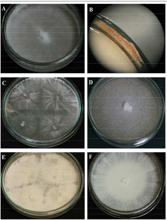
Fig. 1. General aspect of lignicolous basidiomycetes in vitro cultures on adapted media in 9 mm Petri
dish: A – Bjerkandera fumosa; B – Fruiting body of Bjerkandera fumosa; C – Crepidotus applanatus;
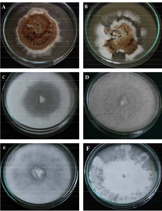
Fig. 2. General aspect of lignicolous basidiomycetes in vitro cultures grown on different media in 9