1271
COPPER-INDUCED CHANGES OF LIPID PEROXIDATION AND HEMATO-BIOCHEMICAL PARAMETERS IN RAT BLOOD: PROTECTIVE ROLE OF FLAVONOIDS
JelenA M. MlADenOvIć1, MIlIcA G. PAunOvIć1, MIlOš M. MAtIć1, veROlJuB S. KneŽevIć2,
BRAnKA I. OGnJAnOvIć1,*, AnDRAš š. štAJn1 and ZORIcA S. SAIčIć3 1 Department of Biology and Ecology, Faculty of Science, University of Kragujevac, Kragujevac, Serbia
2 Laboratory Diagnostics Department, Health Centre of Kragujevac, Kragujevac, Serbia
3 Department of Physiology, Institute for Biological Research “Siniša Stanković”, University of Belgrade, Belgrade, Serbia
corresponding author: [email protected]
Abstract – he efects of subchronic exposure to copper (cu) on lipid peroxidation, hemato-biochemical parameters, and the possible protective role of lavonoids Quercetin and (-)-epicatechin were studied. Male Wistaralbino rats were treated with cu (560 mg/l, p.o. as cucl2·2H2O for 5 weeks) and Quercetin and (-)-epicatechin (40 mg/kg BW each, i.p., every third day during the last 3 weeks) alone or in combination. cu increased the concentration of lipid peroxides, decreased the number of erythrocytes, hemoglobin and hematocrit values and increased the activities of aspartate aminotransferase, alanine aminotransferase and lactate dehydrogenase. coadministration of Quercetin and (-)-epicatechin with cu lowered the process of lipid peroxidation and restored examined hemato-biochemical parameters to control values. Our results indicate that cu induced oxidative damage in erythrocytes, which led to anemia, while Quercetin and (-)-epicatechin showed a protective efect on the hemato-biochemical processes in the blood of rats.
Keywords: copper; erythrocyte; lavonoids; hematological parameters; biochemical parameters; lipid peroxidation
IntRODuctIOn
copper (cu) is an essential trace element involved in many processes responsible for normal growth and development, as an integral part of specialized cupro-proteins, such as ceruloplasmin (cP), cytochrome c oxidase, dopamine β-hydroxylase, superoxide dis-mutase and tyrosinase (Ferenci, 2004; Kodama and Fujisawa, 2009). However, the accumulation of cu in amounts that exceed the metabolic requirements of the organism or cu homeostasis disorders can lead to the manifestation of its toxic efects (Fuentealba and Aburto, 2003). It is well known that redox-active metals, such as cu, are capable of inducing oxidative stress by increasing the production of reactive
oxy-gen species (ROS) which causes peroxidative degra-dation of polyunsaturated fatty acids in membrane lipids, and leads to the damage of biomolecules (Hal-liwell and Gutteridge, 2007).
with a genetic disorder of cu metabolism (Brewer, 2000). Increased cu accumulation in humans oc-curs mainly due to metabolic disorders of genetic origin, as in Wilson’s disease, which causes hepatitis and liver cirrhosis, and nervous system and kidney disorders (Brewer, 2000; Patil et al., 2013). In addi-tion, an excess of cu adversely afects the cardio-vascular system, leading to high blood pressure and promoting atherosclerosis (Iskra and Majewski, 2000).
Quercetin (Qe) and (-)-epicatechin (ec) belong to a group of plant polyphenolic lavonoids present in the daily diet of humans. he main sources of Qe are apples, citrus fruits, broccoli, onion, berries, tea and red wine (nutrient Data, laboratory, 2011). ec is the most abundant lavonoid in green and black tea, cocoa products, red wine and berries (nutrient Data, laboratory, 2011). A diet rich in lavonoids can reduce blood pressure, the risk of cardiovascular dis-ease, improves the liver antioxidant defense system and has a beneicial efect on symptoms of neuro-degenerative disorders in Parkinson’s and Alzheim-er’s disease (Middleton et al., 2000; Schroeter et al., 2006; larson et al., 2012). Apart from plant sources, these lavonoids are components of supplements in an alternative therapy for the treatment of allergies, asthma, bacterial infections, arthritis, gout, eye dis-orders, hypertension and neurodegenerative disor-ders (larson et al., 2012).
Flavonoids exhibit a wide range of biological ac-tivities, including oxidative, allergic, anti-viral, neuroprotective and cancer inhibiting, in vitro
or in animal tissues (verma et al., 1988; Deschner et al., 1991; Middleton et al., 2000; Ishisaka et al., 2011). In recent years attention has been devoted to their antioxidant activity. Due to their structure, lavonoids exhibit the ability to chelate transition metal ions and to “capture” and neutralize free radi-cals, acting as chain-breaking antioxidant (Bors et al., 1990).
he aim of this study is to investigate the efects of cu and the protective capacity of these particular lavonoids, Qe and ec, on hematological and
bio-chemical parameters in the blood of rats subchroni-cally exposed to cu in excess.
MAteRIAlS AnD MetHODS
Chemicals
chemicals for this study were obtained from Sig-ma-Aldrich chemie GmbH (Germany) and Merck (Darmstadt, Germany). Quercetin and (-)-epicate-chin were purchased from Sigma-Aldrich chemie GmbH (Germany). Solutions were prepared with double-distilled water. All reagents and chemicals were of analytical grade or higher purity.
Experimental animals
he study included male adult Wistar albino rats, 8 weeks old, weighing 230±20 g at the beginning of the experiment. he animals were maintained in indi-vidual cages under standard laboratory conditions (temperature 22°c±2°c; 12 h light-dark cycle). he animals had unlimited access to drinking water or a solution of cucl2 and standard rodent laboratory
diet. he concentration of cucl2 was determined
based on the oral median lethal dose for rats (lD50).
he amount of water and solutions they drank was measured every third day. At the end of experimental period, the animals were anesthetized with ether and sacriiced by decapitation. he experimental proce-dures were approved by the university ethics com-mittee.
Experimental design
Ater a period of adaptation for one week prior to the experiment, the animals were randomly divided into 4 groups, 7 animals per group: Group 1 (control) received saline (0.3 ml/kg BW); Group 2 (cu) was treated with copper (as cucl2·2H2O at a
injec-tions were administered; Group 4 (cu+Qe+ec) was treated with copper (as cucl2·2H2O at a
concentra-tion of 560 mg/l, p.o.) via drinking water for 5 weeks, and with quercetin coadministered with epicatechin (40 mg Qe/kg BW + 40 mg ec/kg BW, in 0.3 ml double-distilled water) i.p., every third day for the last 3 weeks of the experiment. A total of seven injec-tions were administered.
Analytical procedures
he animals were measured, anesthetized with ether and decapitated 24 h ater the last injection. Blood samples were collected in K-eDtA tubes for hema-tological analysis or in tubes without anticoagulants for other analyses. Hematological and biochemi-cal parameters were measured on the day of sacri-ice. Hematological analysis included the number of erythrocytes (RBc), hemoglobin (Hb), hematocrit (Hct) values, as well as hematological indices (mean corpuscular volume (Mcv), mean corpuscular he-moglobin (McH), mean corpuscular hehe-moglobin concentration (McHc) and red cell distribution width (RDW)) in whole blood, and was performed by standard methods on an automated hematology analyzer (Horiba Medical ABx Micros 60, Japan).
Measurements of biochemical parameters, se-rum total protein (tP), albumin (Alb), and activi-ties of aspartate aminotransferase (ASt), alanine aminotransferase (Alt) and lactate dehydrogenase (lDH) were performed on BioSystem BtS 330 (Spain), and serum concentrations of ceruloplasmin (cP) was performed on a Roche Hitachi 911 (Swiss) analyzer.
to measure lipid peroxidation (lPO), whole blood samples with eDtA were centrifuged at 1 000 × g (+4°c) for 10 min and the plasma was removed. he erythrocytes were washed three times with an equal volume of cold saline (0.9 %, v/v). One millilit-er of washed millilit-erythrocytes was lysed on ice in 3 ml of dH2O (4°c) for 30 min. lPO in the hemolysate was
determined using the method described by Ohkawa et al. (1979), based on the reaction of lipid peroxida-tion products (MDA-malondialdehydes) with
thio-barbituric acid (tBA) (tBARS analysis). Hemolysate samples were extracted with 28% trichloroacetic acid and centrifuged at 1 000 × g for 10 min. he color re-action was carried out by adding 1% tBA and incu-bation of the samples in a warm bath at 90°c for 15 min. he absorbances were measured with a uv-vis Spectrophotometer (JenWAy 6105, Stafordshire, uK) and results were expressed in nmol MDA/ml erythrocytes, using a molar extinction coeicient for MDA of 1.56 × 10⁵ M−1·cm−1.
Statistical analysis
All data were evaluated using SPSS for Windows (version 13.0) sotware (SPSS Inc., chicago, Il, uSA). he results are expressed as mean ± standard error of the mean (SeM). comparisons were made using either factorial analysis of variance (AnOvA) with a post hoc Bonferroni/Dunnett’s multiple analysis or Kruskal-Wallis test (for comparison across several groups) and Mann-Whitney test (for comparison be-tween two groups). Diferences at p<0.05 were con-sidered statistically signiicant.
ReSultS
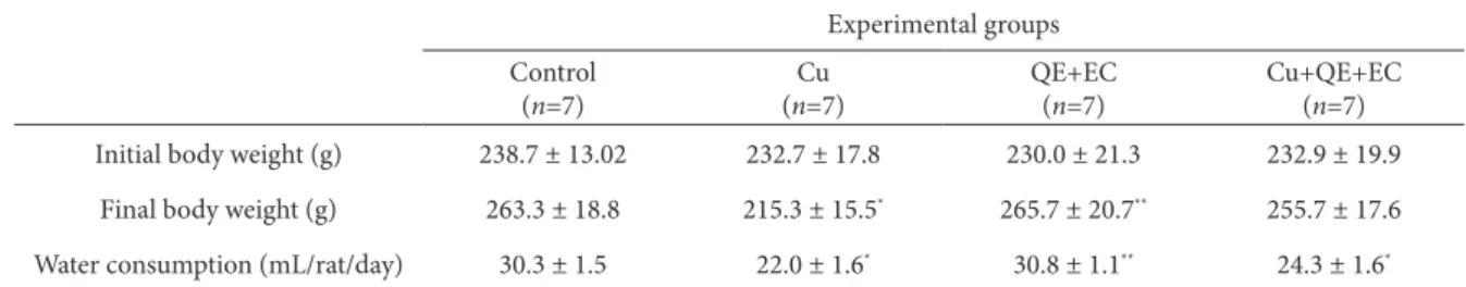
he efects of treatment with cu and the lavonoids Qe and ec on average animal weight are shown in table 1. he animals in group 1 (control), as well as groups 3 (Qe+ec) and 4 (cu+Qe+ec) gained in body weight during the 5-week experiment, while in group 2 (cu) the body weight decreased signiicant-ly. he average amount of cucl2 solution emptied in
the experimental group 2 (22.0 ml/rats/day) and in group 4 (24.3 ml/rats/day), was signiicantly lower than the volume of tap water drunk by the control animals (30.3 ml/rats/day) throughout the expo-sure (table 1). he calculated average intake of cu2+
per rat was about 5 mg/day. here was no mortal-ity among the animals, despite a reduction in body weight that was due to the avoidance of water and food intake and probable dehydration.
Table 1. Body weights and water consumption of control and treated groups of rats ater 5 weeks of treatment.
experimental groups control
(n=7)
cu (n=7)
Qe+ec (n=7)
cu+Qe+ec (n=7) Initial body weight (g) 238.7 ± 13.02 232.7 ± 17.8 230.0 ± 21.3 232.9 ± 19.9
Final body weight (g) 263.3 ± 18.8 215.3 ± 15.5* 265.7 ± 20.7** 255.7 ± 17.6
Water consumption (ml/rat/day) 30.3 ± 1.5 22.0 ± 1.6* 30.8 ± 1.1** 24.3 ± 1.6*
n: number of animals in the group; cu: copper; Qe: quercetin; ec: epicatechin. values are given as mean ± SD. *p<0.05, signiicantly diferent from
con-trol; **p<0.05, signiicantly diferent from cu group.
Table 2. changes in haematological parameters of control and treated groups of rats ater 5 weeks of treatment.
experimental groups
Parameters control
(n=7)
cu (n=7)
Qe+ec (n=7)
cu+Qe+ec (n=7)
RBc (×1012/l) 6.32 ± 0.89 5.16 ± 0.77* 7.19 ± 0.42** 6.87 ± 0.35**
Hb (g/l) 135.3 ± 4.22 101.1 ± 4.27* 144.0 ± 7.79** 141.3 ± 6.8**
Hct (%) 38.41 ± 4.86 29.52 ± 4.46* 41.05 ± 2.51** 38.35 ± 2.89**
Mcv (fl) 59.21 ± 2.08 56.80 ± 1.91 57.03 ± 2.96 56.10 ± 1.83
McH (pg) 21.1 ± 2.11 19.7 ± 2.46 20.02 ± 1.24 20.57 ± 0.82
McHc (g/l) 352.75 ± 12.62 347.87 ± 12.27 350.83 ± 7.78 368.75 ± 16.07**
RDW (%) 14.83 ± 1.73 15.95 ± 0.88 14.73 ± 1.67** 13.73 ± 0.4**
n: number of animals in the group; cu: copper; Qe: quercetin; ec: epicatechin; RBc: red blood cell; Hb: haemoglobin; Hct: haemat-ocrit; Mcv: mean corpuscular volume; McH: mean corpuscular haemoglobin; McHc: mean corpuscular haemoglobin concentration; RDW: red cell distribution width. values are given as mean ± SD. *p<0.05, signiicantly diferent from control; **p<0.05, signiicantly
diferent from cu group.
Table 3. changes in biochemical parameters of control and treated groups of rats ater 5 weeks of treatment.
experimental groups
Parameters control
(n=7)
cu (n=7)
Qe+ec (n=7)
cu+Qe+ec (n=7)
tP (g/l) 60.9 ± 2.6 59.0 ± 2.9 61.4 ± 3.5 60.3 ± 5.1
Alb (g/l) 12.7 ± 1.5 12.0 ± 1.4 12.8 ± 1.9 11.5 ± 1.8
cP (mg/l) 54.4 ± 10.7 37.9 ± 12.2* 45.7 ± 11.4 52.7 ± 12.1
ASt (u/l) 141.8 ± 14.7 216.2 ± 18.4* 147.2 ± 13.7** 138.8 ± 18.7**
Alt (u/l) 50.6 ± 6.3 70.1 ± 12.3* 55.2 ± 8.9 61.4 ± 18.1
lDH (u/l) 1084.7 ± 158.3 2012.2 ± 114.2* 1097.7 ± 81.8** 998.3 ± 142.9**
n: number of animals in the group; cu: copper; Qe: quercetin; ec: epicatechin; tP: total protein; Alb: albumin; ASt: aspartate amin-otransferase; Alt: alanine aminamin-otransferase; lDH: lactate dehydrogenase; cP: ceruloplasmine. values are given as mean ± SD. *p<0.05,
ec with cu signiicantly increased the values of the examined parameters compared to the cu group. We noticed a slight a decrease in the values of the hema-tological indices Mcv, McH and McHc in the cu group compared to the control. However, the McHc value was signiicantly higher in the cu+Qe+ec group compared to the cu group.
table 3 shows the efects of treatment on bio-chemical parameters. exposure to cu led to a slight decrease of tP and Alb, and a signiicant decrease in cP serum level compared to the control. coadmin-istration of Qe+ec with cu raised cP to the level of the control.
he cu treatment caused a signiicant increase in the activities of ASt, Alt and lDH as compared to the control. coadministration of Qe+ec with cu reversed these changes to the values measured in control.
he data in Fig. 1 show that subchronic treat-ment with cu led to a signiicant increase in lPO in the rats’ erythrocytes when compared to the control.
co-treatment of Qe+ec with cu restored the lPO level to nearly those measured in the control group.
DIScuSSIOn
Although cu is an essential element for a number of biological processes, prolonged exposure to elevated concentrations may have adverse efects (Fuentealba and Aburto, 2003). In this study, we investigated the efects of cu and the inluence of the lavonoids Qe and ec on hematological and biochemical param-eters in the blood of rats ater subchronical exposure to cu in excess.
Our results show that in animals subchronically exposed to cu their body weight decreased, which can be a predictor of poor general health. he de-crease in body weight may indicate an excessive breakdown of tissue proteins. Similarly, the results of Bataineh et al. (1998) showed that long-term con-sumption of cu salts causes growth disorders and weight loss in animals. On the other hand, animals co-treated with ec, Qe and cu continuously gained weight.
Fig. 1. lPO values in erythrocytes of control and treated groups of rats ater 5 weeks of treatment cu: copper; Qe: quercetin; ec:
epi-catechin. values are given as mean ± SD(n = 7); *p<0.05, signiicantly diferent from control; #p<0.05, signiicantly diferent from cu
exposure to cu signiicantly decreased RBc, Hb and Hct and led to anemia. Similar results were ob-tained in other studies on animals (Bozynski et al., 2009; nikolic et al., 2013). he research of Fernandes et al. (1988) showed that the incubation of an eryth-rocyte suspension with cu2+ causes lipid peroxidation
and hemolysis as a consequence of oxyhemoglobin oxidation by cu. he erythrocytes are more suscep-tible to oxidative damage than other cells because they are constantly exposed to ROS and their mem-branes are rich in unsaturated fatty acids (clemens and Waller, 1987). It was found that toxic cu con-centrations reduce deformability of erythrocytes and increase membrane permeability and osmotic fragil-ity of cells, resulting in reduced erythrocyte survival (Adams et al., 1979). he observed reduced number of RBc, Hb and Hct ater cu exposure in our study is probably due to the oxidative damage of erythrocytes and increased erythrocyte destruction.
coadministration of Qe+ec with cu showed protective efects on hematological parameters. In the study of chouhan et al. (2011), Qe exerted ben-eicial efect on hematological parameters in rats treated with luoride. Martinez et al. (2012) found that the presence of ec prevents protein oxidation and positively afects membrane luidity and eryth-rocyte morphology, thereby preventing hemolysis resulting from peroxidation. hese results suggest that the protective efects of Qe and ec are likely due to their ability to capture ROS and thereby prevent erythrocyte damage and hemolysis.
Metabolic processes in rats exposed to cu were also afected, which is relected in the disturbance of biochemical parameters. cP, a cuproprotein that is synthesized in the liver, is the major carrier of cu in the blood. Reduced levels of cP are typically as-sociated with a reduced level of cu in the serum of patients with Wilson’s disease, but reduced cP with normal or increased levels of serum cu may suggest increased levels of cu not bound to cP (the so-called free copper or non-ceruloplasmin-bound copper) (Kodama and Fujisawa, 2009). An elevated level of cu in circulation may be the result of a sudden cu release from the hepatocytes, caused by liver damage
or intoxication with cu (Patil et al., 2013). treatment with cu in the present study signiicantly decreased cP but did not lead to signiicant changes in the se-rum level of cu (data not shown) as compared to the control, which may indicate the release of non-cer-uloplasmin-bound cu from damaged hepatocytes. coadministration of Qe+ec with cu reversed the changes in protein levels and cP, suggesting protec-tive efects of these lavonoids on hepatocytes.
cu treatment increased the activities of ASt, Alt and lDH in the serum. transaminases are widely distributed in tissues, while their concentra-tions in the serum are low. Since the liver is the or-gan of cu accumulation, these changes may indicate liver damage and, due to the distortion of functional integrity of hepatocyte cell membranes, the release of these enzymes in serum and their enhanced ac-tivity. Increased transaminase (ASt and Alt) activi-ties in serum serve as parameters in the diagnosis of hepatitis and hemolytic anemia in humans and also indicate liver damage in animal (Fuentealba and Aburto, 2003; Gaetke and chow, 2003). George and chandrakasan (1997) reported a signiicant increase in lDH activity in the serum of rats as a result of hepatic ibrosis.
Qe+ec administered alone did not cause sig-niicant diferences in the activities of these enzymes compared to the control group. However, coadmin-istration of Qe+ec with cu decreased Alt and sig-niicantly reduced ASt and lDH activities compared to the cu group. his is consistent with the results of other studies in which Qe inhibited the increase of ASt, Alt and lDH activities in serum by reducing the lPO in the liver (tokyol et al., 2006) and myocar-dium of animals (Matouk et al., 2013; Milton et al., 2013). hese results indicate that Qe and ec are able to preserve the functional integrity of hepatocyte cell membranes and to prevent leakage of these enzymes into the blood.
shown, an excess of cu in rat diet may cause lPO and damage of the membrane (Dillard and tappel, 1984; Burkitt, 2001; Gaetke and chow, 2003). Fernandes et al. (1988) reported increased lPO ater incubation of human erythrocyte suspension with cu2+ ions. he
cu+ ion generated by the reduction of cu2+ in the
presence of superoxide anion (O2•-) catalyzes the
for-mation of hydroxyl radicals (•OH) that readily en
-ter into further reactions (Halliwell and Gut-teridge, 2007). O2•-, •OH, or lipid peroxyl radicals can cause
lPO, a decrease in membrane potential, and an in-crease in permeability to H+ and other ions, which
eventually leads to the release of contents from cells (Halliwell and Gutteridge, 2007). cu in our study signiicantly increased lPO in erythrocytes, indicat-ing oxidative damage of cell membranes and the oc-currence of oxidative stress in erythrocytes.
Our data indicate that Qe+ec inhibited cu-induced lPO. Previous studies have shown that lavonoids can block lPO due to their structure that enables them to chelate metal ions or scavenge ROS as hydrogen- or electron-donating compounds (Middleton et al., 2000; Mira et al., 2002). hey thus can be helpful in conditions and diseases caused by increased metal concentrations or in conditions that are the result of oxidative stress. Mira et al. (2002) showed that Qe and catechin chelate cu2+ ions, due
to their hydroxyl groups with interaction probably between the 5-hydroxyl and 4-oxo groups. It has also been reported that catechins have an antioxidant ef-fect on iron-induced lPO due to iron chelation (Sug-ihara et al., 2001).
lPO could be inhibited by ROS scavenging, and Qe as well as ec appears to be an extremely eicient radical scavenger (Middleton et al., 2000). he anti-oxidant properties of Qe and ec may contribute to the improvement of cells’ antioxidant defense. Filipe et al. (2001) showed that Qe inhibited cu-induced lPO in human plasma. hese authors have also shown that there was a synergy of Qe with endog-enous urate in antioxidant action against cu-induced lPO, and that the lavonoids were able to protect urate from oxidative degradation. Dietrich-Muszal-ska et al. (2012) have shown that ec was more
efec-tive than Qe in reducing lPO in human plasma in vitro. In an in vitro study of Boadi et al. (2003), it was shown that in reducing lPO, combinations of lavo-noids provide better antioxidant protection than the individual treatments.
According to Bors et al. (1990), three structural group of lavonoids are responsible for metal chelat-ing and radical scavengchelat-ing: i) the O-dihydroxy struc-ture in the B-ring, which is the radical target site for all lavonoids with a saturated c-2/c-3 bond; ii) the c-2/c-3 double bond in conjugation with a 4-oxo function, which is responsible for electron delocali-zation from the B-ring; and iii) the additional pres-ence of both 3- and 5-hydroxyl groups for strongest radical absorption.
Our data suggest that cu in excess exerted prooxidant efects on hematological and biochemi-cal processes in the blood and caused oxidative damage in erythrocytes. treatment with Qe and ec could protect against oxidative damage in the blood of rats subchronically exposed to cu in excess. he cytoprotective role of Qe and ec can be ascribed to their ability to chelate the ions of transition metals and scavenge ROS. hey managed to inhibit lPO, thus demonstrating a signiicant role in alleviating the manifestations of cu toxicity.
Acknowledgments – his work was supported by the Ministry
of education, Science and technological Development of the Republic of Serbia, Grant no 173041.
ReFeRenceS
Adams, K. F., Johnson, G. Jr., Hornowski, K. E. and Lineberger, T. H. (1979). he efect of copper on erythrocyte deform-ability. A possible mechanism of hemolysis in acute cop-per intoxication. Biochim. Biophys. Acta – Biomembranes.
550, 279-287.
Araya, M., Olivares, M., Pizarro, F., Llanos, A., Figueroa, G. and
Uauy, R. (2004). community-based randomized double-blind study of gastrointestinal efects and copper exposure in drinking water. Environ. Health Perspect. 112, 1068-1073.
adult male rat following long-term ingestion of four in-dustrial metals salts. Hum. Exp. Toxicol.17, 570-576.
Boadi, W. Y., Iyere, P. A. and Adunyah, S. E. (2003). efect of quercetin and genistein on copper- and iron-induced lipid peroxidation in methyl linolenate. J. Appl. Toxicol.23, 363-369.
Bors, W., Heller, W., Michel, C. and Saran, M. (1990). Flavonoids as antioxidants: determination of radical scavenging ei-ciencies. Methods Enzymol.186, 343-355.
Bozynski, C. C., Evans, T. J., Kim, D. Y., Johnson, G. C., Hughes-Hanks, J. M., Mitchell, W. J., et al. (2009). copper toxicosis with hemolysis and hemoglobinuric nephrosis in three adult Boer goats. J. Vet. Diagn. Invest.21, 395-400.
Brewer, G. J. (2000). Is heterozygosity for a Wilson’s disease gene defect and important underlying cause of infantile and childhood copper toxicosis syndromes? J.Trace Elem. Exp. Med. 13, 249-254.
Burkitt, M. J. (2001). A critical overview of the chemistry of cop-per-dependent low density lipoprotein oxidation: roles of lipid hydroperoxides, alpha-tocopherol, thiols and cerulo-plasmin. Arch. Biochem. Biophys.394, 117-135.
Chouhan, S., Yadav, A., Kushwah, P., Kaul, R. K. and Flora, S. J. S. (2011). Silymarin and quercetin abrogates luoride in-duced oxidative stress and toxic efects in rats. Mol. Cell. Toxicol.7, 25-32.
Clemens, M. R. and Waller, H. D. (1987). lipid peroxidation in erythrocytes. Chem. Phys. Lipids. 45, 251-268.
Deschner, E. E., Ruperto, J., Wong, G. and Newmark, H. L. (1991). Quercetin and rutin as inhibitors of azoxymethanol-in-duced colonic neoplasia. Carcinogenesis12, 1193-1196.
Dietrich-Muszalska, A., Kontek, B., Olas, B. and Rabe-Jabłońska, J. (2012). epicatechin inhibits human plasma lipid peroxi-dation caused by haloperidol in vitro. Neurochem. Res.37, 557-562.
Dillard, C. J. and Tappel, A. L. (1984). lipid peroxidation and copper toxicity in rats. Drug Chem. Toxicol.7, 477-487.
Ferenci, P. (2004). Pathophysiology and clinical Features of Wil-son Disease. Metab. Brain Dis.19, 229-239.
Fernandes, A., Mira, M. L., Azevedo, M. S. and Manso, C. (1988). Mechanisms of hemolysis induced by copper. Free Radic. Res. Commun.4, 291-298.
Filipe, P., Lanç, V., Silva, J. N., Morlière, P., Santus, R. and Fer-nandes, A. (2001). Flavonoids and urate antioxidant inter-play in plasma oxidative stress. Mol. Cell. Biochem.221, 79-87.
Fuentealba, C. and Aburto, E. M. (2003). Animal models of cop-per-associated liver disease. Comparative Hepatology2, 5.
Gaetke, L. M. and Chow, C. K. (2003). copper toxicity, oxidative stress and antioxidant nutrients. Toxicology189, 147-163.
George, J. and Chandrakasan, G. (1997). lactate dehydrogenase isoenzymes in dimethylnitrosamine-induced hepatic i-brosis in rats. J. Clin. Biochem. Nutr.22, 51-62.
Halliwell, B. and Gutteridge, J. M. C. (2007). Oxygen is a toxic gas – an introduction to oxygen toxicity and reactive species. In: Free Radicals in Biology and Medicine. (eds. B. Hal-liwell and J. M. c. Gutteridge), 1-29. Oxford university Press, Oxford.
Ishisaka, A., Ichikawa, S., Sakakibara, H., Piskula, M. K., Naka-mura, T., Kato, Y., et al. (2011). Accumulation of orally ad-ministered quercetin in brain tissue and its antioxidative efects in rats. Free Radical Bio. Med.51, 1329-1336.
Iskra, M. and Majewski, W. (2000). copper and zinc concentra-tions and the activities of ceruloplasmin and superoxide dismutase in atherosclerosis obliterans. Biol. Trace Elem. Res.73, 55-65.
Kodama, H. and Fujisawa, C. (2009). copper metabolism and inherited copper transport disorders: molecular mecha-nisms, screening and treatment. Metallomics1, 42-52.
Larson, A. J., Symons, J. D. and Jalili, T. (2012). herapeutic Po-tential of Quercetin to Decrease Blood Pressure: Review of eicacy and Mechanisms. Adv. Nutr.3, 39-46.
Martínez, V., Ugartondo, V., Vinardell, M. P., Torres, J. L. and
Mitjans, M. (2012). Grape epicatechin conjugates prevent erythrocyte membrane protein oxidation. J. Agric. Food Chem.60, 4090-4095.
Matouk, A. I., Taye, A., Heeba, G. H. and El-Moselhy, M. A.
(2013). Quercetin augments the protective efect of losar-tan against chronic doxorubicin cardiotoxicity in rats. En-viron. Toxicol. Pharmacol.36, 443-450.
Middleton, E. J. R., Kandaswami, C. and heoharides, T. C. (2000). he efects of plant lavonoids on mammalian cells: impli-cations for inlammation, heart disease and cancer. Phar-macol. Rev.52, 673-751.
Milton, P. S., Muthumani, M. and Shagirtha, K. (2013). Quercetin potentially attenuates cadmium induced oxidative stress mediated cardiotoxicity and dyslipidemia in rats. Eur. Rev. Med. Pharmacol. Sci.17, 582-595.
Mira, L., Fernandez, M. T., Santos, M., Rocha, R., Florêncio, M. H.
Nikolic, R., Krstic, N., Jovanovic, J., Kocic, G., Cvetkovic, T. P.
and Radosavljevic-Stevanovic, N. (2013). Monitoring the toxic efects of Pb, cd and cu on hematological pa-rameters of Wistar rats and potential protective role of lipoic acid and glutathione. Toxicol. Ind. Health. DOI: 10.1177/0748233712469652.
Nutrient Data, Laboratory, uSDA Database for the Flavonoid content of Selected Foods, Release 3.0. u.S. Department of Agriculture, Agricultural Research Service, Beltsville Human nutrition Research center, uSDA; 2011.
Ohkawa, H., Ohishi, N. and Yagi, K. (1979). Assay for lipid per-oxides in animal tissues by thiobarbituric acid reaction.
Anal. Biochem.95, 351-358.
Patil, M., Sheth, K. A., Krishnamurthy, A. C. and Devarbhavi, H.
(2013). A Review and current Perspective on Wilson Dis-ease. J. Clin. Exp. Hepatol.3, 321-336.
Schroeter, H., Heiss, C., Balzer, J., Kleinbongard, P., Keen, C. L., Hollenberg, N. K., et al. (2006). (-)-epicatechin mediates beneicial efects of lavanol-rich cocoa on vascular func-tion in humans. Proc. Natl. Acad. Sci. U S A.103, 1024-1029.
Sugihara, N., Ohnishi, M., Imamura, M. and Furuno, K. (2001). Diferences in Antioxidative eiciency of catechins in various Metal-Induced lipid Peroxidations in cultured Hepatocytes. J. Health Sci.47, 99-106.
Tokyol, C., Yilmaz, S., Kahraman, A., Cakar, H. and Polat, C.
(2006). he efects of desferrioxamine and quercetin on liver injury induced by hepatic ischemia-reperfusion in rats. Acta chir. Belg.106, 68-72.
Verma, A. K., Johnson, J. A., Gould, M. N. and Tanner, M. A.