Single transverse extended incision for radical neck dissection
Esvaziamento cervical radical por incisão transversa única estendida
José Francisco sales chagas2; Maria Beatriz nogueira Pascoal2; José luís Braga aquino, tcBc-sP2; luís antônio Brandi Filho2;
evandro von zuBen Previtale2; ana soFia Pontes trillo2; otávio alBerto curioni, tcBc-sP1; aBrão raPoPort, tcBc-sP1; rogério
aParecido dedivitis, tcBc-sP1.
INTRODUCTION
T
he neck dissection is a risky procedure due to its per-formance in an anatomically complex region, with multiple key structures involved in lymph node dissection, beyond the extent of detachment of the flaps and the fre-quent combination with extensive surgery for resection of the primary tumor. Complications range from dehiscence of cervical suture flap to serious complications such as rupture of large vessels.The patient’s nutritional status, concomitant diseases such as diabetes, anemia and cardiovascular disease, the extent of resection and previous treatments such as radiotherapy and chemotherapy tend to increase the risk of systemic or local complications, the latter being mainly related to the cervical flap1.
Traditionally, surgeons perform neck dissec-tions through two or more associated cervical incisions, to expose widely and easily all levels of the cervical lymph nodes. This reduces the technical difficulty, which could
influence the complete resection of the cervical lymph nodes, leading to regional recurrence and worse prog-nosis. These often combine a transverse incision with a vertical one, which usually intersect at the level of the carotid bulb. Due to the necessity of decoupling the skin flap from the mandible to the clavicle radical dissections, there is a great possibility of interference with vascular-ization of these flaps, and consequently increased risk of dehiscence of the suture and exposure of large vessels at risk of rupture by dehydration of their walls and damage to the vasa vasorum. Thus, a minor complication such as suture dehiscence can become life-threatening for pa-tients, especially those undergoing previous treatments such as radiotherapy and/or chemotherapy. In addition to the wide exposure of the cervical lymph nodes, neck dissection planning must also address the need for an en bloc associated resection of the primary lesion, usually when the primary tumor is larger and resection includes communication between the high aero-digestive tract and the neck.
1 - Department of Head and Neck Surgery and Otorhinolaryngology, Heliópolis Hospital, São Paulo, SP, Brazil. 2 - Department of Head and Neck Surgery, Hospital of the Pontifical Catholic University of Campinas, Campinas, SP, Brazil.
A B S T R A C T
Objective: to assess the efficacy of the single transverse extended cervical incision in radical neck dissection. Method: we conducted a prospective study, from January 2008 to January 2009, with 18 patients undergoing surgical treatment of malignant tumors of the upper aero-digestive tract. The primary lesion was located in the oral cavity in eight cases, in the oropharynx in three, in the hypopharynx in three, in the larynx in two, in the maxillary sinus, and in one case, the primary injury was hidden. There were 29 neck dissections, eight bilateral and 10 unilateral (26 radical and three selective). Staging revealed nine patients with T4 tumor, one T3, six T2, one T1 and one Tx. Five patients were N0, nine N2b, one N2c and three N3. The average number of dissected lymph nodes was 34.25. We performed the neck dissection through a single incision located in the middle neck, coincident with the skinfold, with a length of about 2 to 3 cm behind the an-terior edge of the trapezius muscle and 3 to 4 cm from the midline for the unilateral neck dissections. Results: as complications, there were myocutaneous flap necrosis in one patient with prior radiation therapy, one lymphatic fistula, one dehiscence of the tracheostomy, one cer-vical abscess, one salivary fistula and one suture dehiscence. Conclusions: the single extended incision provides adequate exposure of the neck structures, without compromising surgical time, even in bilateral dissections. It does not compromise the resection of all cervical lymph nodes; it has excellent aesthetic and functional results and is easily associated with other approaches to resection of the primary tumor.
In addition to the inadequate aesthetic appear-ance of incisions that run in oppositions to the lines of Langer, ie, the skinfolds in the case of the neck, the other complications from neck dissection are the scar retrac-tions, which can result in chronic pain, besides restricting the movement the muscles of the shoulder girdle and neck. These complications interfere with patients’ work-ing capacity and therefore in their quality of life2.
In order to reduce such complications, improve the functional aspects and post-surgical aesthetics, and based on our experience with the McFee incision, rou-tinely used for female patients3, we introduced the single and extended cervical incision for unilateral or bilateral cervical dissections from a literature time series4.
METHODS
We conducted a prospective study to assess the viability of the single extended cervical incision for radical neck dissection, and to analyze its interference in post-operative complications in terms of functional and esthetic aspects. In the Service of Head and Neck Sur-gery of the Hospital of the Pontifical Catholic University of Campinas and at the Department of Head and Neck Surgery and Otorhinolaryngology, Heliópolis Hospital, 18 consecutive patients with squamous cell carcinoma of the
aerodigestive tract upper underwent surgical treatment from January 2008 to January 2009, with a minimum fol-low-up period of one year. There were eight patients with primary tumors of the oral cavity, three of the orophar-ynx, three of the hypopharorophar-ynx, two of the larorophar-ynx, one of the maxillary sinus and one hidden primary lesion. With respect to the neck dissections performed, there were 19 cases, eight bilateral and 10 unilateral (26 radical and three supraomo-hyoid). The staging of the primary tu-mors revealed: Nine T4, one T3, six T2, one T1 and one Tx, and in relation to the cervical lymph nodes, we found five N0, nine N2b, one N2c and three N3, with prior radi-ation treatment in one case.
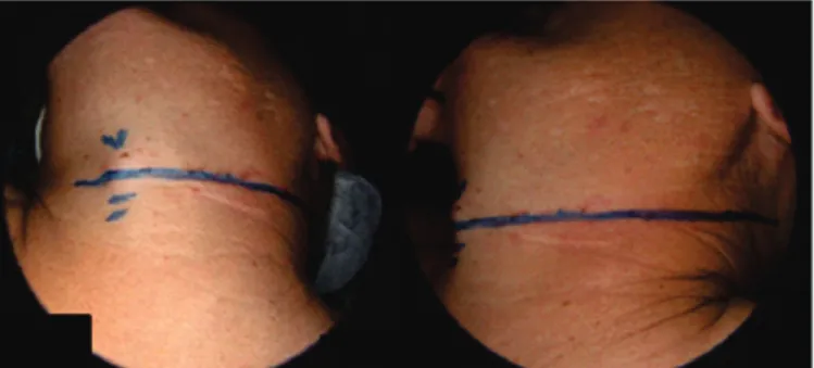
The eligibility criterion was the indication of radical or selective neck dissection. All patients under-went neck dissection through a single incision in the mid-dle third of the neck, coinciding with the skin fold and extending about 2 to 3 cm beyond the anterior edge of the trapezius muscle and exceeding 3 to 4 cm from the midline in unilateral dissections (Figure 1). In bilateral dis-sections, the length of the incision went beyond the front edge of the contralateral trapezius muscle.
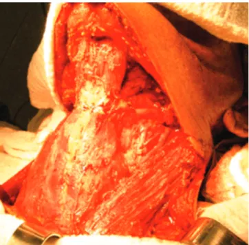
The skin flaps were raised under the platysma muscle, to help preserve vascularity, and extended to the limits of neck dissection, ie, the lower edge of the jaw and upper edge of the clavicle unilaterally or bilaterally on
radical dissections, and lower edge of the jaw and middle third of the neck in supraomo-hyoid ones (Figure 2). We dissected the neck dissection product, with separation of the lymph nodes by levels and chains, and noted the sur-gical time.
We monitored patients for at least one year, with quarterly reviews for detection of complications related to the surgical approach, such as cervical pain, dehiscence or skin necrosis, scar retraction, presence of keloids, hypertrophic scar and limited motion of neck and shoulder girdle (Figure 3).
This study was approved by the Ethics in Re-search Committee of the Pontifícia Universidade Católica de Campinas, under number 108/09.
RESULTS
Surgical time ranged from two to six hours and the average of dissected lymph nodes was 34.25.
The following complications occurred: one lym-phatic fistula, one tracheostomy dehiscence, one necrosis of the pectoralis major myocutaneous flap, one cervical abscess, one salivary fistula, one dehiscence of skin su-ture and scar retraction. The skin dehiscence was treat-ed conservatively and occurrtreat-ed in one patient with prior radiation treatment. The patient who had scar retraction
underwent surgical approach under local anesthesia, in which we evidenced fibrosis and retraction of infrahyoid muscles. The surgical treatment occurred after failure of physical therapy and also due to decrease in cervical movements and severe pain.
DISCUSSION
Cervical lymph node dissection was first described in 19065 and remains the same as the standard treatment for cervical lymph node metastases, with a large number of success reports in medical literature. In addition, mor-bidity and surgical mortality decreased to very acceptable levels due to advances in anesthesia, antibiotics, blood transfusions and surgical technique. Despite these advan-tages, there is still some reluctance to accept a surgery considered mutilating or disfiguring, especially in young female patients7,8.
Numerous incisions are normally used for the surgical approach, and most of them combine a trans-verse neck incision with a vertical one7-9. The vertical component is in opposition to the skin force lines, which increases the risk of scar contraction or keloid formation. Scarring retractions can lead to limitation of neck
move-Figure 2. Bilateral cervical lymph node emptying with exposure of all lymph node levels.
ment and the emergence of pain conditions, ranging from moderate to severe. These complications are able to interfere with labor or leisure activity, reducing patients’ quality of life9,10.
Another complication that may occur in the post-operative period of neck dissection is the skin flaps necro-sis, usually at their intersection, which generally happens at the level of the carotid bulb, leading to its exposure and dryness. This necrosis has a higher incidence in the vertical component of skin incisions, since the blood sup-ply is less, for it reduces the skin flap pedicle11,12. The skin incisions with a single component in the shape of “J” or “U” also oppose to the skin force lines, enabling the same functional and aesthetic complications of incisions with two components.
Numerous cervical surgical techniques have been described13, none of which fully meeting the criteria for cervical access, thus described: 1) adequate surgical field exposure; 2) adequate blood supply to the resulting flaps; 3) acceptable relation of the incision with large neck ves-sels; 4) easy conversion or conjunction with other inci-sions to approach the primary tumor; 5) convenience for preparation of stomas; 6) compatibility with reconstruc-tive techniques; and 7) functional characteristics and ac-ceptable cosmetic. In addition, the technique needs to be easy to learn and to reproduce. Based on these criteria, the ideal surgical approach would be in transverse direc-tion, and which could be associated with pathways for resection of the primary lesion.
The use of two parallel incisions for performing cervical lymph node dissection3 presents risk of skin ne-crosis because it interferes with the viability of the cuta-neous blood supply, especially in bilateral cases13. Thus, the optimal surgical approach is one transverse incision, provided that exposure of all cervical lymph node levels is possible. Based on the experience with the McFee3 and Altie4 incisions, we introduced the single and extended incision for neck dissection.
To facilitate the access to the surgical areas of greatest difficulty, such as submentonian lymph nodes (Ia level) and posterior portion of the supraclavicular fossa (Vb level), we extended the incision to 3 to 4 cm from midline and 2 to 3 cm beyond the front edge of the
trape-zius muscle. This allowed the complete exposure of lymph node chains of these areas and facilitated the exposure of the level IV and distal ligation of the internal jugular vein. In longilineal patients, there may also be greater difficulty in dissection of level IIb (junctional lymph nodes), and a small upper curvature of the posterior end of the incision solves this problem.
The advantages of the cervical transverse inci-sions are evident, since the natural skin folds are in the same direction, which provides quick, firm healing and excellent aesthetics11,12 . The rational argument for their use is that the blood supply to the neck has vertical sense, running from bottom to top in the lower cervical limits and from top to bottom in upper cervical limits13-16. The biggest fear with the single and extended transverse incision approach for cervical lymph node dissection is the possibility of technical difficulty inducing greater lymph node recurrence rate. According to our results of lymph nodes dissected in the surgical specimens (aver-age 34.25), which do not differ from that found in the literature nor from our average in combined incisions, we can confirm the effectiveness of this surgical approach to cervical lymph node dissections.
Another aspect one must consider when plan-ning a surgical approach for cervical lymph node dissec-tion is the possibility of associadissec-tion with other incisions to treat the primary tumor17. In patients with primary tu-mors of the posterior portions of the oral cavity or oro-pharynx and undergoing unilateral neck dissection, the maneuver used for surgical exposure was to extend the cervical incision in the opposite neck and associate a pre-auricular access. In this way, we avoid incision through the mentonian region and the lower lip, which provides great aesthetic gain. Extending the incision to the oppo-site neck side demands more surgical handling and hence could lead to increased surgical time. However, when comparing the surgical time with the surgeries performed with a combination of incisions, we found that it did not happen. Moreover, the aesthetic gain was large, since it avoided a vertical incision in a visible region, as is the sub-mental region.
radical neck dissection, whether classic, modified or se-lective. Without increasing the surgical time, it provided good surgical exposure, little technical difficulty, the pos-sibility of proper handling of primary lesions associated or not with other incisions, and especially good aesthetic and functional results. This led us to standardize this ap-proach in our service as a routine when handling neck dissections. As for the aesthetic and functional aspect, we could observe the benefit of the transverse incision in the postoperative follow-up, with aesthetic gains in conceal-ing the incision with the natural skin folds. In addition, in the functional aspect, although the follow-up time is still small, there were no complaints of the longer follow-up patients regarding scarring retractions. The dysfunction
of the shoulder girdle does not depend on skin incision, so we did not consider this aspect in our study.
Since the patients studied were consecutive, there were no exclusion criteria for indicating the surgical incision. Due to the low number of complications related to this surgical approach, we consider that the staging had no influence on them.
In conclusion, transverse and extended skin inci-sions for radical neck dissections provided: (1) good sur-gical exposure and little technical difficulty; (2) feasibility when dissecting all lymph node levels; (3) no interference with the surgical procedure; (4) the possibility of proper handling of primary lesions, associated or not with other incisions; and (5) good aesthetic and functional results.
REFERENCES
1.
Magrin J, Kowalski LP, Correia LMC. Esvaziamen-tos cervicais. In: Kowalski LP Afecções cirúrgicas do pescoço. 1a ed. São Paulo: Atheneu; 2005. p.183-99.2.
Becker GD. Extended single transverse incision for composite resections: an update of technique and re-sults. Laryngoscope. 1984;94(5 Pt 1):605-7.3.
McFee WF. The surgical treatment of carcinoma of the thyroid gland, with special reference to the metasta-sis. Surg Clin North Am. 1953;361-87.4.
Attie JN. A single transverse incision for radical neck dissection. Surgery. 1957;41(3):498-502.5.
Crile G. Excision of cancer of the head and neck, with special reference to the plan of dissection based on 132 patients. JAMA. 1906;47:1780-86.6.
Chagas JFS, Pascoal MBN, Aquino JLB. Incisão trans-versa estendida e única para esvaziamento linfonod-al cerviclinfonod-al radiclinfonod-al. Rev Bras Cir Cabeça Pescoço. 2006;35(1):45-7.7.
Brown JB, McDowell F. Neck dissections for metastatic carcinoma. Surg Gynec Obst. 1944;79:115-24.8.
Myssiorek D, Becker GD. Extended single transverse neck incision for composite resections: does it work? J Surg Oncol. 1991;48(2):101-5.9.
Acar A, Dursun G, Aydin O, Akbas Y. J incision in neck dissections. J Laryngol Otol. 1998;112(1):55-60.10.
Woods JE, Yugueros P. A safe and rapid tech-nique for modified neck dissection. Ann Plast Surg. 1999;43(1):90-5.11.
Omura S, Bukawa H, Kawabe R, Aoki S, Fujita K. Comparison between hockey stick and reversed R E S U M OObjetivo: verificar a eficácia da incisão cervical única, transversa e estendida, para o esvaziamento cervical radical. Método: estudo prospectivo, de janeiro de 2008 a janeiro de 2009, de 18 pacientes submetidos a tratamento cirúrgico de tumores malignos da via ae-ro-digestiva superior. A lesão primária se situava na cavidade oral em oito casos, na orofaringe em três, no seio piriforme em três, na laringe em dois, no seio maxilar em um e em um caso a lesão primária era oculta. Houve 29 esvaziamentos, sendo oito bilaterais e 10 unilaterais (26 radicais e três seletivos). O estadiamento revelou nove pacientes com tumor T4, um T3, seis T2, um T1 e um Tx. Cinco pacientes eram N0, nove N2b, um N2c e três N3. A média de linfonodos dissecados foi de 34,25. O esvaziamento cervical foi realizado por meio de uma única incisão localizada no terço médio do pescoço, coincidente com dobra cutânea, com extensão de cerca de 2 a 3 cm para trás da borda anterior do músculo trapézio e 3 a 4 cm da linha média para os esvaziamentos cervicais unilaterais. Resultados: como complicações houve necrose de retalho miocutâneo em um paciente com radioterapia prévia, uma fistula linfática, uma deiscência do traqueostoma, um abscesso cervical, uma fístula salivar e uma deiscência de sutura. Conclusões: a incisão única e estendida propor-ciona exposição adequada das estruturas do pescoço, sem comprometer o tempo cirúrgico, mesmo em esvaziamentos bilaterais. Não compromete a ressecção de todos os linfonodos cervicais, apresenta excelentes resultados estéticos e funcionais e é facilmente associada com outras abordagens para ressecção do tumor primário.
hockey stick incision: gently curved single linear neck incisions for oral cancer. Int J Oral Maxillofac Surg. 1999;28(3):197-202.
12.
Dissanayaka L. A modified single flap for neck dis-section in oral cancer. Head Neck. 1990;12(1):74-6.13.
Appiani E, Delfino MC. Plastic incisions for facial and neck tumors. Ann Plast Surg. 1984;13(4):335-52.14.
Daniell CH, Fee WE Jr. MacFee incisions: dispelling the myth of cervical flap vascular inadequacy. Head Neck Surg. 1987;9(3):167-71.15.
Doberneck RC. An evaluation of wound compli-cations after neck dissection and composite resec-tion utilizing transverse cervical incisions. Surgery. 1973;73(2):261-5.16.
Kademani D, Dierks EJ. A straight-line incision forneck dissection: technical note. J Oral Maxillofac Surg. 2005;63(4):563-5.
17.
Becker GD. The extended single transverse neck incision for composite resections. Trans Sect Otolaryngol Am Acad Ophthalmol Otolaryngol. 1977;84(5):816-7.Received in: 02/04/2016
Accepted for publication: 11/07/2016 Conflict of interest: none.
Source of funding: none.
Mailing address:
Rogerio Aparecido Dedivitis