Mirizzi syndrome grades III and IV: surgical treatment
Síndrome de Mirizzi graus III e IV: tratamento cirúrgico
Ronald ReveRdito1; andRé de MoRicz, tcBc-SP2; téRciode caMPoS, tcBc-SP2; adheMaR MonteiRo Pacheco JúnioR, tcBc-SP2; RodRigo altenfeldeR Silva, tcBc-SP2.
INTRODUCTION
M
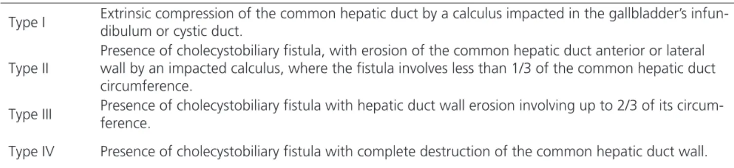
irizzi Syndrome (MS) is a rare cause of benign obstruc-tive jaundice triggered by impacted calculus in Hart-mann bag or cystic duct, causing extrinsic obstruction of the common hepatic duct. In cases of intense inflammation, it is difficult to treat. According to Csendes et al., responsible for one of its classifications, more advanced degrees include pa-tients with cholecystobiliary fistula, with common bile duct wall erosion of two-thirds of its circumference (grade III) and complete erosion (grade IV)1-3. Subtotal cholecystectomyassociated with choledocoplasty and tube “T” drainage or biliodigestive anastomosis are the main surgical alternatives, there being, however, no standard procedure.
This study aims to assess the epidemiology and outcomes of surgical treatment of patients with more ad-vanced degrees of Mirizzi Syndrome (III and IV).
METHODS
We conducted a retrospective, cross-sectional study through the review of records from 13 cases
oper-ated from December 2001 to September 2013 in the De-partment of Surgery of the Faculty of Medical Sciences of the São Paulo Holy Home (FCMSCSP) and Brotherhood of the São Paulo Holy Home (ISCMSP). We classified the cases according to Csendes et al. and selected grades III and IV. We assessed current age, gender, time of disease, diagno-sis, treatment performed and postoperative outcome.
RESULTS
Of the 3,691 patients admitted for treatment of gallstones from December 2001 to September 2013, 23 (0.6%) had MS. Disease in grades III and IV occurred in 13 patients (0.35%), ten being women (76.9%) and three men (23.1%). The mean age was 55.6 years ranging from 31 to 89. Two patients had hypertension and diabetes mellitus type II. The average time of disease was 375 days, ranging from one to 60 months. Seven patients were admitted to the emergency room and six, at an outpatient basis
The most prevalent symptom was abdominal pain (92%), followed by jaundice in 84.6%; fever was present in two of the seven patients with cholangitis. Of
1 - Department of Surgery, Brotherhood of the São Paulo Holy Home, São Paulo, SP, Brazil. 2 - Biliary and Pancreas Surgery Group, Brotherhood of the São Paulo Holy Home, São Paulo, SP, Brazil.
A B S T R A C T
Objective: to evaluate the epidemiology and outcomes of surgical treatment of patients with Mirizzi Syndrome (MS) grades III and IV, the most advanced according to Csendes classification. Methods: we conducted a retrospective, cross-sectional study by reviewing records of thirteen patients with grades III and IV MS operated from December 2001 to September 2013, among the 3,691 cholecystectomies per-formed in the period. Results: the incidence of MS was 0.6% (23 cases) and grades III and IV amounted to 0.35% of this number. There was a predominance of type IV (12 cases). The preoperative diagnosis was possible in 53.8% of cases. The preferred approach was biliary-diges-tive derivation (10 cases), and “T” tube drainage with suture of the bile duct was the choice in three special occasions. Three patients had biliary fistula resolved with clinical management, and one coliperitoneum case required reoperation. In the outpatient follow-up of patients who underwent biliodigestive anastomosis (eight), 50% are asymptomatic, 25% had anastomotic stricture and 25% lost follow-up. The mean follow-up was 41.8 months. Conclusion: MS in advanced degrees has low incidence, preoperative diagnosis in only half of cases, and has the biliodigestive anastomosis as the best conduct, but not without morbidity.
the 11 individuals with history of jaundice, 63.6% had a total bilirubin greater than 3 mg/dl in the first evalua-tion. At least one of the canalicular enzymes was altered in 92.3% of cases. Only one patient had both enzymes normal at admission. Alkaline phosphatase levels ranged between 142 and 1,942 U/L, and gamma glutamyl trans-ferase, 31-2,836 U/L.
Among imaging tests, abdominal ultrasound was performed in all patients and suggested the diagnosis in 23% of cases; five patients underwent computed to-mography of the abdomen, which showed the diagnosis in two cases and only one case underwent Magnetic res-onance cholangiography, which was suggestive of MS. The main differential diagnosis was isolated choledocho-lithiasis. Endoscopic retrograde cholangiopancreatogra-phy (ERCP) was performed preoperatively in seven cases (53.8%) and suggested the diagnosis in 57.1% of cases. Of these, it identified choledocholithiasis in six cases, three had cholangitis and in two a prosthesis was inserted in the bile duct. All of them undergone endoscopic papillotomy.
The preoperative diagnosis was suspected in 53.8% of cases. In two situations surgery started by laparoscopy, but due to the difficulties of dissection and anatomy doubt, conversion proved the best option. The intraoperative finding was MS grade III in one case (7.7%) and grade IV in 12 (92.3%). Choledocholithiasis was as-sociated in 61.5% and cholangitis in 38.4%. There was one case of cholecystoduodenal fistula and one case of cholecystogastric fistula.
All patients underwent cholecystectomy and the preferred surgical approach was Roux-en-Y hepatico-jejunal anastomosis, performed in nine patients (62.2%). Tube “T” drainage was used in three cases (23%) and
choledocoduodenal anastomosis in one (7.7%). In all cas-es, the bile duct was explored and the abdominal cavity drained. The average operative time was 318 minutes, ranging from 180 to 420. Three patients had to receive blood transfusion during surgery.
Local postoperative complications occurred in 53.8% of cases, the most frequent being biliary fistula (30.7%), followed by stenosis of the biliodigestive anas-tomosis (15.3%) and transpapillary bleeding (one case). Reoperation was required in one case, on the seventh day after surgery, due to choleperitoneum. This patient pre-sented the two main complications, fistula and stenosis. Patients who developed stenosis required sessions of trans-parietohepatic dilatation; one presented with good results and one with secondary biliary cirrhosis due to initial treat-ment dropout. In patients for whom tube “T” drainage was used, this remained for four to six months3-7 (Table 2).
Two patients died (15.3%). One previously sub-mitted to preoperative ERCP presented sudden death on the second day after surgery. The necropsy showed a not externalized papillae bleeding. The other had septic shock secondary to cholangitis on the third day after surgery.
Pathological examination showed benign dis-ease in all cases (calculi cholecystitis, whether chronic or in an acute outbreak). The mean postoperative follow-up was 41.8 months, ranging from one to 132; 15.3% of patients lost follow-up and 53.8% are asymptomatic, with normal exams.
DISCUSSION
The results of this review confirm the low inci-dence of MS (0.6%) in patients undergoing
cholecystec-Table 1. Description of the stages of development of Mirizzi Syndrome and colecistobiliar fistula described by Csendes et al. (1989)2
Type I Extrinsic compression of the common hepatic duct by a calculus impacted in the gallbladder’s infun-dibulum or cystic duct.
Type II
Presence of cholecystobiliary fistula, with erosion of the common hepatic duct anterior or lateral wall by an impacted calculus, where the fistula involves less than 1/3 of the common hepatic duct circumference.
Type III Presence of cholecystobiliary fistula with hepatic duct wall erosion involving up to 2/3 of its circum-ference.
Type IV Presence of cholecystobiliary fistula with complete destruction of the common hepatic duct wall.
tomy due to gallbladder calculi disease. In the literature there is incidence of 0.05% to 2.7%8,9. The advanced
cases, the object of this study, had an even lower inci-dence (0.35%). Due to low frequency, the experience with this type of condition is limited and restricted to work series.
There was a predominance of women and a mean age of 55.6 years, consistent with literature values that show approximate incidence of 70% in women and aged 53-70 years10.
The main symptoms of abdominal pain (92%) and jaundice (84.6%) were present in most cases, asso-ciated with a long mean time of disease, more than one year. In the literature, the most common clinical presenta-tion includes obstructive jaundice (60% to 100%) accom-panied by abdominal right upper quadrant pain (50% to 100%), and fever1,2,6-14.
Canalicular enzymes were altered in 92.3% of tests; bilirubin was above 3mg/dl in 53.8%. Hyper-bilirubinemia is common, and alkaline phosphatase and gamma GT levels may be high according to other re-searchers1,2,8-10. Leukocytosis can be seen associated with
complications such as acute cholecystitis, cholangitis and acute pancreatitis8. Elevated levels of CA 19.9 have been
found in patients with MS grade II or higher8,15.
Preoperative diagnosis is ideal, but not always done. The incidence of intraoperative bile duct injury in previously undiagnosed cases may be more than 17%8.
In our sample, preoperative diagnosis was made in 53% of cases. Abdominal ultrasound, done in all cases, sug-gested diagnosis in only 23% of the time. Computed
tomography, magnetic resonance cholangiography and endoscopic retrograde cholangiopancreatography (ERCP) complemented the diagnostic armamentarium.
The accuracy of abdominal ultrasound in MS is 29% and the sensitivity varies from 8.3% to 27%6,8.
The CT scan is not specific, but can exclude neoplastic lesions6,7,9-13. Magnetic resonance cholangiography has
an accuracy of 50% and ERCP is the most sensitive im-aging modality, with 50% to 100%, and can be thera-peutic through biliary calculi extraction and insertion of prosthesis8.
Surgery began laparoscopically twice and on both occasions, the intense inflammatory process prompt-ed conversion. Work on laparoscopic cholecystectomy in MS show high levels of conversion (31% to 100%), com-plication rates of zero to 60%, biliary damage rates of zero to 22%, and mortality ranging from zero to 25%10.
With the technical advances, new materials and greater surgeon experience, this modality may become more se-cure. However, the conventional approach is still the gold standard6-14.
Considering MS with biliary fistula of more than a third of choledocus diameter, the cases studied comprise on grade III case (7.7%) and 12 grade IV cas-es (92.3%) of the 23 MS cascas-es diagnosed in the period, differing from most work in which the lower grades pre-dominate6-14.
In fact, this aspect reflects the delay in the treatment of patients with cholelithiasis, most often with a long disease time and the difficulty of access to health services.
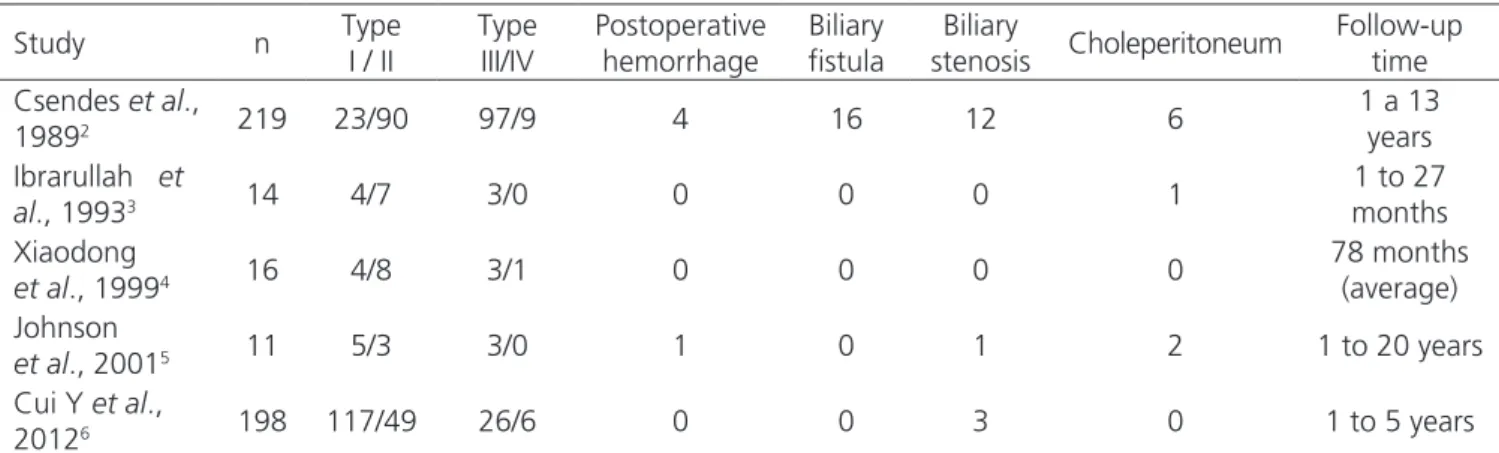
Table 2. Results of the surgical management of Mirizzi Syndrome.
Study n Type I / II
Type III/IV
Postoperative hemorrhage
Biliary fistula
Biliary
stenosis Choleperitoneum
Follow-up time Csendes et al.,
19892 219 23/90 97/9 4 16 12 6
1 a 13 years Ibrarullah et
al., 19933 14 4/7 3/0 0 0 0 1
1 to 27 months Xiaodong
et al., 19994 16 4/8 3/1 0 0 0 0
78 months (average) Johnson
et al., 20015 11 5/3 3/0 1 0 1 2 1 to 20 years
Cui Y et al.,
20126 198 117/49 26/6 0 0 3 0 1 to 5 years
As for the procedure performed, all patients were submitted to cholecystectomy, exploration of bile duct and cavity drainage. Surgical times were long, with an average of five hours. Choledocholithi-asis was found in 61.5% of cases and cholangitis in 38.4%.
The preferred therapeutic approach was Roux-enY hepaticojejunal anastomosis. One case with partial gastrectomy for peptic ulcer disease underwent a cho-ledocoduodenal anastomosis, since this segment was al-ready excluded from intestinal transit.
When using the “T” tube drainage, it re-mained for four to six months, aiming to shape the bile duct and prevent stenosis. This approach was ad-opted on three occasions associated with the suture of the biliary orifice over the drain: One patient in the second trimester of pregnancy who remained with the drain till the late postpartum period; one individual who had verticalized hepatic ducts, hampering the biliodigestive anastomosis; and one patient with chol-angitis, displaying a bile duct with thickened wall and diminished lumen.
There were two cases of fistula with the di-gestive tract: the duodenum and stomach. Fistulas is not unusual due to the same pathophysiological mech-anism, ie, intense inflammatory process, leading to adhesions between the viscera triggered by vesicular calculation.
Pathological examination showed benign dis-ease in all cases. The association between MS and gall-bladder cancer is well established. Some studies suggest the incidence of up to 5.3%, but series with the largest
number of patients do not bring any report of associated cancer6,15,16.
Postoperative local complications occurred in half the cases, mainly the biliary fistula (30.7%). Usual-ly benign, it presents resolution in a few days. OnUsual-ly one case required reoperation for better drainage of the cav-ity. Comparative data of postoperative complications of different works are shown in Table 2.
Mortality was 15.3% (2 cases) and was due to postoperative, not externalized transpapillary bleed-ing in a patient who had undergone preoperative endo-scopic papillotomy and presented sudden death on the second day after surgery. The diagnosis was confirmed at autopsy. The other death was in one patient admitted with cholangitis who had multiple organ failure preop-eratively.
In the outpatient follow-up of patients with biliary-enteric anastomosis (8 cases), 50% are asymp-tomatic, 25% had anastomotic stricture and 25% were lost to follow-up early in the process. Among those who underwent drainage of the bile duct with “T” tube (three cases), all remained asymptomatic after tube re-moval.
Being a low incidence condition and display-ing preoperative diagnosis in only half of the cases, Mi-rizzi Syndrome is a challenge. The preferred approach in advanced cases is biliodigestive anastomosis, especially when there are fistulas wider than two-thirds of the di-ameter of the common hepatic duct, when both morbid-ity and mortalmorbid-ity have considerable rates. Among the late complications, the stenosis of the bile duct stands out, the long-term follow-up being imperative.
R E S U M O
Objetivo: avaliar a epidemiologia e os resultados do tratamento cirúrgico de doentes portadores de graus III e IV, mais avançados, da Síndrome de Mirizzi (SM) de acordo com a classificação de Csendes. Métodos: estudo retrospectivo, de corte transversal através da revisão de prontuários de 13 pacientes portadores de graus III e IV da SM operados de dezembro de 2001 a setembro de 2013, entre 3691 colecistectomias realizadas neste período. Resultados: a incidência da SM foi 0,6% (23 casos) e os graus III e IV perfizeram 0,35% deste número. Houve um predomínio de tipo IV (12 casos). O diagnóstico pré-operatório foi possível em 53,8% dos casos. A conduta preferencial foi derivação biliodigestiva (10 casos) e foi optado por drenagem com tubo ”T” e sutura da via biliar em três ocasiões espe-ciais. Três pacientes apresentaram fístula biliar resolvida com conduta expectante e um caso de coleperitônio necessitou reoperação. No seguimento ambulatorial dos pacientes que realizaram a anastomose biliodigestiva (oito), 50% estão assintomáticos, 25% apresentaram estenose da anastomose e 25% perderam seguimento. O tempo médio de acompanhamento foi 41,8 meses. Conclusão: de incidência baixa e de diagnóstico pré-operatório em apenas metade dos casos, a SM em graus avançados tem na anastomose biliodigestiva sua melhor conduta, porém não isenta de morbimortalidade.
REFERENCES
1. Leopardi LN, Maddern GJ. Pablo Luis Mirizzi: the man behind the syndrome. ANZ J Surg. 2007;77(12):1062-4. 2. Csendes A, Diaz JC, Burdlies P, Maluenda F, Nava O.
Mirizzi syndrome and cholecystobiliary fistula: a uni-fying classification. Br J Surg. 1989;76(11):1139-43. 3. Ibrarullah M, Saxena R, Sikora SS, Kapoor VK,
Saras-wat VA, Kaushik SP. Mirizzi’s syndrome: identifica-tion and management strategy. Aust N Z J Surg. 1993;63(10):802-6.
4. Xiaodong H, Hongsheng L, Chaoji Z, Zhenhuan Z, Jianxi Z. Diagnosis and treatment of the Mirizzi syn-drome. Chin Med Sci J. 1999;14(4):246-8.
5. Johnson LW, Sehon JK, Lee WC, Zibari GB, McDonald JC. Mirizzi’s syndrome: experience from a multi-insti-tucional review. Am Surg. 2001; 67(1):11-4.
6. Cui Y, Liu Y, Li Z, Zhao E, Zhang H, Cui N. Appraisal of diagnosis and surgical approach for Mirizzi syndrome. ANZ J Surg. 2012;82(10):708-13.
7. Lai EC, Lau WY. Mirizzi syndrome: history, present and future development. ANZ J Surg. 2006;76(4):251-7. 8. Erben Y, Benavente-Chenhalls LA, Donohue JM, Que
FG, Kendrick ML, Reid-Lombardo KM, et al. Diagno-sis and treatment of Mirizzi syndrome: 23-year Mayo Clinic experience. J Am Coll Surg. 2011;213(1):114-9. 9. Waisberg J, Corona A, de Abreu IW, Farah JF, Lupinac-ci RA, Goffi FS. Benign obstruction of the common he-patic duct (Mirizzi syndrome): diagnosis and operative management. Arq Gastroenterol. 2005;42(1):13-8. 10. Beltrán MA. Mirizzi syndrome: history, current
knowledge and proposal of a simplified
classifica-tion. World J Gastroenterol. 2012;18(34):4639-50. 11. Akeely MH, Alam MK, Bismar HA, Khalid K,
Al-Teimi I, Al-Dossary NF. Mirizzi syndrome: ten years experience from a teaching hospital in Riyadh. World J Surg. 2005;29(12):1687-92.
12. Karademir S, Astarciolu H, Sökmen S, Atila K,
Tankurt E, Akpinar H, et al. Mirizzi’s syndrome: di-agnostic and surgical considerations in 25 patients. J Hepatobiliary Pancreat Surg. 2000;7(1):72-7.
13. Wichmann C, Wildi S, Clavien PA. The relationship of Mirizzi syndrome and cholecystoenteric fistula: validation of a modified classification. World J Surg. 2008;32(10):2244-5.
14. Shah OJ, Dar MA, Wani MA, Wani NA. Manage-ment of Mirizzi syndrome: a new surgical approach. ANZ J Surg. 2001;71(7):423-7.
15. Redaelli CA, Büchler MW, Schilling MK, Krähenbühl L, Ruchti C, Blumgart LH, et al. High coincidence of Mirizzi syndrome and gallblandder carcinoma. Sur-gery. 1997;121(1):58-63.
16. Prasad TL, Kumar A, Sikora SS, Saxena R, Kapoor VK. Mirizzi syndrome and gallbladder cancer. J Hepato-biliary Pancreat Surg. 2006;13(4):323-6.
Received in: 24/02/2016
Accepted for publication: 06/06/2016 Conflict of interest: none.
Source of funding: none.
Mailing address:
Ronaldo Reverdito