689 Rev Soc Bras Med Trop 50(5):689-692, September-October, 2017 doi: 10.1590/0037-8682-0025-2017
Short Communication
Corresponding author: Dra. Adriene Siqueira de Melo.
e-mail: [email protected]
Received 30 March 2017
Accepted 26 June 2017
Ex vivo
T-lymphocyte chemokine receptor phenotypes
in patients with chronic Chagas disease
Matheus Barbosa de Miranda
[1], Adriene Siqueira de Melo
[2], Mariana Silva Almeida
[1],
Silvia Martins Marinho
[3],[4], Wilson Oliveira Junior
[3],[4]and Yara de Miranda Gomes
[1],[4][1]. Centro de Pesquisas Aggeu Magalhães, Fundação Oswaldo Cruz, Recife, PE, Brazil. [2]. Faculdade Pernambucana da Saúde, Recife, PE, Brazil.
[3]. Ambulatório de Doença de Chagas e Insuiciência Cardíaca, Pronto Socorro Cardiológico de Pernambuco, Universidade de Pernambuco, Recife, PE, Brazil.
[4]. Programa Integrado de Doença de Chagas, Instituto Oswaldo Cruz, Fundação Oswaldo Cruz, Rio de Janeiro, RJ, Brazil.
Abstract
Introduction: Elucidating the molecules involved in the inlammatory process of chronic Chagas disease may allow identiication of treatment targets. Methods: The ex vivo phenotypic expression of chemokine receptors CCR1, CCR3, CCR4, CCR5, CXCR2,
CXCR3, CXCR4, and CXCR5 on the CD4+ and CD8+ T-cells of patients with chronic Chagas cardiomyopathy of varying severity was evaluated using low cytometry. Results: Differential expression of CD4+CCR3+ and CD8+CCR4+ T-cells was observed in patients with mild cardiac involvement compared, respectively, with patients with severe cardiac and asymptomatic forms of Chagas disease. Conclusions: These receptors are possibly involved in the pathogenesis of chronic Chagas cardiomyopathy.
Keywords: Chronic Chagas disease. Cardiomyopathy. Chemokine receptors.
Chagas disease, caused by the hemolagellate protozoan
Trypanosoma cruzi, presents with systemic features and
follows a chronic evolution; it is a serious public health problem throughout Latin America1. The chronic phase of this
disease comprises three main clinical forms: asymptomatic or indeterminate, cardiac, and digestive forms2. The cardiac form
is associated with high morbidity and mortality, occurring mostly in symptomatic patients in the chronic phase of the disease. One of the main immunologic features of intracellular protozoan infections such as T. cruzi is polarization to a
TH1 response. The TH1 response profile is characterized by a repertoire of proinlammatory cytokines, mainly tumor necrosis factor-α and interferon-γ, that play a role in parasite elimination and host survival3. However, if the inlammatory
environment is modulated immunologically by the expression of anti-inlammatory cytokines such as interleukin-10, this may trigger a balanced response with consequent attenuation of inflammation4. Therefore, modulatory mechanisms are
essential to prevent exacerbated inlammatory responses against the parasite, with consequent tissue injury. Evaluation of the inlammatory milieu in patients with chronic Chagas disease, by quantifying chemokine production and the expression chemokine receptors, could further our understanding of the mechanisms causing heart damage and they could help to
explain the absence of clinical alterations among individuals with the indeterminate form of the disease.
Patients with chronic Chagas disease attending the
Ambulatório de Doença de Chagas e Insuiciência Cardíaca
do Pronto Socorro Cardiológico de Pernambuco (PROCAPE),
Universidade de Pernambuco, Brazil, were selected. Patients were included according to following criteria: 1) positive serology for Chagas disease on enzyme-linked immunosorbent assay and immunofluorescence reaction (RIFI); 2) results of clinical tests and complementary examinations (physical examination, electrocardiography, chest radiography, Doppler echocardiography, and esophagography) were available to allow characterization of cardiac stages A, B (B1 and B2), C and D, according to the I Latin American Guidelines for the diagnosis and treatment of Chagas cardiomyopathy5; 3) absence
of gastrointestinal symptoms and comorbidities (hypertension, diabetes, thyroid disease, and inlammatory and/or infectious diseases); and 4) absence of previous etiological treatment with benznidazole.
In total, 33 individuals (13 men and 20 women) were included in the study. The group of patients classiied as having the indeterminate form of the disease (n = 12, aged ~56 years)
comprised those with stage A cardiac involvement, i.e. without current or previous symptoms of heart failure and whose clinical tests were normal. Patients were classiied as having a mild cardiac form of the disease (n = 10, aged ~68 years) if they
690
Miranda MB et al. - Chemokines in chronic Chagas disease
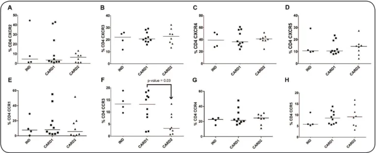
FIGURE 1 -Ex vivo phenotype expression of chemokine receptors on the surface of CD4+ T-cells of patients with chronic Chagas disease. A: %CD4 CXCR2. B: %CD4 CXCR3. C: %CD4 CXCR4. D: %CD4 CXCR5. E: %CD4 CCR1. F: %CD4 CCR3. G: %CD4 CCR4. H: %CD4 CCR5. IND: indeterminate form (n = 12); CARD1: mild cardiac form (n = 10); CARD2: severe cardiac form (n = 11). CD4: cluster of differentiation 4; CXCR2, CXCR3, CXCR4, CXCR5, CCR1, CCR3, CCR4, CCR5: chemokine receptors. Median values are indicated by the horizontal lines. The Kruskal-Wallis test was performed, followed by
Dunn’s test. Signiicant differences are indicated by arrows and exact p-values.
cardiac form of the disease (n = 11, age ~58 years) if they were
in stage C, i.e. a previous or current clinical diagnosis of heart failure according to the Framingham criteria (simultaneous presence of two major criteria or one major criterion and two minor criteria)6 and evidence of ventricular dysfunction (systolic
heart failure with a left ventricular ejection fraction <50%, as determined by Doppler echocardiography).
After collecting 10mL samples of blood in Becton Dickinson Vacutainers® containing sodium heparin, 100µL were used directly in the execution of the phenotypic expression protocol. Samples were immunostained by incubation for 30 min with luorescein isothiocyanate-conjugated anti-CD8 (clone: 3B5, Caltag, Burlingame, CA, USA ); peridinin-chlorophyll protein complex-conjugated anti-CD4 (clone: S3.5, Invitrogen™, Carlsbad, CA, USA); Alexa Fluor-conjugated anti-CCR1 (clone: 5354), anti-CCR3 (clone: SF8), and anti-CXCR5 (clone: PF8B2; Becton Dickinson, Franklin Lakes, NJ, USA); and phycoerythrin-conjugated anti-CCR4 (clone: 161), anti-CCR5 (clone: 2D7/ CCR5), anti-CXCR2 (clone: 6C6), anti-CXCR3 (clone: 1C6/ CXCR3), and anti-CXCR4 (clone: 1D9; Becton Dickinson, Franklin Lakes, NJ, USA). After incubation, erythrocytes were lysed (FACS Lysing Solution; Becton Dickinson Bioscience), followed by washing in phosphate buffered saline containing 0.5% bovine serum albumin and 0.1% sodium azide (Sigma-Aldrich) and centrifugation (300 × g for 5 min).
Samples were processed using a BD FACSCalibur™ low cytometer (Becton Dickinson, Franklin Lakes, NJ, USA), available on the Core Technology Platforms (NPT1) of
Centro de Pesquisas Aggeu Magalhães/Fundação Oswaldo Cruz (CPqAM/FIOCRUZ). Sample acquisition and analysis
were performed using BD Cell Quest™ Pro software. For each analysis, 20,000 events were acquired, gated on cluster of differentiation 4 (CD4+) and cluster of differentiation 8+
(CD8+) T-cell subpopulations expressing the chemokine receptors. The analysis was conducted by obtaining two-dimensional graphics of luorescent punctual distributions.
The graphics were developed and the statistical tests performed using GraphPad Prism 6.0 software. Bartlett’s test for homogeneity of variance was irst applied. To compare the median values of the phenotypic expression of chemokine receptors on the surface of T-cells among the groups of patients, the Kruskal-Wallis test was applied, followed by the multiple-comparison Dunn’s test when differences between medians were demonstrated. Statistical signiicance was set at 5% (α = 0.05).
The group of patients with mild cardiac disease had higher levels of CD4+ T-cells expressing the CCR3 receptor than did the group with severe cardiac disease (p = 0.03; Figure 1). In a study investigating associations between Chagas disease morbidity and the expression of cytokines and cytokine receptors, Gomes et al.7 described the CCR3 receptor as being
691
Rev Soc Bras Med Trop 50(5):689-692, September-October, 2017
FIGURE 2 - Ex vivo phenotype expression of chemokine receptors on the surface of CD8+ T-cells of patients with chronic Chagas disease. A: %CD8 CXCR2. B: %CD8 CXCR3. C: %CD8 CXCR4. D: %CD8 CXCR5. E: %CD8 CCR1. F: %CD8 CCR3. G: %CD8 CCR4. H: %CD8 CCR5. IND: indeterminate form (n = 12); CARD1:, mild cardiac form (n = 10); CARD2: severe cardiac form (n = 11). CD8: cluster of differentiation 8; CXCR2, CXCR3, CXCR4, CXCR5, CCR1, CCR3, CCR4, CCR5: chemokine receptors. Median values are indicated by the horizontal lines. The Kruskal-Wallis test was performed, followed by
Dunn’s test. Signiicant differences are indicated by arrows and exact p-values.
The mild cardiac disease group had higher levels of CD8+ T-cells expressing the CCR4 receptor than did the indeterminate group (p = 0.02; Figure 2). Induction of cellular immunity and immune responses, mediated by CD8+ T-cells, is essential for the control of T. cruzi proliferation, as shown in murine
models and in human infections8. The concomitant inding of the presence of Chagas disease and high numbers of CD8+ T-cells in experimental models has led some researchers to suggest that CD8+ T-cells cause the observed tissue damage9.
Although CCR4 is a predominant receptor on TH2 cells, the fact that it has been observed to have greater expression in CD8+ T-cells suggests that CD8+ T-cells might use CCR4 for migrating to sites of inlammation. Hence, it is necessary to evaluate the production of cytokines as well as the production of perforins and granzymes in this particular subpopulation, to determine their possible role in the establishment of the disease their role is likely to be proinlammatory. Additionally Figure 2, demonstrates an apparent increase in the expression of CD8+CCR4+ T-cells in patients with the mild cardiac form of Chagas disease, compared with patients with the severe cardiac form. In this group of patients, with the severe cardiac form, especially the older patients, it is not uncommon to observe immune response dysfunction and exhaustion, possibly as a result of chronic antigen exposure and myocardial remodeling10.
Therefore, the level of some immunological markers, notably memory cells, could be in lower in patients with the severe cardiac form of Chagas disease, supporting the hypothesis that the severity of disease is inversely correlated with the quality of immune responses to T. cruzi11. Thus, with further analysis,
this marker may also provide a useful tool for identifying patients who are likely to progress from the mild to a more severe cardiac form of the disease. The expression of other
chemokine receptors on the surface of CD8 + T-cells did not vary signiicantly among the different groups (Figure 2).
To conclude, our indings suggest that in patients with the cardiac form of chronic Chagas disease, the chemokine receptors CCR3 and CCR4 are involved in the mechanisms of anti-inlammatory and pro-inlammatory immune responses, respectively.
Ethical considerations
The approaches conducted in this study were approved by the Ethics Committee on Research in Human Beings of CPqAM/ FIOCRUZ (CAEE: 0032.0.095.000-10) as per the 1975 Declaration of Helsinki, revised in 1983. All subjects provided written informed consent prior to their inclusion in the study.
Acknowledgements
The authors thank the Programa de Desenvolvimento Tecnológico em Insumos para Saúde (PDTIS)/FIOCRUZ for the use of its facilities.
Financial support
This research study received inancial support from the Conselho Nacional de
Desenvolvimento Cientíico e Tecnológico [(CNPq) (CNPq/Papes VI/Fiocruz No. 407763/2012-0] and Coordenação de Aperfeiçoamento de Pessoal de
Nível Superior (CAPES). During the study, Y.M. Gomes was research fellow of CNPq (No. 304543/2012-8), A.S. Melo received a CAPES PhD scholarship (064.074.324-21), and M.B. Miranda received a scholarship from CNPq/
Programa Institucional de Bolsas de Iniciação Cientíica (PIBIC) /Fiocruz.
Conlict of interest
692
REFERENCES
1. Coura JR, Dias JCP. Epidemiology, control and surveillance of
Chagas disease - 100 years after its discovery. Mem Inst Oswaldo Cruz. 2009;104(Suppl 1):31-40.
2. World Health Organization. Chagas Disease (American
trypanosomia-sis); 2015. http://www.who.int/mediacentre/factsheets/fs340/en/
3. Cunha-Neto E, Teixeira PC, Nogueira LG, Mady C, Ianni B, Stolf N,
et al. Novos conceitos sobre em patogenia da cardiopatia chagásica crônica: peris de expressão gênica e protéica do miocárdio. Rev Soc Bras Med Trop. 2006;39(Suppl III):59-62.
4. Dutra WO, Menezes CAS, Magalhães LMD, Gollob KJ.
Immunoregulatory networks in human Chagas disease. Parasite Immunol. 2014;36(8):377-87.
5. Andrade JP, Marin-Neto JA, Paola AAV, Vilas-Boas F, Oliveira GMM, Bacal F, et al. I Diretriz Latino Americana para o Diagnóstico e Tratamento da Cardiopatia Chagásica. Arq Bras Cardiol. 2011;97(no. 2 supl. 3):1-48.
6. McKee PA, Castelli WP, McNamara PM, Kannel WB. The natural
history of congestive heart failure: the Framingham study. N Engl J Med. 1971;285(26):1441-6.
7. Gomes JA, Bahia-Oliveira LM, Rocha MO, Busek SC, Teixeira
MM, Silva JS, et al. Type 1 chemokine receptor expression in Chagas' disease correlates with morbidity in cardiac patients. Infec Immunity. 2005;73(12):7960-6.
8. Egui A, Thomas MC, Carrilero B, Segovia M, Alonso C, Marañón C, et al. Differential phenotypic and functional proiles of TcCA-2-speciic cytotoxic CD8+ T cells in the asymptomatic versus cardiac phase in chagasic patients. PLoS One. 2015;10(3):e0122115. 9. Martin D, Tarleton R. Generation, speciicity, and function of
CD8+ T cells in Trypanosoma cruzi infection. Immun Rev.
2004;201(1):304-17.
10. Albareda MC, Laucella SA, Alvarez MG, Armenti AH, Bertochi
G, Tarleton RL, et al. Trypanosoma cruzi modulates the proile
of memory CD8+ T cells in chronic Chagas' disease patients. Int Immunol. 2006;18(3):465-71.
11. Laucella SA, Postan M, Martin D, Hubby Fralish B, Albareda MC,
Alvarez MG, et al. Frequency of interferon-gamma-producing T cells speciic for Trypanosoma cruzi inversely correlates with
disease severity in chronic human Chagas disease. J Infect Dis. 2004;189(5):909-18.