774
INTRODUCTION
www.scielo.br/rsbmt Revista da Sociedade Brasileira de Medicina Tropical 45(6):774-776, Nov-Dec, 2012
Case Report
Address to: Dr. Pedro Silva Vaz. Serviço de Cirurgia Geral/Hospital Amato Lusitano/Unidade Local de Saúde de Castelo Branco. Av. Pedro Álvares Cabral, 6000-085 Castelo Branco, Portugal.
Phone: 00351 966 498 337 e-mail: [email protected] Received in 19/10/2011 Accepted in 27/01/2012
Hepaic hydaid cyst: a non-surgical approach
Pedro Silva Vaz
[1], Eduardo Pereira
[2], Sergiu Usurelu
[1], Ana Monteiro
[1], Ana Caldeira
[2],
Gina Melo
[1], Rui Sousa
[2], António Gouveia
[1]and Arnandina Loureiro
[1][1]. Serviço de Cirurgia Geral, Hospital Amato Lusitano, Unidade Local de Saúde de Castelo Branco, Castelo Branco, Portugal. [2]. Serviço de Gastroenterologia, Hospital Amato Lusitano, Unidade Local de Saúde de Castelo Branco, Castelo Branco, Portugal.
ABSTRACT
Echinococcosis/hydaidosis is common in socieies where agriculture and livestock are frequent, and represents a public health problem. The therapeuic management depends on the cyst’s characterisics, the paient, and surgical contraindicaions. Endoscopic retrograde cholangiopancreatography is a valuable tool in the diagnosis and treatment of complicated hepaic hydaid disease. Ultrasonography is a useful diagnosic, therapeuic and follow-up tool. The authors report a case of a 56 years old paient who was diagnosed with a hepaic hydaid cyst in the IVa/VIII segments, describe the therapeuic opions and 50 months of disease-free follow-up.
Keywords: Hepaic hydaid cyst. Endoscopic retrograde cholangiopancreatography. Ultrasonography.
Echinococcosis/hydatidosis is common in societies where agriculture and livestock are frequent, and it coninues to represent a serious public health problem, especially in the Mediterranean, Middle East, South America, New Zeland and Turkey1-5. In Portugal, Cartucho et al. point to a higher incidence in Alentejo, Setúbal, Vale do Tejo, and Castelo Branco6.
Hydaid cysts are most oten hepaic (50-80%) and pulmonary (5-30%) but they may also be found in other locaions1-4,7. Treatment depends on the cystic lesion’s characteristics, the patient, and whether there are any contraindicaions to surgery, which is sill the therapeuic gold-standard5,8. Medical treatment is characterized by the use of benzimidazoles1,7. Puncture, aspiration, injection and re-aspiration (PAIR) has been shown to be effective in the presence of non-complicated hydaid cysts. Endoscopic retrograde cholangiopancreatography (ERCP) has been described as the best approach in complicated hepatic hydatid disease, being both a diagnosic and therapeuic tool2.
Case RepORT
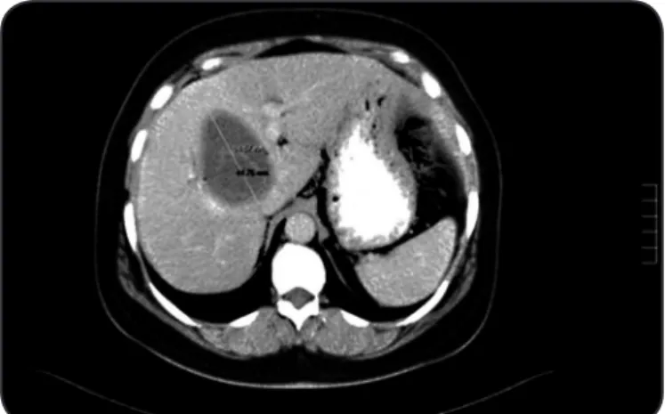
A 56 year old female paient, born and residing in the City of Castelo Branco, Portugal, underwent abdominal ultrasonography in the clinical context of recently diagnosed diabetes mellitus associated with dyspepic complaints. The ultrasonography showed a predominantly liquid heterogeneous formaion of about 75mm, apparently capsulated, located in the IVa/VIII segments of the liver, with characteristics which suggested a hydatid cyst. Abdomino-pelvic computerized tomography (CT) revealed a 80x62x42mm cysic formaion, adjacent the gallbladder, with incipient wall calciicaion
and roughly nodular endoluminal images. It extended until the VIII segment in strict associaion with the inferior vena cava (IVC) (Figure 1). Serology tests for hydaidosis were posiive. Endoscopic retrograde cholangiopancreatography showed a contrast filled collecion, which proved a communicaion between the cyst and the biliary tree. However, it was not possible to determine the istulous trajectory. Therapy with albendazole (15mg/kg/day) was iniiated. The following therapeuic opions were proposed in this clinical context: surgical intervenion in a specialist hepaic surgery center, due to the locaion of the cysic lesion as well as the proximity to the vascular structures, such as the IVC, or non-surgical treatment in our
FIGURE 1 - Abdomino-pelvic computerized tomograph revealing a cysic formaion with incipient wall calciicaion and roughly nodular endoluminal images.
insituion with the support of the Gastroenterology Department. Aware of the risks involved with the surgical opion, the paient chose the non-surgical treatment. Ater 6 months of treatment, an elevaion of the hepaic enzymology and cholestasis was veriied. This was atributed to albendazole hepatotoxicity, and a decision was made to suspend medical treatment. At this point, imaging showed a signiicant reducion of the size of the lesion, and the serologic tests were negaive. The paient abandoned the follow-up in light of the clinical improvement.
775 Vaz PS et al - Hepaic hydaid cyst: a non-surgical approach
DIsCUssION revealed a 60x40mm hydaid cyst located between the hepaic hilus
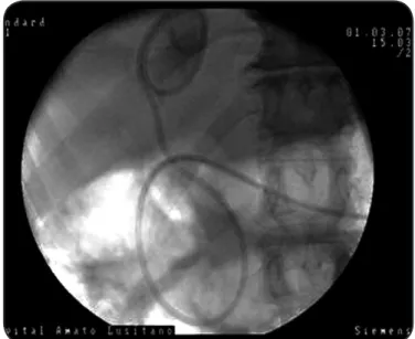
IVC, with no alteraions of the gallbladder and with slight dilaion and wall thickening of the common bile duct (CBD). Serology was again posiive and therefore albendazole (10mg/kg/day) was iniiated. She performed ERCP (Figure 2) which showed contrast-illed cyst with easy cannula access. A basket was used to clean the biliary duct and there was evidence of the removal of hydaids. At the end of the procedure, a double pig’s tail tube was placed in the cyst (Figure 3). The paient was again given a choice of therapeuic opions and sill refused surgical treatment. Ten days later, she performed another ERCP to replace the double pig’s tail tube by a nasocysic catheter. Three lavages with 20% hypertonic soluion were carried out over the following 5 days unil the liquid drained from the catheter was clear. Irrigaion was done under clinical and ultrasonographic control which allowed the direct visualizaion of the cysic cavity illing. The nasocysic catheter was removed ater the third cyst lavage.
Five months later she presented elevated liver funcion tests again. Ultrasonography showed a residual cyst. The serology study was repeated and was negaive. The albendazole was therefore suspended ater one month of serologic evaluaion.
Currently the paient is under annual ultrasonographic control. The last appointment, at 50 months, showed a residual cyst. The paient is asymptomaic and liver funcion tests are normal.
FIGURE 2 - Endoscopic retrograde cholangiopancreatography showing contrast cyst formaion with easy cannula access.
FIGURE 3 - Double pig’s tail tube in the cyst formaion for draining.
The indicaions for ERCP in hepaic hydaid cyst paients during the pre-surgical period include the characterizaion of the relaion between the hydaid cyst and the biliary tree, which aid in the drawing up of a surgical plan, the evaluaion of acute clinical condiions (cholangiis and obstrucion of CBD), the decrease of the incidence of post-surgical istulas, or even the curaive treatment of intrabiliary rupture cases if the cysic cavity or the biliary duct are accessible through this method.
In the presented clinical case, ERCP was used both for characterizaion of the biliary tree (diagnosis of intrabiliary rupture), and in the context of cholangiis for the placement of a nasocysic catheter for irrigaion and draining (therapeuic).
The diagnosis of hepaic hydaidosis using imaging is based on the CT, abdominal ultrasonography, both described as the ideal exams for diagnosis and for evaluaion of therapeuic eicacy. CT and magneic resonance imaging (MRI) provide addiional structural details and show more precisely the locaion and depth of the cyst within the liver, data essenial for planning treatment. According to Gharbi, ultrasonography is the preferred method for the classiicaion of hydaid cysts1,3,7-12. In some cases, ultrasonography is pathognomonic, allowing the deiniion of the parasite’s stage of evoluion (WHO classiicaion)3.
In the majority of publicaions, the therapeuic opions for the hepaic hydaid disease include surgery and benzimidazoles.
Surgery is sill the primary treatment opion and promotes the inacivaion of the scolex, the prevenion of the disseminaion of the cysic content into the peritoneal cavity, the eliminaion of all the cyst’s viable elements, and the handling of the residual cavity. Nevertheless, the main surgical complications are postoperative biliary fistula (5-25%) and relapse (2-16%)1,2.
776
Rev Soc Bras Med Trop 45(6):774-776, Nov-Dec, 2012
ReFeReNCes As a therapeuic method, ERCP was essenial in the context of the
paient’s cholangiis, as well as in the treatment of the hydaid disease with placement of a nasocysic tube that allowed for the controlled draining of hydaids and irrigaion with a scolicidal agent. Simsek et al state that endoscopic procedures, namely sphincterotomy (ETE), stent inserion or nasobiliary draining have a high success rate of approximately 90% in cysic hydaid disease2.
Therapy with ERCP for hepaic hydaid cysts has been described by some authors as a irst choice procedure in selected paients.
The use of ultrasonography as a tool to control each lavage has also been described here. In this case, ultrasonography has assumed a fundamental role in therapeuics, allowing a direct visualizaion of the 20% hypertonic soluion illing of the cysic cavity leading to a control of the possible side efects of the scolicide (sclerosing cholangiis and pancreaiis2). The control, both ultrasonographical and clinical, allowed us to deine of the safe quanity of scolicidal agent to be injected at each lavage.
The use of the ultrasonography as an aid in the non-surgical treatment of hydaid cysts is innovaive.
The paient remains under imaging and clinical follow-up, the last of which reveals a residual image of a cyst. The 50 month follow-up with no relapse indicates therapeuic success.
Endoscopic retrograde cholangiopancreatography with ETE ofers an excellent opion with immediate results both in diagnosis and in therapeuics of hepaic hydaid disease with intrabiliary rupture. It could be an alternaive treatment to be considered for high surgical risk paients.
Ultrasonography, used in this innovaive manner, proved a useful aid in the non-surgical treatment of a hydaid cyst by allowing the safe quaniicaion of scolicidal agent to be injected, thereby decreasing the side efects with which these substances are associated.
1. Pawlowski ZS, Eckert J, Vuitton RW, Ammann PK, Craig PS, Dar KF, et al. Echinococcosis in humans: clinical aspects, diagnosis and treatment.
In: Eckert J, Gemmell MA, Meslin FX, Pawlowski ZS, editors. WHO/OIE Manual on Echinococcosis in Humans and Animals: a Public Health Problem of Global Concern. Paris, France: World Health Organizaion; 2001. p. 20-62.
2. Simsek H, Ozaslan O, Sayek I, Savas C, Bbasoglu O, Soylu AR, et al. Hepaic hydaid disease: diagnosic and therapeuic ERCP. Gastrointest Endosc 2003; 58:384-389. 3. Menezes-da-Silva A, Caldeira J, Nunes JL. PAIR - Alternaiva terapêuica do quisto
hidáico do ígado. J Port Gastrenterol 2001, 8:113-120.
4. Palacios-Ruíz JA, Ramirez-Solis ME, Moreno-Moller M, Cardenas-Mejia A, Gonzalez-Monroy LE, Diaz-Pizarro JI, et al. Seguridad y eicacia de la solución salina hipertónica al 17.7% durante el tratamiento laparoscópico de un quiste hidáico hepáico. Assoc Mex Cirugía Endoscópica 2001; 2:206-210.
5. Brunicardi FC, Andersen DK, Billiar TR, Dunn DL, Hunter JG, Pollock RE. Liver. Sxwartz’s Principles of Surgery. 8th ed. New York, USA: McGraw-Hill; 2005. 6. Cartucho D, Marrão G, Vaiadas G, Rocha G, Santana A, Patrício J. Caracterização
das esirpes de Quisto Hidáico em Portugal: onde estamos nós? Rev Saúde Amato Lusitano 1999; 9:13-17.
7. María Luisa C, Lloeznaly O, Julio V, Noya B, Carmen Z. Quiste hidaídico hepáico: a propósito de un caso. Rev Soc Venezolana Gastrenterol 2007; 6:206-209. 8. Moreno-González E, Meneu Diáz JC, Moreno Elola A. Echinococcal Cyst. In:
Baker RJ, Fischer JE, editors. Mastery of Surgery. 5th ed. Philadelphia: Lippincot Williams Wilkins Inc; 2007. p. 1043-1075.
9. Reddy DN. Endoscopic diagnosis and management of biliary parasitosis [Internet]. Up to Date online 2008; Available from: htp://www.uptodate.com/contents/ endoscopic-diagnosis-and-management-of-biliary-parasitosis/.
10. Özaslan E. Therapeutic endoscopic retrograde cholangiopancreatography and related modalities have many roles in hepatobiliary hydatid disease. World J Gastroenterol 2006; 12:4930-4931.
11. Manouras A, Genetzakis M, Antonakis PT, Lagoudianakis E, Patas M, Papadima A, et al. Endoscopic management of a relapsing hepaic hydaid cyst with intrabiliary rupture: A case report and review of the literature. Can J Gastroenterol 2007; 21:249-253. 12. Dandan IS, Soweid AM, Abiad F. Hydaid cyst. Drugs, Diseases & Procedures 2008; [Cited 2011 January 11]. Available from: htp://emedicine.medscape.com/ aricle/178648-overview/.
Cisto hidáico hepáico: abordagem não cirúrgica
A equinococose/hidaidose é comum em sociedades onde predominam a agricultura e a criação de gado, sendo um problema de saúde pública. As várias opções terapêuicas dependem das caraterísicas do quisto, do doente e da eventual presença de contraindicações cirúrgicas. A colangiopancreatograia retrógrada endoscópica consitui uma válida ferramenta no diagnósico
aBsTRaCT IN pORTUgUese
e tratamento da doença hidáica hepáica complicada. A ecograia é um instrumento úil no diagnósico, na terapêuica e seguimento. Os autores apresentam um caso de uma doente de 56 anos a quem foi diagnosicado um quisto hidáico hepáico nos segmentos IVa/VIII, descrevem as opções terapêuicas e o seguimento de 50 meses livres de doença.