Prolonged cardioprotective effect of pyridostigmine encapsulated in liposomes
Alessandra Teixeira Vidal
a, Homero Nogueira Guimarães
b, Danielle Cristiane Correa de Paula
a,
Frederic Frezard
c, Neila Márcia Silva-Barcellos
a, Andrea Grabe-Guimarães
a,⁎
aCiPharma, Escola de Farmácia, Universidade Federal de Ouro Preto (UFOP), Ouro Preto, Minas Gerais, Brazil
bDepartamento de Engenharia Elétrica, Escola de Engenharia, Universidade Federal de Minas Gerais (UFMG), Belo Horizonte, Minas Gerais, Brazil cDepartamento de Fisiologia e Biofísica, Instituto de Ciências Biológicas, Universidade Federal de Minas Gerais (UFMG), Belo Horizonte, Minas Gerais, Brazil
a b s t r a c t
a r t i c l e
i n f o
Article history:
Received 28 May 2009 Accepted 20 October 2009
Keywords:
Pyridostigmine Liposomes
Sympathetic stimulation QT interval, ECG Rats
Aims:The purpose of the present work was to investigate the ability of pyridostigmine encapsulated in long-circulating liposomes, to protect against ECG (electrocardiogram) alterations induced by sympathetic stimulation in rats.
Main methods:The encapsulation of pyridostigmine was carried out by freeze–thaw and extrusion. Blood pressure and ECG (limb lead II) were monitored in anaesthetized male Wistar rats. The formulation containing pyridostigmine was intravenously administrated in 0.1, 0.3 and 1.0 mg/kg doses, and sympathetic stimulation was conducted by administration of 1 or 3μg of noradrenaline (NA) after 1, 2, 4 or 6 h. The obtained cardiovascular parameters were compared to animals that received intravenous injection of pyridostigmine in free form or saline.
Key findings:After saline, NA induced a significant increase in QT interval (22.3% after 3.0μg). Previous administration of free pyridostigmine significantly prevented the increase of QT interval after sympathetic stimulation and the most prominent effect was observed after 1h for the dose of 0.3 mg/kg (6.8% after 3.0μg of NA) and was no longer observed after 2 h of the treatment. On the other hand, the maximum effect of pyridostigmine in liposomal formulation preventing QT interval increase was observed 2 h after treatment (9.7% after 3.0μg of NA) and was still present until 6 h when 1 mg/kg was previous administrated.
Significance:The results of the present study, beyond to confirm the cardioprotective action of pyridostigmine, suggest that liposomal pyridostigmine may be a potential therapeutic alternative to prevent cardiovascular disturbances resulting from sympathetic hyperactivity.
© 2009 Elsevier Inc. All rights reserved.
Introduction
Several studies had demonstrated the relationship between the
autonomic nervous system (ANS) and cardiovascular diseases (Francis
1988; Porter et al. 1990; Tai et al. 2002; Hoyer et al. 2008). The
misbalance of this system, characterized by an increase of sympathetic
and depression of parasympathetic activities, may lead to arrhythmias
and sudden death, mainly in patients with myocardial infarction and
heart failure (Porter et al. 1990; Hoyer et al. 2008). Among the changes
caused by ANS on cardiovascular system, the in
fl
uence on QT interval of
electrocardiogram (ECG) has particular importance (Ahnve and Vallin
1982; London et al. 1998; Magnano et al. 2002). In fact, prolongation of
QT interval is an independent risk factor for sudden death due to cardiac
arrest (Schwartz and Wolf 1978; Algra et al. 1991; Schouten et al. 1991).
The effectiveness of adrenergic blockade in preventing fatal
arrhyth-mias, by reducing the sympathetic effects, has been extensively
characterized (Hjalmarson 1997; Catelli et al. 2003; Taggart et al. 2003).
Although the benefits of parasympathetic stimulation in the prevention of
arrhythmias have been de
fi
ned (De Ferrari et al. 1992), there is not a
pharmacological option available. In this context, the pyridostigmine
bromide, a reversible cholinesterase inhibitor, has been demonstrated as
a promissory agent on cardiac ischemic disease (Grabe-Guimarães et al.
1999; Sant'anna et al. 2003). Patients with exercise induced myocardial
ischemia (Castro et al. 2004) or heart failure (Serra et al. 2009) presented
improvements in autonomic and hemodynamic pro
fi
les during exercise
after oral administration of pyridostigmine. However, its half-life is short
(Aquilonius et al. 1980), the side effects related to cholinergic stimulation
are frequent and a limitation for safety clinical use.
The importance of liposomes to target the cardiovascular system is
widely studied (Torchilin 1995). It was shown that liposomes coated with
polyethylene glycol (PEG) prolong its life time in the circulation (Klibanov
et al. 1990). Additionally, PEGylated liposomes were found to accumulate
in infarcted myocardium (Torchilin et al. 1992) and are useful to protect
the myocardium from the ischemia damage (Verma et al. 2005).
The main goal of the present work was to investigate the ability of
pyridostigmine encapsulated in long-circulating liposomes to prolong
its cardioprotective action, by analysing ECG parameters, particularly
⁎Corresponding author. Universidade Federal de Ouro Preto, Escola de Farmácia, RuaCosta Sena, 171, Centro Ouro Preto, 35400-000 Minas Gerais, Brazil. Tel./fax: +55 31 3559 1628.
E-mail address:grabe@ef.ufop.br(A. Grabe-Guimarães).
0024-3205/$–see front matter © 2009 Elsevier Inc. All rights reserved. doi:10.1016/j.lfs.2009.10.011
Contents lists available at
ScienceDirect
Life Sciences
QT interval, and arterial blood pressure changes in anaesthetized
Wistar rats, followed by IV (intravenous) injection of a single dose of
noradrenaline (NA).
Materials and methods
Drugs and reagents
Pyridostigmine bromide and cholesterol were purchased from
Sigma (USA). L-
α
-Distearoylphosphatidylcholine (DSPC) and PEG
(2000)-distearoylphosphatidyl-ethanolamine (DSPE-PEG) were
pur-chased by Lipoid GmbH (Ludwigshafen, Germany). The solvents were
of analytical grade and all other chemicals were commercially
available. Water was puri
fi
ed by reverse osmosis (Symplicity System
185, Millipore, USA).
PEGylated liposomes preparation
The liposome system consisted of DSPC, cholesterol and DSPE-PEG at
a molar ratio of 5:4:0.3. Pyridostigmine encapsulation was carried out by
freeze
–
thaw and extrusion (Nayar et al. 1989). Control liposomes were
similarly prepared, using only PBS (150 mM NaCl, 10 mM phosphate,
pH 7.2).
Liposomes characterization
Pyridostigmine content of the liposomes was measured by UV
(
λ
=270 nm) (Hegazy et al. 2002) against a pyridostigmine-solution
standard curve. Particle size, polydispersity index and zeta potential of
the liposome population by photon correlation spectrometry were
carried out using Zetasizer 3000 HS (Malvern Instruments, UK).
Animals
Male Wistar rats were supplied by Universidade Federal de Ouro
Preto and maintained with water and food ad libitum at constant
humidity and temperature with a light/dark cycle of 12 h. Six animals
were used in each experimental group. All procedures related to the
use of animals in these studies were reviewed and conform to the
Ethical Principles of Animal Experimentation (Brazilian College of
Animal Experimentation) and were approved by the UFOP Ethics
Committee under number 11/2009.
Determination of in vivo cardiovascular parameters
Experimental procedures
Rats were anaesthetized with intraperitoneal sodium pentobarbital
(60 mg/kg). Intravenous (IV) injections of free pyridostigmine,
pyridos-tigmine in liposomes (Lipo-Pyr) or empty liposomes were performed via
a catheter inserted into the femoral vein. Free pyridostigmine and
pyridostigmine in liposomes were dissolved in saline to give the desired
dose (0.1, 0.3 or 1.0 mg/kg). All formulations were administered in bolus,
at maximum volume of 0.2 ml. Control rats received a corresponding
volume of saline or empty liposomes. In order to simulate an adrenergic
discharge, the animals received a single in bolus dose of NA solution (1.0
or 3.0
μ
g), 1, 2, 4 or 6 h after the treatment.
Arterial blood pressure (AP) was continuously recorded using a
polyethylene catheter,
filled with heparinised saline, inserted into
femoral artery and joined to a disposable pressure transducer
(TruWave; Edwards Life Sciences); the pressure transducer was
connected to a bridge ampli
fi
er. Limb lead II ECG was continuously
recorded using subcutaneous stainless steel needle electrodes
connected by a shielded cable to a biopotencial ampli
fi
er with a
band-pass of 0.5
–
100 Hz. These signals from ampli
fi
ers were simultaneously
sampled at a rate of 1200 Hz per channel, with an A/D conversion board
of 16 bits resolution (DaqBoard/2001, IOtech).
Data analyses
The ECG parameters measured were QT (interval between the
beginning of the
Q
-wave and the end of the
T
-wave), RR (interval
between two successive
R
-waves and used to obtain the heart rate:
HR =60/RR), PR (interval between the beginning of the
P
-wave and
the end of the
R
-wave) and QRS (interval from the beginning of the
Q
-wave to the end of the
S
-wave) intervals.
As the QT interval on standard ECG is in
fl
uenced by a variety of
physiological and pathological factors, such as heart rate, autonomic
nervous system activity, day time, age, gender, drugs, hormone
concentrations, electrolyte variations, heart disease or ventricular
dysfunction (Hodges 1997), several mathematical formulae have been
proposed to derive a heart rate corrected QT interval (QTc), or at least
minimize their dependence on heart rate.
Simonson et al. (1962)
enumerate 9 formulae for these purpose, but the best known are
Bazett's
(QTc=QT/RR
1/2) (1920) and Fridericia's (QTc=QT/RR
1/3) (1920)
formulae, commonly used in clinical trials and pre-clinical studies.
There is an almost centenary unsolved controversy about the rate
adjustment methods of QT intervals (Puddu et al. 1988; Hayes et al. 1994;
Malik et al. 2002; Koga et al. 2007). Embedded in a plethora of formulae
and methods on the strength of this controversy, we based our choice on
the work of
Abernethy et al. (2001), who suggest the Fridericia correction
for the QTc for the cases of RR
b
500 ms, as was consistently observed in
our experiments.
The systolic (SAP) and diastolic arterial pressure (DAP) were also
determined from the same segments. These parameters were
measured before and after injection of pyridostigmine formulations
followed by adrenergic stimulation at different times.
Statistical analysis
The Kolmogorov
–
Smirnov method was used to determine whether
continuous variables were normally distributed. Results are expressed
as mean±standard error of the mean (S.E.M.). Statistical comparisons
were made using ANOVA and Tukey post-hoc test. Signi
fi
cance was
accepted when
P
b
0.05.
Results
Particle size, polydispersity index and zeta potential
The freezing thawing/extrusion method resulted in vesicles with a
calibrated size smaller than 200 nm, an encapsulation ef
fi
ciency of
15.5%, and narrow size distribution for both empty and
pyridostig-mine in liposomes. Zeta-potential values were similar between both
formulations (Table 1).
Determination of cardiovascular parameters
Tables 2 and 3
reports in details the absolute values of ECG
parameters measured at different times after treatment with saline,
free pyridostigmine or liposomal pyridostigmine. The IV
administra-tion of 1.0
μ
g NA in Wistar rats that previously received saline, did not
caused signi
fi
cant increase of PR and QRS intervals of ECG when
compared to the control period. A slight increase of these parameters
was caused by 3.0
μ
g of NA, such 5.7% and 6.0% of increase to PR and
QRS, respectively. The analysis of QT interval showed increases of
20.1% and 22.3% after 1.0 and 3.0
μ
g of NA, respectively. The
pre-Table 1
Characterization of liposomes formulations.
Diameter (nm) Polydispersity index Zeta potential (mV) Lipo-Pyr 174.3 ± 6,01 0.061 ± 0.0127 −50.6 ± 2.5
Empty liposomes 158.5 ± 4,42 0.070 ± 0.0133 −41.2 ± 0.9
Each data represents the mean ± e.s.m. of three preparations.
treatment with free pyridostigmine at 0.1 and 0.3 mg/kg and with the
liposomal form at 0.1, 0.3 and 1.0 mg/kg did not change the baseline
values, but attenuated signi
fi
cantly the increases of the ECG
parameters produced by IV injection of NA compared to the control
group. After previous treatment with pyridostigmine, changes in PR
and QRS intervals caused by 1.0
μ
g of NA were also not observed. All
formulations prevented the increase of these parameters caused by
3.0
μ
g of NA. Free pyridostigmine and liposomal pyridostigmine in all
used doses inhibited the QT interval increase after NA injection. The
maximum effect of free pyridostigmine to inhibit the QT interval
increase was observed after 1h for the dose of 0.3 mg/kg (6.5% and
6.8% after 1.0 and 3.0
μ
g of NA, respectively) but it was not observed
after 2 h. The maximum effect of QT interval after liposomal
pyridostigmine was observed after 2 h (8.5% and 9.7% after 1.0 and
3.0
μ
g of NA, respectively) and this effect was also observed after 4
and 6 h of its administration at 1.0 mg/kg.
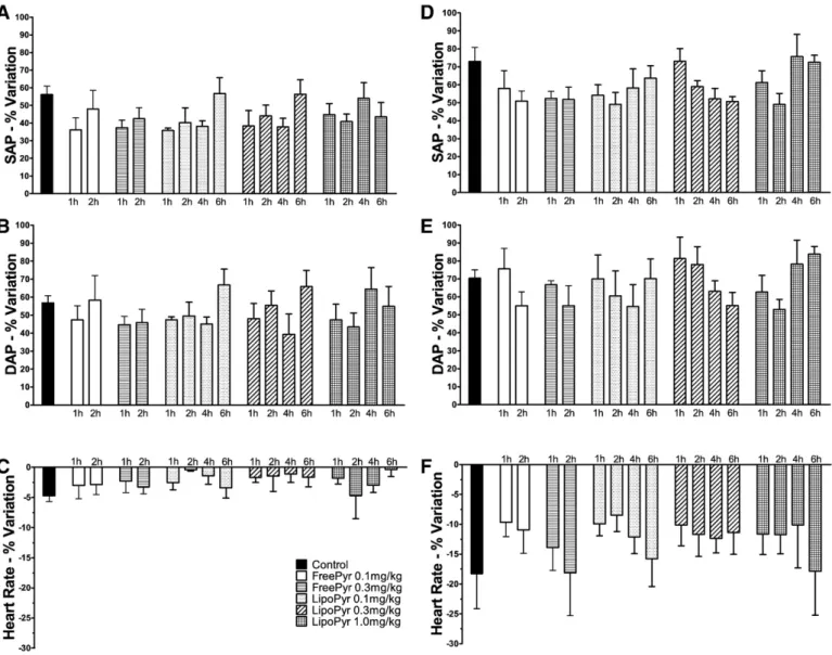
Fig. 1
shows the percentage
variation of ECG parameters induced by NA after the administration of
each formulation of pyridostigmine. The maximum effect of NA to
increase all parameters was observed between 15 and 25 s after its
administration and the return to baseline values occurred after about
3 min.
Fig. 2
shows representative ECG segments of animals that
received saline, free pyridostigmine or liposomal pyridostigmine,
before and after 3.0
μ
g NA.
Fig. 3
shows the percentage variation of AP and HR induced by NA
after the administration of each formulation of pyridostigmine. The
SAP and DAP were both similarly increased, about 55% after 1.0
μ
g and
70% after 3.0
μ
g NA. The administration of free pyridostigmine and
liposomal pyridostigmine did not change the baseline AP values and
was not able to inhibit the increases of AP. The maximum effect of NA
to increase AP was observed between 10 and 25 s after
administra-tion. The IV administration of 1.0
μ
g of NA did not cause signi
fi
cant
changes of HR when compared to the control period. The dose of
3.0
μ
g of NA decreased 18% the HR. Previous treatment with free
pyridostigmine or liposomal pyridostigmine did not change HR.
In animals subjected to sympathetic stimulation with 1.0
μ
g of NA,
as no relevant changes were observed in HR, the QTc index showed
similar pro
fi
les compared to the QT interval. Thus, signi
fi
cant
decreases of QTc prolongation were observed in animals treated 1 h
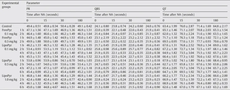
Table 2Means of absolute values of ECG parameters measured before and after IV injection of NA 1μg to animals previously treated with free or liposomal pyridostigmine at different times.
Experimental groups
Parameter
PR QRS QT
Time after NA (seconds) Time after NA (seconds) Time after NA (seconds)
0 15 30 180 0 15 30 180 0 15 30 180
Control 49.4± 0.51 49.8± 0.34 50.4 ±0.28 49.1 ± 0.42 24.1 ± 0.80 23.8 ± 0.74 24.2± 0.60 24.0± 0.76 63.4 ±1.99 76.0 ± 2.87 71.4 ± 1.69 64.8 ± 2.17 FreePyr
0.1 mg/kg
1 h 47.1± 1.56 47.1± 1.69 46.9 ±1.36 46.9 ± 1.51 22.0 ± 0.39 22.1 ± 0.48 22.0± 0.43 21.9± 0.41 65.1 ±1.46 71.1 ± 2.07 70.8 ± 2.03 65.3 ± 1.54 2 h 46.4± 1.60 46.6± 1.66 46.2 ±1.49 46.3 ± 1.64 21.4 ± 0.84 21.4 ± 0.97 21.3± 0.85 21.3± 0.87 62.0 ±1.32 70.3 ± 2.24 71.6 ± 1.90 63.5 ± 1.65 FreePyr
0.3 mg/kg
1 h 44.9± 1.48 45.0± 1.42 44.9 ±1.53 45.0 ± 1.43 23.1 ± 2.19 23.3 ± 2.22 23.2± 2.12 23.1± 2.32 71.7 ±1.10 76.3 ± 1.18 75.6 ± 1.02 72.1 ± 1.24 2 h 49.9± 1.88 50.0± 1.89 49.7 ±1.91 49.9 ± 1.91 22.1 ± 0.50 22.2 ± 0.32 22.2± 0.35 21.9± 0.36 69.5 ±0.95 77.6 ± 1.31 77.7 ± 0.93 70.6 ± 0.70 LipoPyr
0.1 mg/kg
1 h 46.2± 1.33 46.5± 1.32 46.3 ±1.28 46.2 ± 1.35 21.7 ± 0.45 21.9 ± 0.39 22.0± 0.46 21.6± 0.41 67.6 ±1.31 76.8 ± 2.52 78.0 ± 1.34 69.0 ± 1.62 2 h 53.4± 0.93 53.5± 1.19 53.1 ±1.12 53.1 ± 0.92 25.8 ± 0.98 25.6 ± 0.89 25.7± 0.77 25.4± 0.82 67.2 ±1.30 72.7 ± 1.34 72.5 ± 1.97 68.1 ± 1.46 4 h 47.0± 1.43 47.1± 1.45 47.3 ±1.40 47.0 ± 1.44 21.9 ± 0.35 22.1 ± 0.33 22.2± 0.35 22.0± 0.38 64.9 ±1.82 73.5 ± 3.04 75.5 ± 1.92 65.7 ± 2.23 6 h 45.5± 1.51 45.9± 1.54 45.4 ±1.43 45.6 ± 1.52 22.3 ± 0.35 22.5 ± 0.24 22.7± 0.31 22.3± 0.38 66.4 ±1.61 77.3 ± 1.92 76.6 ± 1.62 67.6 ± 1.81 LipoPyr
0.3 mg/kg
1 h 53.8± 0.96 53.9± 0.86 54.1 ±0.70 54.0 ± 1.03 23.6 ± 0.17 23.1 ± 0.14 23.1± 0.13 23.1± 0.18 67.9 ±1.02 74.1 ± 1.80 78.4 ± 1.66 68.4 ± 0.95 2 h 54.6± 1.67 54.0± 1.91 53.6 ±1.90 53.4 ± 1.21 24.7 ± 0.65 24.7 ± 0.51 24.8± 0.36 25.1± 0.44 62.7 ±1.77 65.8 ± 1.51 67.6 ± 1.50 63.8 ± 2.06 4 h 47.2± 1.18 47.5± 1.07 47.0 ±0.99 47.3 ± 1.18 21.8 ± 0.25 22.1 ± 0.25 22.2± 0.25 21.9± 0.27 65.4 ±2.05 73.7 ± 3.04 75.3 ± 1.94 65.7 ± 2.25 6 h 45.4± 0.99 45.2± 1.10 45.4 ±0.89 45.2 ± 1.01 21.0 ± 0.79 20.9 ± 0.86 21.0± 0.81 21.0± 0.80 64.7 ±1.39 69.5 ± 1.92 70.7 ± 1.77 64.5 ± 1.41 LipoPyr
1.0 mg/kg
1 h 46.8± 1.44 46.8± 1.36 46.4 ±1.29 46.9 ± 1.44 21.4 ± 0.47 21.7 ± 0.46 21.6± 0.50 21.5± 0.41 66.2 ±1.77 73.3 ± 2.34 73.2 ± 2.06 66.6 ± 2.00 2 h 42.4± 0.88 42.4± 0.95 42.8 ±0.77 42.4 ± 0.90 22.8 ± 0.24 23.1 ± 0.24 23.2± 0.23 22.9± 0.23 66.9 ±1.47 72.5 ± 1.59 72.2 ± 1.45 67.3 ± 1.63 4 h 46.1± 1.32 45.9± 1.40 46.5 ±1.46 46.2 ± 1.39 21.1 ± 0.36 20.9 ± 0.31 20.9± 0.36 21.0± 0.32 68.9 ±0.30 76.1 ± 0.49 76.4 ± 0.80 69.2 ± 0.33 6 h 45.0± 1.68 44.8± 4.67 44.6 ±1.51 44.9 ± 1.68 21.3 ± 0.88 21.5 ± 0.92 21.5± 0.92 21.4± 0.90 62.6 ±1.48 67.0 ± 1.79 67.1 ± 1.63 63.2 ± 1.69 Each value represents the mean ± e.s.m. of six animals.
Table 3
Means of absolute values of ECG parameters measured before and after IV injection of NA 3μg to animals previously treated with free or liposomal pyridostigmine at different times.
Experimental groups
Parameter
PR QRS QT
Time after NA (seconds) Time after NA (seconds) Time after NA (seconds)
0 15 30 180 0 15 30 180 0 15 30 180
Control 46.7± 0.92 49.4 ± 0.99 48.6 ± 1.07 47.0± 0.89 22.4 ± 0.87 23.5 ± 0.86 23.6± 1.02 22.5 ± 0.84 71.2 ± 0.90 84.5± 1.68 85.0± 1.52 73.3 ± 1.20 FreePyr
0.1 mg/kg
1 h 45.6± 1.52 45.4 ± 1.60 45.8 ± 1.61 45.3± 1.50 23.1 ± 0.93 23.1 ± 0.82 23.3± 0.89 23.1 ± 0.98 70.9 ± 0.81 80.7± 1.49 78.5± 1.54 71.3 ± 1.04 2 h 45.8± 2.02 46.3 ± 2.12 45.8 ± 1.89 45.3± 1.88 24.8 ± 1.24 25.0 ± 1.41 24.9± 1.25 24.6 ± 1.26 71.4 ± 0.98 84.9± 1.60 80.4± 1.03 72.4 ± 1.06 FreePyr
0.3 mg/kg
1 h 46.0± 1.57 46.0 ± 1.37 46.0 ± 1.39 46.1± 1.54 21.5 ± 0.52 21.9 ± 0.51 21.8± 0.63 21.5 ± 0.46 69.8 ± 1.35 76.1± 1.61 75.8± 1.46 70.0 ± 1.36 2 h 45.4± 1.82 45.6 ± 1.76 45.0 ± 1.84 45.2± 1.80 22.5 ± 0.35 22.6 ± 0.41 22.7± 0.38 22.4 ± 0.30 68.3 ± 1.18 74.5± 2.04 76.2± 1.37 68.9 ± 1.92 LipoPyr
0.1 mg/kg
1 h 47.4± 1.50 47.6 ± 1.47 47.1 ± 1.41 47.4± 1.61 21.9 ± 0.40 22.0 ± 0.40 21.8± 0.46 21.9 ± 0.37 65.4 ± 1.69 72.8± 2.34 73.3± 1.84 65.8 ± 1.79 2 h 46.0± 1.91 45.9 ± 1.96 46.3 ± 1.82 45.9± 1.94 22.1 ± 0.46 22.2 ± 0.47 22.2± 0.44 22.2 ± 0.46 66.8 ± 2.08 75.4± 2.69 76.2± 2.92 67.0 ± 1.89 4 h 46.0± 1.33 46.4 ± 1.39 46.8 ± 1.47 46.2± 1.37 22.1 ± 0.37 22.1 ± 0.22 22.6± 0.32 22.1 ± 0.38 67.9 ± 1.34 77.8± 2.46 78.1± 1.24 69.1 ± 1.65 6 h 47.8± 0.97 48.5 ± 0.60 48.0 ± 0.77 48.0± 1.02 21.6 ± 0.31 21.8 ± 0.16 21.8± 0.24 21.6 ± 0.18 65.7 ± 1.88 74.6± 2.54 78.5± 2.31 66.6 ± 2.32 LipoPyr
0.3 mg/kg
1 h 45.7± 1.89 45.8 ± 1.87 46.0 ± 1.75 45.6± 1.87 23.3 ± 0.76 23.3 ± 0.84 23.2± 0.73 23.2 ± 0.69 67.0 ± 2.28 76.9± 3.02 76.3± 2.94 67.8 ± 2.29 2 h 46.3± 1.69 46.2 ± 1.68 45.9 ± 1.58 45.9± 1.62 21.9 ± 0.43 22.3 ± 0.34 22.2± 0.40 21.8 ± 0.42 66.9 ± 1.65 75.9± 2.62 75.1± 1.97 67.0 ± 1.82 4 h 46.1± 1.95 45.9 ± 1.92 46.3 ± 1.84 45.9± 1.95 22.2 ± 0.44 22.3 ± 0.48 22.3± 0.46 22.2 ± 0.45 66.3 ± 1.80 74.2± 2.81 75.7± 2.40 66.7 ± 1.93 6 h 46.9± 1.45 47.2 ± 1.47 46.7 ± 1.26 46.9± 1.45 21.9 ± 0.35 22.1 ± 0.35 22.3± 0.33 21.9 ± 0.37 65.1 ± 2.11 72.9± 3.18 75.3± 1.90 65.8 ± 2.22 LipoPyr
1.0 mg/kg
before sympathetic stimulation with free pyridostigmine in doses 0.1
and 0.3 mg/kg. For liposome formulations, signi
fi
cant differences
were observed at 2 h of treatment with doses 0.3 and 1.0 mg/kg, and
these effects occurred up to 6 h after treatment with the dose of
1.0 mg/kg. The signi
fi
cant decrease in HR caused by 3.0
μ
g NA changed
the pro
fi
le of variation of QTc index for all protocols of treatment. No
signi
fi
cant differences for this parameter between groups untreated
and treated with pyridostigmine were observed. The alterations of AP
and ECG intervals induced by NA in animals that previously received
saline were similar to the animals that received empty liposomes
(data not shown).
Discussion
Our
fi
ndings suggest that encapsulation of pyridostigmine in
liposomes was able to extend its protective effects under sympathetic
hyperactivity. Several studies showed that the autonomic misbalance
with adrenergic hyperactivity could be accompanied by vagal
hypoac-tivity (Goldstein et al. 1975; Porter et al. 1990; Padley et al. 2005; Lahiri
et al. 2008). It was identi
fi
ed the bene
fi
ts of electrical vagal stimulation in
dogs (Henning et al. 1990), cats (Zuanetti et al. 1987), rats (Li et al. 2004)
and in patients with cardiovascular diseases (Zamotrinsky et al. 2001),
thus encouraging the search for alternative therapies that could
modulate the parasympathetic system, particularly enhancing the
acetylcholine activity. Taking into account that cholinergic agonists, as
oxitremorine, have characteristics of cardiac protection (De Ferrari et al.
1992, 1993), the effect of transdermal scopolamine in patients with
advanced congestive heart failure was evaluated and the main observed
bene
fi
cial effect was the increase of heart rate variability (Casadei et al.
1996; Venkatesh et al. 1996). This effect, however, was only observed
with low doses of the drug and, at high concentrations, the scopolamine
acts as a cholinergic blocker, limiting the performance of prolonged
studies (Hayano et al. 1999). The pyridostigmine is a reversible
cholinesterasic inhibitor that increases the concentration of endogenous
acetylcholine. It is clinically used in patients with myasthenia gravis by
increasing the concentration of acetylcholine at the synaptic cleft,
reducing the de
fi
cit in muscle strength. Its cardiovascular action is
usually considered a side effect (Castro et al. 2000). Previous studies in
normal rats (Grabe-Guimarães et al. 1999) and in humans (Nóbrega et al.
1996) evaluated the therapeutic potential of pyridostigmine as a
cardioprotective drug and its potential use in congestive heart failure
(Androne et al. 2003) and in neurogenic orthostatic hypotension (Singer
et al. 2006). Although the advantages of pyridostigmine has been shown
both in health (Nóbrega et al. 1996, 1999; Castro et al. 2000; Sant'anna
et al. 2003) and in patients with heart failure (Castro et al. 2002, 2004,
2006; Nóbrega et al. 2008; Serra et al. 2009), side effects characterized
mainly by intestinal distress were observed with a daily oral dose of
5 mg/kg for 3 months, and the dose of 20 mg/kg was lethal to dogs when
given for up to 14 days (Kluwe et al. 1989). Furthemore, combined
exposure of mice to 10 mg/kg/day of pyridostigmine bromide and
shaker stress for 7 days resulted in neurobehavioral changes such as
sensorimotor alterations and decreased locomotor activity (Dubovicky
et al. 2007). That same dose of pyridostigmine caused to male mice
adverse in
fl
uence on cardiac growth and vascular structure, speci
fi
cally
reduction of the aortic wall thickness/diameter ratio and reduced
relative heart weight (Bernátová et al. 2006). In our experiments, the
administration of free pyridostigmine at 1.0 mg/kg caused toxic effects
characteristic of cholinergic hyperstimulation, and for this reason, we
discontinued the study for this dose on free form. As expected, it was not
observed the toxic effects for the same dose of liposomal pyridostigmine.
This
fi
nding suggests a potential ability of liposomes to reduce the
incidence of adverse effects of pyridostigmine, which should be further
studied.
The most important
fi
nding of the present study is the
cardiopro-tective effect of pyridostigmine by inhibiting the increases of the QT
interval caused by sympathetic hyperstimulation in rats. The utility of
the QT interval measurement as a tool to evaluate the cardiotoxic
activity of drugs was previously demonstrated in our laboratory (Leite
et al. 2007). The autonomic tone has its signature on the QT interval,
which is primarily determined by the parasympathetic branch, since
the cholinergic blockade was impressively related to QT prolongation
(Ahnve and Vallin 1982). In coronary disease, when the vagal activity
is reduced, the QT interval prolongation is a predictor of arrhythmias
(Zuanetti et al. 1987; London et al. 1998) and sudden death (Schwartz
Fig. 1.Percentage variation of QT interval and QTc index measured after the sympathetic stimulation with NA in animals pre-treated with saline, free or liposomal pyridostigmine at different times. (A) and (B) changes after 1μg of NA. (C) and (D) changes after 3μg of NA. FreePyr: free form of pyridostigmine; LipoPyr: liposomal form of pyridostigmine. Eachvalue represents the mean of the maximum variation of six animals, compared to the control group. *Pb0.05—ANOVA, Tukey post-hoc test.
and Wolf 1978; Ahnve 1991). Therefore, pyridostigmine at the doses
used (0.3 and 1.0 mg/kg), presented potential cardioprotective effect
in regard to its ability to prevent increases in the QT interval induced
by NA. As expected, PEGylated liposomes were able to prolong
(Takahama et al. 2009) the circulation of pyridostigmine, augmenting
its cardioprotective effects. Thus, the use of pyridostigmine, especially
in liposomal form, could prevent the occurrence of ventricular
arrhythmias and sudden death, as the encapsulation was capable of
promoting its slow release prolonging its protective effects on the QT
interval up to 6 h after administration, providing maintenance of
acetylcholine in the synaptic cleft. Furthermore, the density or af
fi
nity
of muscarinic receptors are increased on the atria in rats with
sinoaortic denervation (Soares et al. 2006) and dogs with heart failure
(Dunlap et al. 2003), indicating a natural mechanism to control the
decrease on the parasympathetic activity. In this context,
pyridos-tigmine encapsulated in PEGylated liposomes may be a powerful
formulation to release the drug into the ischemic heart (Torchilin
1995; Lukyanov et al. 2004).
The administration of the higher dose of NA caused signi
fi
cant
bradycardia, a factor that interfered with the evaluation of the bene
fi
cial
effects of pyridostigmine when using the Fridericia's correction formula
(QTc). According to
Indik et al. (2006), this calculation may
underesti-mate the values of the QT interval when HR decreased, as observed in our
experiments. Moreover, studies showed that drugs that modulate the
ANS can alter the absolute values of the QT interval independently of HR
(Browne et al. 1982), and the autonomic conditions that directly affect
the ventricular myocardium of healthy subjects, causing variations in QT
are also independent of HR (Magnano et al. 2002).
The absence of cardiodepression, characterized by the normal
baselines values observed after administration of IV pyridostigmine in
free and liposomal forms is in conformity with previous studies
(Soares et al. 2004). These observations favor its use in patients with
cardiovascular diseases. Cholinergic substances may slow the
con-duction of the cardiac electrical impulse, inducing bradycardia
(Pontes et al. 1999), what was not observed in the present study in
anesthetised rats. In this study, it was shown that the cardiac
protection by IV administration of pyridostigmine in rats involves
the modulation of the QT interval under conditions of sympathetic
hyperactivity. Moreover, despites of the de
fi
nition of probable
mechanisms of cardioprotection promoted by pyridostigmine, it is
known that increased vagal tone is related to the good prognosis in
ischemic heart disease and heart failure (Osterziel and Dietz 1996),
since both diseases are characterized by an increase in sympathetic
tone and a decrease in cardiac vagal activity.
Conclusion
The most important result emerging from this work is the ability of a
liposomal system to prolong the cardioprotective effect of
pyridostig-mine when compared to the free drug, mainly by its ability to prevent the
prolongation of the QT interval under conditions of sympathetic
hyperactivity. It can be speculated that pyridostigmine in liposomes
Fig. 2.Representative traces of ECG showing the effects of pre-treatment with pyridostigmine to prevent the alterations of the QT interval induced by 3μg of NA, compared to themay be a potential therapeutic alternative to prevent cardiovascular
disturbances resulting from sympathetic hyperactivity in patients with
ischemic heart disease.
Acknowledgements
We wish to thank FAPEMIG (Rede Nanobiomg and CDS574) and
CNPq for their grants, the scholarship from CNPq and CAPES and our
Universities, UFOP and UFMG, for the complete support.
References
Abernethy DR, Wesche DL, Barbey JT, Ohrt C, Mohanty S, Pezzullo JC, Schuster BG. Stereoselective halofantrine disposition an effect: concentration-related QTc prolongation. British Journal of Pharmacology 51 (3), 231–237, 2001.
Ahnve S. Is QT interval prolongation a strong or weak predictor for cardiac death? Circulation 84 (4), 1862–1865, 1991.
Ahnve S, Vallin H. Influence of heart rate and inhibition of autonomic tone on the QT interval. Circulation 65 (3), 435–439, 1982.
Algra A, Tijssen JG, Roelandt JR, Pool J, Lubsen J. QTc prolongation measured by standard 12-lead electrocardiography is an independent risk factor for sudden death due to cardiac arrest. Circulation 83 (6), 1888–1894, 1991.
Androne AS, Hryniewicz K, Goldsmith R, Arwady A, Katz SD. Acetylcholinesterase inhibition with pyridostigmine improves heart rate recovery after maximal exercise in patients with chronic heart failure. Heart 89 (8), 854–858, 2003.
Aquilonius SM, Eckernäs SÅ, Hartvig P, Lindström B, Osterman PO. Pharmacokinetics and oral bioavailability of pyridostigmine in man. European Journal of Clinical Pharmacology 18 (5), 423–428, 1980.
Bazett HC. An analysis of the time-relations of the electrocardiogram. Heart 7, 353–370, 1920.
Bernátová I, Babál P, Grubbs RD, Morris M. Acetylcholinesterase inhibition affects cardiovascular structure in mice. Physiology Research 55 (Suppl 1), S89–S97, 2006. Browne KF, Zipes DP, Heger JJ, Prystowsky EN. Influence of the autonomic nervous system on the QT interval in man. The American Journal of Cardiology 50 (5), 1099–1103, 1982.
Casadei B, Conway J, Forfar C, Sleight P. Effect of low doses of scopolamine on RR interval variability, baroreflex sensitivity, and exercise performance in patients with chronic heart failure. Heart 75 (3), 274–280, 1996.
Castro RRT, Serra SM, Nóbrega ACL. Reduction of Qtc interval dispersion. Potential mechanism of cardiac protection of pyridostigmine bromide. Arquivos Brasileiros de Cardiologia 75 (3), 210–213, 2000.
Castro RTT, Porphirio G, Serra SM, Nóbrega ACL. Cholinergic stimulation with pyridostigmine reduces the QTc interval in coronary artery disease. Brazilian Journal of Medical and Biological Research 35 (6), 685–689, 2002.
Castro RRT, Porphirio G, Serra SM, Nóbrega ACL. Cholinergic stimulation with pyridostigmine protects against exercise induced myocardial ischaemia. Heart 90 (10), 1119–1123, 2004.
Castro RTT, Serra SM, Porphirio G, Mendes F, Oliveira L, Nóbrega A. Pyridostigmine reduces QTc interval during recovery from maximal exercise in ischemic heart disease. International Journal of Cardiology 107 (1), 138–139, 2006.
Catelli M, Feldman J, Bousquet P, Tibiriçá E. Protective effects of centrally acting sympathomodulatory drugs on myocardial ischemia induced by sympathetic overactivity in rabbits. Brazilian Journal of Medical and Biological Research 36 (1), 85–95, 2003.
Fig. 3.Percentage variation of SAP, DAP and HR measured after the sympathetic stimulation with NA in animals pre-treated with saline, free or liposomal pyridostigmine at different times. (A), (B) and (C) after 1μg of NA. (D), (E) and (F) after 3μg of NA. FreePyr: free form of pyridostigmine; LipoPyr: liposomal form of pyridostigmine. Each value represents the
mean of the maximum variation of six animals, compared to the control group.
De Ferrari GM, Vanoli E, Curcuruto P, Tommasini G. Prevention of life-threatening arrhythmias by pharmacologic stimulation of the muscarinic receptors with oxotremorine. American Heart Journal 124 (4), 883–890, 1992.
De Ferrari GM, Salvati P, Grossoni M, Ukmar G, Vaga L, Patrono C, Schwartz PJ. Pharmacologic modulation of the autonomic nervous system in the prevention of sudden cardiac death. A study with propranolol, methacholine and oxotremorine in conscious dogs with a healed myocardial infarction. Journal of the American College of Cardiology 22 (1), 283–290, 1993.
Dubovicky M, Paton S, Morris M, Mach M, Lucot JB. Effects of combined exposure to pyridostigmine bromide and shaker stress on acoustic startle response, pre-pulse inhibition and openfield behavior in mice. Journal of Applied Toxicology 27 (3), 276–283, 2007.
Dunlap ME, Bibevski S, Rosenberry TL, Ernsberger P. Mechanisms of altered vagal control in heart failure: Influence of muscarinic receptors and acetylcholinesterase activity. American Journal of Physiology - Heart and Circulatory Physiology 285 (4), H1632–H1640, 2003.
Francis G. Modulation of peripheral sympathetic nerve transmission. Journal of the American College of Cardiology 12, 250–254, 1988.
Fridericia LS. Die Systolendauer im Elektrokardiogramm bei normalen Menschen und bei Herzkranken. Acta Medica Scandinavica 53, 469–486, 1920.
Goldstein RE, Beiser GD, Stampfer M, Epstein SE. Impairment of autonomically mediated heart rate control in patients with cardiac dysfunction. Circulation Research 36 (5), 571–578, 1975.
Grabe-Guimarães A, Alves LM, Tibiriçá E, Nóbrega ACL. Pyridostigmine blunts the increases in myocardial oxygen demand elicited by the stimulation of the central nervous system in anesthetized rats. Clinical Autonomic Research 9 (2), 83–89, 1999. Hayano T, Shimizu A, Ikeda Y, Yamamoto T, Yamagata T, Ueyama T, Furutani Y,
Matsuzaki M. Paradoxical effects of pirenzepine on parasympathetic activity in chronic heart failure and control. International Journal of Cardiology 68 (1), 47–56, 1999.
Hayes E, Pugsley MK, Penz WP, Adaikan G, Walker MJA. Relationship between QaT and RR intervals in rats, guinea pigs, rabbits, and primates. Journal of Pharmacological and Toxicological Methods 32 (4), 201–207, 1994.
Hegazy N, Demirel M, Yazan Y. Preparation and in vitro evaluation of pyridostigmine bromide microparticles. International Journal of Pharmaceutics 242 (1–2), 171–174, 2002.
Henning RJ, Khalil IR, Levy MN. Vagal stimulation attenuates sympathetic enhancement of left ventricular function. American Journal of Physiology - Heart and Circulatory Physiology 258 (5), H1470–H1475, 1990.
Hjalmarson A. Effects of beta blockade on sudden cardiac death during acute myocardial infarction and the postinfarction period. The American Journal of Cardiology 80 (9B), 35J–39J, 1997.
Hodges M. Rate correction of the QT interval. Cardiac Electrophysiology Review 1 (3), 360–363, 1997.
Hoyer D, Maestrib R, La Rovere MT, Pinna GD. Autonomic response to cardiac dysfunction in chronic heart failure: a risk predictor based on autonomic informationflow. Pacing and Clinical Electrophysiology 31 (2), 214–220, 2008.
Indik JH, Pearson EC, Fried K, Woosley RL. Bazett and Fridericia QT correction formulas interfere with measurement of drug-induced changes in QT interval. Heart Rhythm 3 (9), 1003–1007, 2006.
Klibanov AL, Maruyamal K, Torchilin VP, Huang L. Amphipathic polyethyleneglycols effectively prolong the circulation time of liposomes. FEBS Letters 268 (1), 235–237, 1990.
Kluwe WM, Page JG, Toft JD, Ridder WE, Chung H. Pharmacological and toxicological evaluation of orally administered pyridostigmine in dogs. Toxicological Sciences 14 (1), 40–53, 1989.
Koga T, Kuwano K, Kito G, Kanefuji K. Evaluation of QT interval using a linear model in individual cynomolgus monkeys. Journal of Pharmacological and Toxicological Methods 55 (3), 265–270, 2007.
Lahiri MK, Kannankeril PJ, Goldberger JJ. Assessment of autonomic function in cardiovascular disease. Physiological basis and prognostic implications. Journal of the American College of Cardiology 51 (18), 1725–1733, 2008.
Leite EA, Grabe-Guimarães A, Guimarães HN, Machado-Coelho GLL, Barratt G, Mosqueira VCF. Cardiotoxicity reduction induced by halofantrine entrapped in nanocapsule devices. Life Sciences 80 (14), 1327–1334, 2007.
Li M, Zheng C, Sato T, Kawada T, Sugimachi M, Sunagawa K. Vagal nerve stimulation markedly improves long-term survival after chronic heart failure in rats. Circulation 109 (1), 120–124, 2004.
London B, Jerona A, Zhou J, Buckett P, Han X, Mitchell GF, Koren G. Long QT and ventricular arrhythmias in transgenic mice expressing the N terminus andfirst transmembrane segment of a voltage-gated potassium channel. Proceedings of the National Academy of Sciences 95 (6), 2926–2931, 1998.
Lukyanov AN, Hartner WC, Torchilin VP. Increased accumulation of PEG–PE micelles in the area of experimental myocardial infarction in rabbits. Journal of Controlled Release 94 (1), 187–193, 2004.
Magnano AR, Holleran S, Ramakrishnan R, Reiffel JA, Bloomfield DM. Autonomic nervous system influences on QT interval in normal subjects. Journal of the American College of Cardiology 39 (11), 1820–1826, 2002.
Malik M, Farbom P, Camm AJ, Hnatkova K, Batchvarov V. Relation between QT and RR intervals is highly individual among healthy subjects: implications for heart rate correction of the QT interval. Heart 87 (3), 220–228, 2002.
Nayar R, Hope MJ, Cullis PR. Generation of large unilamellar vesicles from long-chain saturated phosphatidylcholines by extrusion technique. Biochimica et Biophysica Acta 986, 200–206, 1989.
Nóbrega ACL, Carvalho ACG, Bastos BG. Resting and reflex heart rate response during cholinergic stimulation with pyridostigmine in humans. Brazilian Journal of Medical and Biological Research 29 (11), 1461–1465, 1996.
Nóbrega ACL, Carvalho ACG, Santos KB, Soares PPS. Pyridostigmine blunts the cardiac response to mental stress. Clinical Autonomic Research 9 (1), 11–16, 1999. Nóbrega ACL, Loures DL, Pontes PV, Sant'Anna ID, Mesquita ET. Cholinergic stimulation
with pyridostigmine prevents the impairment in ventricular function during mental stress in coronary artery disease patients. International Journal of Cardiology 125 (3), 418–421, 2008.
Osterziel KJ, Dietz R. Improvement of vagal tone by ACE inhibition: a mechanism of cardioprotection in patients with mild-to-moderate heart failure. Journal of Cardio-vascular Pharmacology 27 (Suppl 2), 25–30, 1996.
Padley JR, Overstreet DH, Pilowsky PM, Goodchild AK. Impaired cardiac and sympathetic autonomic control in rats differing in acetylcholine receptor sensitivity. American Journal of Physiology - Heart and Circulatory Physiology 289 (5), H1985–H1992, 2005. Pontes PV, Bastos BG, Romêo FLJM, Mesquita ET, Nóbrega ACL. Cholinergic stimulation with pyridostigmine in healthy subjects, hemodynamic and echocardiographic analysis. Arquivos Brasileiros de Cardiologia 72 (3), 302–306, 1999.
Porter TM, Eckberg DL, Fritsch JM, Rea RF, Beightol LA, Schmedtje Jr JF, Mohanty PK. Autonomic pathophysiology in heart failure patients: sympathetic–cholinergic interrelations. Journal of Clinical Investigation 85 (5), 1362–1371, 1990. Puddu PE, Jouve R, Mariotti S, Giampaoli S, Lanti M, Reale A, Menotti A. Evaluation of 10
QT prediction formulas in 881 middle-aged men from the seven countries study: emphasis on the cubic root Fridericia's equation. Journal of Electrocardiology 21 (3), 219–229, 1988.
Sant'anna ID, Sousa EB, Moraes AA, Loures DL, Mesquita ET, Nóbrega ACL. Cardiac function during mental stress: cholinergic modulation with pyridostigmine in healthy subjects. Clinical Science 105 (2), 161–165, 2003.
Schouten EG, Dekker JM, Meppelink P, Kok FJ, Vandenbroucke JP, Pool J. QT interval prolongation predicts cardiovascular mortality in an apparently healthy popula-tion. Circulation 84 (4), 1516–1523, 1991.
Schwartz PJ, Wolf S. QT interval prolongation as predictor of sudden death in patients with myocardial infarction. Circulation 57 (6), 1074–1077, 1978.
Serra SM, Costa RV, Castro RRT, Xavier SS, Nóbrega ACL. Cholinergic stimulation improves autonomic and hemodynamic profile during dynamic exercise in patients with heart failure. Journal of Cardiac Failure 15 (2), 124–129, 2009.
Simonson E, Cady LD, Woodbury M. The normal Q-T interval. American Heart Journal 63 (6), 747–753, 1962.
Singer W, Opfer-Gehrking TL, Nickander KK, Hines SM, Low PA. Acetylcholinesterase inhibition in patients with orthostatic intolerance. Journal of Clinical Neurophys-iology 23 (5), 477–482, 2006.
Soares PPS, Nóbrega ACL, Ushizima MR, Irigoyen MCC. Cholinergic stimulation with pyridostigmine increases heart rate variability and baroreflex sensitivity in rats. Autonomic Neuroscience 113 (1–2), 24–31, 2004.
Soares PPS, Porto CS, Abdalla FMF, Fuente RN, Moreira ED, Krieger EM, Irigoyen MCC. Effects of rat sinoaortic denervation on the vagal responsiveness and expression of muscarinic acetylcholine receptors. Journal of Cardiovascular Pharmacology 47 (3), 331–336, 2006.
Taggart P, Sutton P, Chalabi Z, Boyett MR, Simon R, Elliott D, Gill JS. Effect of adrenergic stimulation on action potential duration restitution in humans. Circulation 107 (2), 285–289, 2003.
Tai CT, Chiou CW, Chen SA. Interaction between the autonomic nervous system and atrial tachyarrhythmias. Journal of Cardiovascular Electrophysiology 13 (1), 83–87, 2002.
Takahama H, Minamino T, Asanuma H, Fujita M, Asai T, Wakeno M, Sasaki H, Kikuchi H, Hashimoto K, Oku N, Asakura M, Kim J, Takashima S, Komamura K, Sugimachi M, Mochizuki N, Kitakaze N. Prolonged targeting of ischemic/reperfused myocardium by liposomal adenosine augments cardioprotection in rats. Journal of the American College of Cardiology 53 (8), 709–717, 2009.
Torchilin VP. Targeting of drugs and drug carriers within the cardiovascular system. Advanced Drug Delivery Reviews 17 (1), 75–101, 1995.
Torchilin VP, Klibanov AL, Huang L, SO'Donnell, Nossiff ND, Khaw BA. Targeted accumulation of polyethylene glycol-coated immunoliposomes in infarcted rabbit myocardium. The FASEB Journal 6 (9), 2716–2719, 1992.
Venkatesh G, Fallen EL, Kamath MV, Connolly S, Yusuf S. Double blind placebo controlled trial of short term transdermal scopolamine on heart rate variability in patients with chronic heart failure. Heart 76 (2), 137–143, 1996.
Verma DD, Hartner WC, Levchenko TS, Bernstein EA, Torchilin VP. ATP-loaded liposomes effectively protect the myocardium in rabbits with an acute experi-mental myocardial infarction. Pharmaceutical Research 22 (12), 2115–2120, 2005. Zamotrinsky AV, Kondratiev B, De Jong JW. Vagal neurostimulation in patients with coronary artery disease. Autonomic Neuroscience: Basic and Clinical 88 (1–2), 109–116, 2001.