Cho line rgic stimulatio n with
pyrido stigmine re duce s the Q Tc
inte rval in co ro nary arte ry dise ase
1Departamento de Fisiologia e Farmacologia, and 2Programa de Pós-Graduação em Cardiologia,
Universidade Federal Fluminense, Niterói, RJ, Brasil
3Instituto Estadual de Cardiologia Aluísio de Castro, Rio de Janeiro, RJ, Brasil
R.R.T. Castro1,
G. Porphirio3,
S.M. Serra3 and
A.C.L. Nóbrega1,2
Abstract
Parasympathetic dysfunction is an independent risk factor in patients with coronary artery disease; thus, cholinergic stimulation is a poten-tial therapeutic measure that may be protective by acting on ventricular repolarization. The purpose of the present study was to determine the effects of pyridostigmine bromide (PYR), a reversible anticholinester-ase agent, on the electrocardiographic variables, particularly QTc interval, in patients with stable coronary artery disease. In a random-ized double-blind crossover placebo-controlled study, simultaneous 12-lead electrocardiographic tracings were obtained at rest from 10 patients with exercise-induced myocardial ischemia before and 2 h after the oral administration of 45 mg PYR or placebo. PYR increased the RR intervals (pre: 921 ± 27 ms vs post: 1127 ± 37 ms; P<0.01) and, in contrast with placebo, decreased the QTc interval (pre: 401 ± 3 ms vs post: 382 ± 3 ms; P<0.01). No other electrocardio-graphic variables were modified (PR segment, QT interval, QT and QTc dispersions). Cholinergic stimulation with PYR caused bradycar-dia and reduced the QTc interval without important side effects in patients with coronary disease. These effects, if confirmed in studies over longer periods of administration, may suggest a cardioprotection by cholinergic stimulation with PYR.
Co rre spo nde nce
A.C.L. Nóbrega
Rua Cinco de Julho, 318/1001 24220-111 Niterói, RJ Brasil
Fax: + 55-21-2611-7059 E-mail: anobrega@ urbi.com.br
Research supported by CNPq (No. 520660-95.1) and FAPERJ (No. E-26/151.399/97).
Received November 8, 2001 Accepted April 1, 2002
Ke y words
•Pyridostigmine •Q Tc interval •Stable angina •Anticholinesterase
Intro ductio n
The impaired function of the autonomic nervous system increases the risk of arrhyth-mic events and sudden death after acute myo-cardial infarction (AMI) (1,2). The early concept that adrenergic hyperactivity could be deleterious has led to the widespread clinical use of beta-blockers in patients after AMI, with a consequent reduction in overall mortality (3). Decreased parasympathetic
ac-tivity also represents an independent risk factor in post-AMI patients (4,5), but few studies have investigated the therapeutic op-tions against parasympathetic dysfunction (6).
agent, on the surface electrocardiogram of patients with stable angina.
Mate rial and Me thods
Ten patients with stable angina and exer-cise-induced myocardial ischemia, five of them with previous AMI (age: 64 ± 9 years; height: 163 ± 8 cm; weight: 73 ± 10 kg) gave written informed consent to participate in the study after full explanation of the proce-dures and potential risks. The study, in ac-cordance with the Declaration of Helsinki, was approved by the Institutional Research Ethics Committee. A randomized cross-over and double-blind protocol was carried out on two separate days. An electrocardiogram (standard surface 12-lead simultaneous
sig-nal acquisition, ErgoPC® software,
Micro-med, Brasília, DF, Brazil) was recorded at rest in the supine position before and 120 min after the oral administration of 45 mg of
pyridostigmine bromide (Mestinon®, Roche
Pharmaceuticals, São Paulo, SP, Brazil) or placebo on each day. Each subject continued taking his/her usual medication throughout the study period. They were instructed to avoid alcohol, caffeine-containing beverages, and strenuous physical activity the day be-fore the measurements.
The same observer, who was blind to the experimental condition, manually measured
the PR, RR and QT intervals over the 12 simultaneous electrocardiographic leads. The software enables enlargement of the electro-cardiographic recordings, so that the meas-urement can be made with highest resolu-tion.
The QT interval was measured from the beginning of the QRS complex to the end of the T wave where its hind limb joined the baseline. There were no U waves in the traces analyzed. The QTc interval was ob-tained according to Bazett’s formula (QT/
RR) (11), using the latest RR before the
QT interval for calculation. Also, QT and QTc dispersions (maximum-minimum QT or QTc from the 12 simultaneous electrocar-diographic leads, respectively) were derived. We used the mean value from the 12 leads for each variable (QT, QTc, RR intervals and PR segment) for analysis. The occur-rence of adverse reactions was compared by the chi-square test. Each variable obtained from the electrocardiogram was analyzed by two-factor analysis of variance (ANOVA) with repeated measures where time (pre, post) and drug (pyridostigmine, placebo)
were the main factors. If a significant F
value was obtained, ANOVA was followed by the Student-Newman-Keuls test for
pair-wise post hoc comparisons. Statistical
sig-nificance was set at P<0.05. Results are re-ported as mean ± standard error.
Re sults
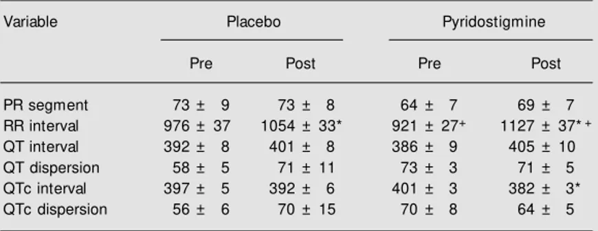
Pyridostigmine increased the duration of the RR intervals (Table 1). These increases corresponded to the following changes in heart rate: placebo - pre: 62 ± 7 bpm, post: 57 ± 7 bpm; pyridostigmine - pre: 65 ± 6 bpm, post: 53 ± 6 bpm. Pyridostigmine also reduced the QTc interval (Table 1; Fig-ure 1). There was no difference between pre-and post-pyridostigmine or placebo values for PR interval, QT interval or QT and QTc dispersions (Table 1).
Pyridostigmine was well tolerated. Two
Table 1. Electrocardiographic variables before (pre) and 2 h after (post) oral administra-tion of placebo or 45 mg pyridostigmine (N = 10).
Variable Placebo Pyridostigmine
Pre Post Pre Post
PR segment 73 ± 9 73 ± 8 64 ± 7 69 ± 7
RR interval 976 ± 37 1054 ± 33* 921 ± 27+ 1127 ± 37*+
QT interval 392 ± 8 401 ± 8 386 ± 9 405 ± 10
QT dispersion 58 ± 5 71 ± 11 73 ± 3 71 ± 5
QTc interval 397 ± 5 392 ± 6 401 ± 3 382 ± 3*
QTc dispersion 56 ± 6 70 ± 15 70 ± 8 64 ± 5
Data are reported as mean ± SEM in ms.
* P<0.01 vs pre - same condition (ANOVA/t-test). +P = 0.02 vs placebo - same moment
subjects complained of mild headache and another subject reported abdominal discom-fort after pyridostigmine, whereas one sub-ject complained of headache and another presented with atrial tachycardia after pla-cebo (P>0.05).
D iscussio n
Pyridostigmine exerts a vagomimetic ac-tion by inhibiting cholinesterase activity and increasing the concentration of acetylcho-line in the synaptic cleft. It has been used extensively to counteract skeletal muscle weakness in patients with myasthenia gravis, but its cardiovascular action has been con-sidered a side effect. Previous studies specif-ically investigating the hemodynamic changes induced by pyridostigmine have shown that a single dose of the drug de-creases resting (12) and exercise (13) heart rate and reduces QTc dispersion (14) in healthy individuals without impairing exer-cise tolerance. When given at 8-h intervals, pyridostigmine elicited sustained 24-h brady-cardia and augmented heart rate variability (15). These effects were obtained without impairment of systolic or diastolic cardiac functions (16). In addition, a single 45 mg dose of pyridostigmine given orally to healthy subjects blunted the double-product eleva-tion during mental stress (17). Finally, pyri-dostigmine inhibited the hemodynamic re-sponse to central adrenergic stimulation pro-duced by intracerebroventricular injection of glutamate in a rat model (18). The present study expands these previous findings dem-onstrating that a single oral dose (45 mg) of pyridostigmine causes bradycardia and re-duces the QTc interval in patients with coro-nary disease, with only mild side effects.
These results may have important clini-cal implications. It is generally accepted that QTc prolongation occurs during the acute phase of AMI (7). Schwartz and Wolf (8) measured the QTc intervals on electrocar-diograms taken every other month for seven
years in survivors of AMI and found a con-stant QTc interval prolongation in those who died suddenly (2.16 times greater risk for sudden death). In addition, Ahnve et al. (9) assessed the first-year prognostic implica-tions of the QTc interval by repeated meas-urements in survivors of AMI under 66 years of age. During the follow-up period, patients who suffered re-infarction or sudden death had significantly longer QTc values (434 ±
35 ms vs 417 ± 42 ms, P<0.001). Also, they
found a weak but significant correlation be-tween QTc and left ventricular dysfunction at the time of discharge as well as one year later. Recently, Okin et al. (10) found that prolonged QTc is a significant predictor of all-cause mortality and cardiovascular mor-tality, even after multivariate Cox regression analyses controlling for other risk factors. Although these studies (7-10) have described a significant correlation between prolonged QTc and worse prognosis, they do not pro-vide epro-vidence that reducing the QTc inter-val, as occurs after pyridostigmine, would reduce morbidity or mortality of post-AMI patients.
A potential limitation of the present study refers to the fact that we have employed a single dose of pyridostigmine administered just once. Thus, further studies using pro-longed administration of pyridostigmine are needed to determine whether QTc shortening
Q
T
c
(
m
s
)
440
420
400
380
360
340
0
Pre Post Pre Post
Pyridostigmine Placebo
Q
T
c
(
m
s
)
440
420
400
380
360
340
0
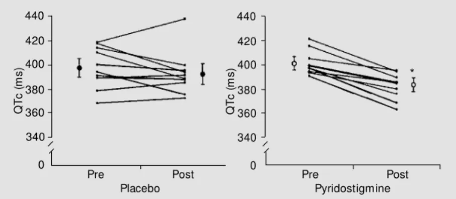
Figure 1. Individual values of QTc intervals before (pre) and 2 h after (post) oral administra-tion of placebo or 45 mg pyridostigmine on different days (N = 10). The means and SEM for each group are also reported on both sides of the pre- and post-data. * P<0.01 vs pre-pyridostigmine (ANOVA/t-test).
will produce and sustain a protective effect. Another issue is the use of Bazett’s for-mula to calculate the QTc interval. There is a complex relationship between heart rate and the QT interval (19), making it impossible to propose a flawless method to correct the duration of ventricular repolarization for the underlying duration of the cardiac cycle. Some studies have investigated the relation-ship between parasympathetic stimulation and QT interval. Davidowski and Wolf (20) and Litovsky and Antzelevitch (21) con-cluded that vagal stimulation and acetylcho-line prolong the QT interval independently of the bradycardia they induce. On the other hand, Cappato et al. (22) found that vagal
tonus increased intrinsic QT dependence on diminishing heart rate. The results of an-other study (23) showed that rapid reflex parasympathetic stimulation does not influ-ence QT interval duration or QT dispersion. Nevertheless, despite the limitations of Bazett’s formula, all the studies investigat-ing the prognostic value of the QTc interval have used it (7-10).
Pyridostigmine decreased resting heart rate and QTc interval. Although not directly investigated in the present study, it is pos-sible that cholinergic stimulation, resulting in bradycardia and QTc reduction, protects the myocardium and decreases mortality in patients with coronary disease.
Re fe re nce s
1. La Rovere M T, Bigger Jr JT, M arcus FI, M ortara A & Schw artz PJ (1998). Barore-flex sensitivity and heart-rate variability in prediction of total cardiac mortality after myocardial infarction. Lancet, 351: 478-484.
2. Schw artz PJ, La Rovere M T & Vanoli E (1992). Autonomic nervous system and sudden death. Experimental basis and clinical observations for post-myocardial infarction risk stratification. Circulation, 85 (Suppl I): I-77-I-99.
3. Bigger JT & Coromilas J (1984). How do beta-blockers protect after myocardial in-farction? Annals of Internal M edicine, 101: 256-258.
4. Kleiger RE, M iller JP, Bigger JTJ & The M ult icent er Post -Inf arct ion Research Group (1987). Decreased heart rate vari-ability and its association w ith increased mortality after acute myocardial infarction.
American Journal of Cardiology, 59: 256-262.
5. Odemuyiw a O, M alik M , Farrell T, Bashir Y, Poloniecki J & Camm J (1991). Com-parison of the predictive characteristics of heart rate variability index and left ventric-ular ejection fraction for all-cause mortal-ity, arrhythmic events and sudden death after acute myocardial infarction. Ameri-can Journal of Cardiology, 68: 434-439. 6. Nóbrega ACL & Castro RRT (2000).
Para-sympathetic dysfunction as a risk factor in
myocardial infarction: What is the treat-ment? American Heart Journal, 140: e20. 7. Doroghazi RM & Childers R (1978). Time-related changes in the QT interval in acute myocardial infarction: possible relation to local hypocalcemy. American Journal of Cardiology, 41: 684-688.
8. Schw artz PJ & Wolf S (1978). QT interval prolongation as predictor of sudden death in patients w ith myocardial infarction. Cir-culation, 57: 1074-1077.
9. Ahnve S, Helm ers C, Lundm an T, Rehnqvist N & Sjögren A (1980). QTc in-tervals in acute myocardial infarction: First-year prognostic implications. Clinical Cardiology, 3: 303-308.
10. Okin PM , Devereux RB, How ard BV, Fabsitz RR, Lee ET & Welty TK (2000). Assessment of QT interval and QT disper-sion for prediction of all-cause and cardiovascular mortality in American Indians -The Strong Heart Study. Circulation, 101: 61-66.
11. Bazett HC (1997). An analysis of the time-relations of electrocardiograms. Annals of Noninvasive Electrocardiology, 2: 177-194.
12. Nóbrega ACL, Carvalho ACG & Bastos BG (1996). Resting and reflex heart rate re-sponses during cholinergic stimulation w ith pyridostigmine in humans. Brazilian Journal of M edical and Biological Re-search, 29: 1461-1465.
13. Serra SM , Vivacqua R, Ramalho SHR, Santos KB, Bastos BG & Nóbrega ACL (2001). Exercise stress testing in healthy subjects during cholinergic stimulation af-ter a single dose of pyridostigmine. Arqui-vos Brasileiros de Cardiologia, 76: 279-284.
14. Castro RRT, Serra SM & Nóbrega ACL (2000). Reduction of QTc interval disper-sion. Potential mechanism of cardiac pro-tection of pyridostigmine bromide. Arqui-vos Brasileiros de Cardiologia, 75: 210-213.
15. Nóbrega ACL, Reis AF, M oraes RS, Bastos BG, Ferlin EL & Ribeiro JP (2001). Enhancement of heart rate variability dur-ing cholinergic stimulation w ith pyridostig-mine in healthy subjects. Clinical Auto-nomic Research, 11: 11-17.
16. Pontes PV, Nóbrega ACL, M esquita ET, Bastos BG, Carvalho ACG & Romêo LJM (1999). Cholinergic stimulation w ith pyri-dostigmine, hemodynamic and echocar-diographic analysis in healthy subjects. Ar-quivos Brasileiros de Cardiologia, 72: 302-306.
17. Nóbrega ACL, Carvalho ACG, Santos KB & Soares PPS (1999). Cholinergic stimula-tion w ith pyridostigmine blunts the car-diac responses to mental stress. Clinical Autonomic Research, 9: 1-6.
blunts the increases in myocardial oxygen demand elicited by the stimulation of the central nervous system in anesthetized rats. Clinical Autonomic Research, 9: 83-89.
19. Funck-Brentano C & Jaillon P (1993). Rate-corrected QT interval: Techniques and limitations. American Journal of Cardiol-ogy, 72: 17B-22B.
20. Davidow ski TA & Wolf S (1984). The QT
interval during reflex cardiovascular adap-tation. Circulation, 69: 22-25.
21. Litovsky SH & Antzelevitch C (1990). Dif-ferences in the electrophysiological re-sponse of canine ventricular subendocar-dium and subepicarsubendocar-dium to acetylcholine and isoproterenol. A direct effect of ace-tylcholine in ventricular myocardium. Cir-culation Research, 67: 615-627. 22. Cappato R, Alboni P, Pedroni P, Gilli G &