Research Article
Synthesis, Structural Characterization, and Thermal
Properties of the Poly(methylmethacrylate)/
𝛿
-FeOOH Hybrid
Material: An Experimental and Theoretical Study
Silviana Corrêa,
1Lívia C. T. Lacerda,
1Maíra dos S. Pires,
1Marcus V. J. Rocha,
1,2Francisco G. E. Nogueira,
3Adilson C. da Silva,
4Marcio C. Pereira,
5Angela D. B. de Brito,
6Elaine F. F. da Cunha,
1and Teodorico C. Ramalho
1,71Department of Chemistry, Federal University of Lavras, No. 37, 37200000 Lavras, MG, Brazil
2Department of Theoretical Chemistry, Vrije Universiteit Amsterdam, De Boelelaan 1083, 1081 HV Amsterdam, Netherlands 3Institute of Chemistry of S˜ao Carlos, University of S˜ao Paulo, Avenue Trabalhador S˜ao Carlense, 400, 13560970 S˜ao Carlos, SP, Brazil 4Institute of Exact Sciences and Biological, Department of Chemistry, Federal University of Ouro Preto, Room 25, ICEB 3 Bauxita,
35400000 Ouro Preto, MG, Brazil
5Institute of Science, Engineering and Technology, Federal University of the Jequitinhonha and Mucuri Valleys,
39803-371 Te´ofilo Otoni, MG, Brazil
6Department of Physics, Federal University of Lavras, No. 37, 37200000 Lavras, MG, Brazil
7Center for Basic and Applied Research, Faculty of Informatics and Management, University of Hradec Kralove,
50000 Hradec Kralove, Czech Republic
Correspondence should be addressed to Teodorico C. Ramalho; [email protected]
Received 31 August 2015; Revised 2 November 2015; Accepted 10 November 2015
Academic Editor: Alessandro Pegoretti
Copyright © 2016 Silviana Corrˆea et al. This is an open access article distributed under the Creative Commons Attribution License, which permits unrestricted use, distribution, and reproduction in any medium, provided the original work is properly cited.
The𝛿-FeOOH/PMMA nanocomposites with 0.5 and 2.5 wt.% of𝛿-FeOOH were prepared by grafting 3-(trimethoxysilyl)propyl methacrylate on the surface of the iron oxyhydroxide particles. The FTIR spectra of the𝛿-FeOOH/PMMA nanocomposites showed that the silane monomers were covalently attached to the𝛿-FeOOH particles. Because of the strong interaction between the PMMA and𝛿-FeOOH nanoparticles, the thermal stability of the𝛿-FeOOH/PMMA nanocomposites was improved compared to the pure PMMA. The SEM analysis conferred the size agglomerate of particles regarding the morphology of samples. The theoretical study enabled a better understanding of the interaction of the polymer with the iron oxyhydroxide. The DFT-based calculations reinforce the radical trapping mechanism of stabilization of nanocomposites; that is, Fe3+species might be able to accept electrons coming from the organic phase that decomposes via radical unzipping. The radical scavenge effect delays the weight loss of polymer.
1. Introduction
Nanocomposites are part of broad family of materials called organic-inorganic hybrid materials. The organic phase is comprised of polymers and the inorganic phase can be constituted of a wide variety of materials, such as metal nanoparticles, oxide nanoparticles, nanotubes, or clays [1, 2]. In nanocomposites, as in other organic-inorganic hybrids, the phases are dispersed at the molecular or nanometric level, while, in microcomposites or conventional composites, inorganic fillers are dispersed at a micrometric
scale. This means that, in conventional composites, the phases are immiscible [3].
The use of the magnetic materials to synthesis organic-inorganic hybrids with polymer matrix has been developed to explore the physical and chemical properties. The poly-mers can be modeled to afford a particular architecture and arrangement of the particles, which could allow the incorporation of inorganics particles [4].
Materials that can appropriately replace living tissues are called biomaterials and must present physical and biological properties consistent with these host tissues, to stimulate an Volume 2016, Article ID 2462135, 7 pages
adequate response. Such property characterizes biocompati-bility [5].
The uses of these materials in controlled release drug to bone regeneration procedures are reported in studies. In keeping with Soundrapandian and collaborators [6], poly-mers and ceramics are applied in nanomaterials for drug delivery in the bones. The carrier materials selected for drug delivery in bones are expected to be affordable and need exhibit predictable release characteristics, biologically and mechanically compatible with local bone tissue.
The influence of the magnetic field on the controlled release of fluorescein isothiocyanate using nanoparticles of magnetite/PMMA—poly(methylmethacrylate)—and cobalt/ PMMA has been studied by Urbina and collaborators [7]. The results showed a higher rate of release material with the magnetite.
In this context, hybrids based on PMMA and iron oxides have been studied in recent years, especially with magnetic iron oxides. However, the magnetic property would only be guaranteed in modified polymers if there was a maximum dispersion of iron oxide on the polymeric matrix. Subsequently, PMMA was considered a suitable dispersant of magnetite nanoparticles [8].
Among the magnetic iron oxides,𝛿-FeOOH has attracted
special attention due to its stability in biochemical media
[9]. 𝛿-FeOOH is a polymorph of several common iron
oxyhydroxides with a structure that is based on a hexagonal
close-packed oxygen lattice similar to that of hematite (𝛼
-Fe2O3) with iron occupying half of the available octahedral
interstices [9]. Due to its superparamagnetic properties [10],
𝛿-FeOOH is a potentially interesting material to be used in
modern medicine. Despite its great importance, surprisingly little detailed computational and experimental work on this subject has appeared.
Thus, the current work aims to develop 𝛿-FeOOH/
PMMA hybrids, characterizing their structure, morphol-ogy, and thermal properties by using several experimental techniques as well as to perform theoretical investigations involving structural and electronic parameters.
2. Materials and Methods
2.1. Synthesis of PMMA/𝛿-FeOOH Films. The synthesis of𝛿 -FeOOH was carried out according to the modified procedure described by Chagas and collaborators [10]. It consists of
precipitating Fe2+alcoholic solution with NaOH followed by
fast oxidation with H2O2, enabling the direct attainment of
the𝛿-FeOOH.
The 3-(trimethoxysilyl)propyl methacrylate (Aldrich) (TMSM) was used to graft the nanoparticles. For that 500 mg of dried nanoparticles was dispersed in a solution of 1 mL of TMSM and 2 mL of tetrahydrofuran (THF, Aldrich). This mixture was kept in an ultrasound bath for 24 hours
at 55∘C. The nanoparticles were washed three times with
toluene (Aldrich) and recuperated by centrifugation (7000 g
for 30 min); then they were dried for 24 hours at 50∘C.
Finally, the grafted nanoparticles were dispersed by ultra-sound during 4 hours in 2 mL of THF and mixed with
2.015 mL of methylmethacrylate (Aldrich) (MMA), 2 mL of THF, and 5 mg of BPO (Benzoyl Peroxide). This mixture was then kept under ultrasound for 2 hours. Polymerization of MMA was made by keeping the samples for 12 hours at
70∘C. The nanocomposites were deposited on Teflon™ sheets
and then dried under air atmosphere for 24 hours at room temperature. The resulting free (not supported) films were
dried for 12 hours at 100∘C.
2.2. Characterization. The nanostructures of the hybrid films were characterized by Fourier transform infrared (FTIR) spectroscopy using Spectrum 2000 PerkinElmer. FTIR mea-surements were performed in attenuated total reflexion (ATR) mode, thus obtaining vibrational absorption spectra
over a spectral range of 4000–400 cm−1.
The crystalline structure was analyzed by X-ray powder diffraction (XRD). Data were obtained using X’Pert Pro multipurpose X-ray diffraction (MPD) system employing Cu
K𝛼radiation (𝜆= 0.154 nm) operated at 40 mA and 45 kV.
Scanning electron microscopy (SEM) was performed on samples using LEO VP 435 scanning electron microscope operating at 20 kV.
The thermogravimetric analyses were performed in trip-licate using a Mettler Toledo equipment (TG/DSC1) Star System, using 10 mg of sample, in the temperatures of 25 to
1000∘C with a heating rate of 10∘C min−1 in a synthetic air
atmosphere.
2.3. Computational Details. All calculations were performed with DFT (Density Functional Theory) method using the ADF-BAND 2009.01 program [11, 12]. The performance for computing the geometries has been done by the PBE density functional. In conjunction with PBE density functional, the TZP basis set has been used, which is a large uncontracted set of Slater-type orbitals containing diffuse functions, which
is of triple-𝜁quality and has been improved with one set of
polarization function: 3d on carbon and silicon, 4f on iron, and 2p on hydrogen. The frozen core approximation was used in the inner cores of O (1s) and Fe (1s2s2p) atoms.
The𝛿-FeOOH structure was built using the parameters
based on previous studies [13], with space group P-3m1. It
has only Fe3+atoms at the octahedral sites “0, 0, and 0” and
“0, 0, and 1/2.” The positions of O and H atoms are defined by their coordinates “1/3, 2/3, and 0.2468” and “1/3, 2/3, and
0.51,” the lattices parameters𝑎 = 2.946A and˚ 𝑐 = 4.552A˚
For the potential surface energy calculation, were varied the
PMMA angles of 80 to 200∘about the iron oxyhydroxide.
3. Results and Discussion
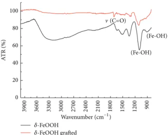
3.1. Structural Characterization. In the present work 𝛿
-FeOOH and the grafted 𝛿-FeOOH/TMSM samples were
characterized with ATR infrared spectroscopy. The obtained
results are summarized in Figure 1. The ATR spectrum of𝛿
-FeOOH (Figure 1) shows a very strong and broad band at
3265 cm−1that can be associated with the stretching modes
of molecules water, present on its surface. The two bands
(Fe-OH)
(Fe-OH)
𝛿-FeOOH
𝛿-FeOOH grafted 0
(C=O)
20 40 60 80 100
A
TR (%)
900
3300 3000 2700 2400 2100 1800 1500 1200
3600
3900
Wavenumber (cm−1)
Figure 1: FTIR spectra of𝛿-FeOOH and grafted𝛿-FeOOH.
vibrations [13]. The most remarkable band is located at
1701 cm−1 and it corresponds to carbonyl (C=O) vibrations
on TMSM structure. The infrared spectrum of functionalized
𝛿-FeOOH showed a band centered at 1096 cm−1, which can
be attributed to Si-O-Fe vibrations [14, 15]. It suggests that
the silane monomers were covalently bonded to𝛿-FeOOH
particles. It should be kept in mind, however, that the infrared bands are attributed to Fe-O-H bending vibrations in the same region. Despite this feature, the functionalization occurs in the formation of the Si-O-Fe bonding, because
the Fe-O-H bending vibration at 908 cm−1 disappears in𝛿
-FeOOH/TMSM infrared spectrum. Probably, one or two
Si-OCH3 groups are broken after functionalization reaction.
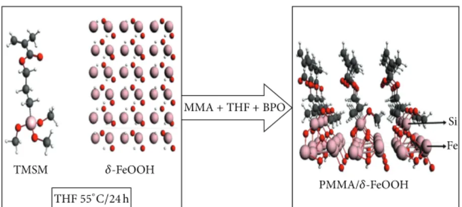
Figure 2 represents the atomic model of𝛿-FeOOH/PMMA
hybrid material.
The loss of two or three methyl groups of TMSM led
to a new chemical bond between oxygen atoms from 𝛿
-FeOOH and silicon. The nucleophilic substitution of Fe-OH
into Si(OCH3) can be described as
Fe-OH+Si-OCH3→Fe-O-Si+CH3OH (1)
In the study of Pereira and collaborators [9], there were found peaks in the X-ray diffraction pattern “100,”
“101,” “102,” and “110” characteristic of𝛿-FeOOH. The lattice
parameters can be indexed on a hexagonal lattice with𝑎 =
2.946A and˚ 𝑐 = 4.552A.˚
The XRD patterns were recorded for the nanoparticles after grafting with TMSM molecules. Figures 3(a) and 3(b)
reveal the presence of peaks2𝜃= 36∘, 56∘, and 63∘,
approx-imately, corresponding to the “100,” “102,” and “110,” to the
grafted𝛿-FeOOH 0.5 wt.% and 2.5 wt.%.
The XRD showed bands characteristic of PMMA such as
in the study of Shobhana and the peaks of𝛿-FeOOH [16]
confirming the presence of PMMA and 𝛿-FeOOH in this
material. Because the main peaks are represented in both XRD patterns, in the second (Figure 3(b)), the peak intensity is smaller; it can be interpreted by the fact that sample
has a higher percentage of feroxyhyte 2.5 wt.%. Already in
Figure 3(a), the intensity is large for the grafted𝛿-FeOOH
0.5 wt.%.
Figures 4(a) and 4(b) show the images obtained by SEM of surface and cross section fractured under liquid
nitrogen of hybrids with 2.5 wt.% of𝛿-FeOOH nanoparticles,
respectively.
A general analysis of the micrographs of all samples shows agglomerates of different sizes and with irregular blocks formats throughout the film. The average size of the
agglomerates was larger than 1𝜇m to 30𝜇m, indicating a
broad size distribution.
3.2. Thermal Properties. The thermal stabilities of the obtained hybrid materials were determined by thermogravi-metric analysis (TGA) and differential thermogravithermogravi-metric (DTA).
Figures 5 and 6 display the TG and DTA curves of PMMA,
𝛿-FeOOH grafted with TMSM, and 𝛿-FeOOH/PMMA
hybrid. These TG and DTA curves indicate that the neat𝛿
-FeOOH and 𝛿-FeOOH/TMSM sample weight loss occurs
in two and three distinct steps, respectively. For both of the materials, the first step of weight loss can be attributed
to the free water in the powder. For neat 𝛿-FeOOH
par-ticles, about 11% weight loss is observed at 273∘C, which
is due to the crystal transition of 𝛿-FeOOH to hematite
[13].
By analyzing the𝛿-FeOOH grafted with TMSM, after the
first step attributed to the free water in the powder, it can be
related to another weight loss at 289∘C, which is due to the
silane groups grafted on the iron oxyhydroxide surface. The
grafting process shifted the second decomposition step of𝛿
-FeOOH at 340∘C to higher values, suggesting higher thermal
stability.
Concerning the thermal decomposition of PMMA, this can take place in three stages: the first step between 130 and
260∘C is related to the decomposition starting at the
head-to-head chain segments. The second step between 260 and
370∘C is attributed to first unzipping starting at unsaturated
polymer end chains, and the third step between 370 and
500∘C corresponds to the random joining of polymeric chains
[17, 18].
The DTA curves shown in Figure 6 indicate that the starting temperature of the second decomposition step is
shifted to higher values for increasing 𝛿-FeOOH loading,
while the end of this step is only slightly affected by 𝛿
-FeOOH content. This implies that the increasing amount
of 𝛿-FeOOH nanoparticles delays the beginning of
poly-mer unzipping. However, during the decomposition the𝛿
-FeOOH nanoparticles confer suppressing effect that seems to be independent of their loading in the nanocomposite, at least within the 0.5 to 2.5 wt.% range presented in this study.
We can also notice, in Figure 6, that the maximum
degradation rate of PMMA occurs at 293∘C, while increasing
𝛿-FeOOH loading this maximum shifts toward higher
tem-peratures, from𝑇= 378∘C for𝛿-FeOOH 0.5 wt.% up to 389∘C
TMSM
Si
Fe
𝛿-FeOOH
PMMA/𝛿-FeOOH
MMA+THF+BPO
THF55∘C/24h
Figure 2: Atomic model of a𝛿-FeOOH nanocrystal embedded in the PMMA.
100
102 110
5 10 15 20 25 30 35 40 45 50 55 60 65 70 75 80 0
2𝜃
0 200 400 600 800 1000 1200 1400 1600 1800 2000 2200
In
ten
si
ty (a.u
.)
(a) Polymer 0.5 wt.%FeOOH
100
102 110
10 15 20 25 30 35 40 45 50 55 60 65 70 75 80 5
2𝜃
0 100 200 300 400 500 600 700 800 900 1000
In
ten
si
ty (a.u
.)
(b) Polymer 2.5 wt.%FeOOH
Figure 3: Powder X-ray diffraction pattern of grafted𝛿-FeOOH 0.5 wt.% (a) and 2.5 wt.% (b) sample.
20 𝜇m
(a)
20 𝜇m
(b)
Figure 4: SEM micrographs of PMMA/𝛿-FeOOH hybrid with (a) 0.5 and (b) 2.5 wt.% of𝛿-FeOOH.
The radical trapping effect might be the responsible for the thermal stability improvement observed for the
increasing amount of 𝛿-FeOOH nanoparticles. This effect
was recently demonstrated for inorganic hybrids with PMMA [19–22]. Therefore the thermal improvement reported above may take place due to a similar effect that may be undergoing
in the presence of𝛿-FeOOH, which acts as radical trappers
and accepts the unpaired electron from the radical polymer chain thus stopping or retarding the unzipping.
𝛿-FeOOH
𝛿-FeOOH/PMMA TMSM
M
ass loss (%)
0 10 20 30 40 50 60 70 80 90 100
500 100 150 200 250 300 350 400 450 50
0
Temperature (∘C)
0.5wt.%
0.25wt.%
Figure 5: Thermogravimetric curves of pure PMMA and PMMA with different𝛿-FeOOH contents.
DT
A
Pure polymer
𝛿-FeOOH0.5%
𝛿-FeOOH2.5% FeOOH Temperature (∘C)
100 200 300 400 500 600 700 800 900 1000
−0.010 −0.008 −0.006 −0.004 −0.002 0.000 0.002
Figure 6: Differential thermogravimetric (DTA) curves of 𝛿 -FeOOH, Polymer, and𝛿-FeOOH/PMMA hybrid 0.5 and 2.5 wt.%.
of𝛿-FeOOH before the functionalization process were
per-formed. From the planes examined, “100,” “101,” “102,” and “110,” the more stable one was “100.”
In the density of states (DOS) graphs, the valence band located in the region of the oxygen 2p and the conduction band situated in the region of the iron 3d (Figure 7) indicate
an electron transfer from O2−anions to Fe3+cations.
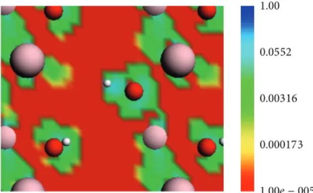
Figure 8 shows areas with high and low electron density in green and red color, respectively. The valence charge density is higher in the volumes close to O atoms. These are preferential regions for nucleophilic or electrophilic substitution, where
Fe-OH reacts with Si-OCH3to originate Fe-O-Si bonds.
3d
Fe Fe
3d 2p
O 0.00 0.28 0.56 0.84
D
OS (a.u
.)
0.00 0.23 0.46 0.69
D
OS (a.u
.)
0.00 0.25 0.50 0.75
D
OS (a.u
.)
−0.35 −0.30 −0.25 −0.20 −0.15 −0.10 −0.05 0.00
−0.40
Energy hartree
−0.35 −0.30 −0.25 −0.20 −0.15 −0.10 −0.05 0.00
−0.40
Energy hartree
−0.35 −0.30 −0.25 −0.20 −0.15 −0.10 −0.05 0.00
−0.40
Energy hartree
Figure 7: Densities of states (DOS) of octahedral Fe and O in the bulk𝛿-FeOOH.
3.2.2. PMMA/𝛿-FeOOH. The potential energy surface vary-ing the PMMA angle in relation to the oxide surface by angles
(I) Si-C-C-C and (II) C-C-O-C from 80 to 200∘ has been
determined. This feature allowed us to evaluate the probable conformation of the monomer of the polymeric matrix close to the surface of feroxyhyte nanoparticles. Our results plotted in Figure 9 indicate that the minimum energy or most stable
conformation of PMMA molecule on the𝛿-FeOOH surface
lies in the range of 155–130∘.
The 𝛿-FeOOH surface interferes in the PMMA angles
so that the system stability increases while angles I and II reduce. This phenomenon can be explained by attractive electrostatic interactions, either between the positive regions
of the PMMA and the surface oxygen or between𝜋-electrons
1.00
0.0552
0.00316
0.000173
1.00e − 005
Figure 8: Electrostatic surface contours of𝛿-FeOOH bulk. Red and green indicate volumes of low electron density and high electron density, respectively. Red = oxygen, white = hydrogen, and pink = iron.
0
−1000
−2000
−3000
−4000
−5000
−6000
200 180
160 140
120 100
80
60
200 180 160 140 120 100 80 60
Ener
g
y (k
ca
l/mo
l)
II (deg.)
I (deg.)
>−1000 <−1000 <−2000
<−3000 <−4000 <−5000
Figure 9: Surface potential energy of 𝛿-FeOOH/PMMA as a function of the (I) Si-C-C-C and (II) C-O-C-C angles. 3D surface: 1 versus 2versus energy. Energy = distance weighted least squares.
4. Conclusions
The hybrid nanocomposites𝛿-FeOOH/PMMA were
success-fully prepared. The results of SEM confirmed the dispersion
of𝛿-FeOOH particles in the polymer matrix.
The TG analysis showed that the thermal stability of𝛿
-FeOOH/PMMA nanocomposites is higher than that of pure PMMA. This trend became more evident by increasing the iron concentration.
The results of FTIR indicate the existence of covalent bonding between silane monomers and atoms located on the
surface of the𝛿-FeOOH nanoparticles. This was confirmed
by surface charge density map, which clearly showed the presence of regions likely to perform this type of interaction. In general, the computational studies, coupled with experimental characterizations, allowed a better understand-ing of the morphology, structure, and electronic properties of
hybrid𝛿-FeOOH/PMMA.
Conflict of Interests
All authors declare that there is no conflict of interests regarding the publication of this paper.
Acknowledgments
The authors thank the Brazilian agencies FAPEMIG, CAPES, and CNPq for funding this work. They are also especially grateful to CNPq and CAPES for the fellowships and schol-arships provided. Teodorico C. Ramalho thanks also the Invited Professor position at the Center for Basic and Applied Research at the Czech Republic.
References
[1] P. Kiliaris and C. D. Papaspyrides, “Polymer/layered silicate (clay) nanocomposites: an overview of flame retardancy,”
Progress in Polymer Science, vol. 35, no. 7, pp. 902–958, 2010. [2] F. Hussain, M. Hojjati, M. Okamoto, and R. E. Gorga, “Review
article: polymer-matrix nanocomposites, processing, manufac-turing, and application: an overview,” Journal of Composite Materials, vol. 40, no. 17, pp. 1511–1575, 2006.
[3] J. Macan, I. Brnardi´c, S. Orli´c, H. Ivankovi´c, and M. Ivankovi´c, “Thermal degradation of epoxy-silica organic-inorganic hybrid materials,”Polymer Degradation and Stability, vol. 91, no. 1, pp. 122–127, 2006.
[4] S. Gross, D. Camozzo, V. Di Noto, L. Armelao, and E. Tondello, “PMMA: a key macromolecular component for dielectric low-K hybrid inorganic-organic polymer films,”European Polymer Journal, vol. 43, no. 3, pp. 673–696, 2007.
[5] G. Toskas, C. Cherif, R. Hund et al., “Chitosan(PEO)/silica hybrid nanofibers as a potential biomaterial for bone regener-ation,”Carbohydrate Polymers, vol. 94, no. 2, pp. 713–722, 2013. [6] C. Soundrapandian, B. Sa, and S. Datta, “Organic-inorganic composites for bone drug delivery,”AAPS PharmSciTech, vol. 10, no. 4, pp. 1158–1171, 2009.
[7] M. C. Urbina, S. Zinoveva, T. Miller, C. M. Sabliov, W. T. Monroe, and C. S. S. R. Kumar, “Investigation of magnetic nanoparticle-polymer composites for multiple-controlled drug delivery,”Journal of Physical Chemistry C, vol. 112, no. 30, pp. 11102–11108, 2008.
[8] S. Kirchberg, M. Rudolph, G. Ziegmann, and U. A. Peuker, “Nanocomposites based on technical polymers and sterically functionalized soft magnetic magnetite nanoparticles: synthe-sis, processing, and characterization,”Journal of Nanomaterials, vol. 2012, Article ID 670531, 8 pages, 2012.
[9] M. C. Pereira, E. M. Garcia, A. Cˆandido Da Silva et al., “Nanostructured𝛿-FeOOH: a novel photocatalyst for water splitting,”Journal of Materials Chemistry, vol. 21, no. 28, pp. 10280–10282, 2011.
AC magnetic field,”Journal of Nanoparticle Research, vol. 15, no. 4, article 1544, 2013.
[11] G. Te Velde, F. M. Bickelhaupt, E. J. Baerends et al., “Chemistry with ADF,”Journal of Computational Chemistry, vol. 22, no. 9, pp. 931–967, 2001.
[12] BAND 2009.01,SCM, Theoretical Chemistry, Vrije Universiteit, Amsterdam, The Netherlands, 2009, http://www.scm.com/. [13] R. M. Cornell and U. Schwertmann,The Iron Oxides,
Wiley-VCH, Weinheim, Germany, 2nd edition, 2003.
[14] U. Schwertmann and H. Thalmann, “The influence of [Fe(II)], [Si], and pH on the formation of lepidocrocite and ferrihydrite during oxidation of aqueous FeCl2 solutions,”Clay Minerals, vol. 11, no. 3, pp. 189–200, 1976.
[15] L. Carlson and U. Schwertmann, “Natural occurrence of ferox-yhite (𝛿-FeOOH),”Clay and Clays Minerals, vol. 28, no. 4, pp. 272–280, 1980.
[16] E. Shobhana, “X-Ray diffraction and UV-visible studies of PMMA thin films,”International Journal of Modern Engineering Research, vol. 2, no. 3, pp. 1092–1095, 2012.
[17] P. R. Westmoreland, T. Inguilizian, and K. Rotem, “Flamma-bility kinetics from TGA/DSC/GCMS, microcalorimetry and computational quantum chemistry,”Thermochimica Acta, vol. 367-368, pp. 401–405, 2001.
[18] T. Kashiwagi, A. Inaba, J. E. Brown, K. Hatada, T. Kitayama, and E. Masuda, “Effects of weak linkages on the thermal and oxidative degradation of poly(methyl methacrylates),” Macro-molecules, vol. 19, no. 8, pp. 2160–2168, 1986.
[19] P. K. Sahoo and R. Samal, “Fire retardancy and biodegradability of poly(methyl methacrylate)/montmorillonite nanocompos-ite,”Polymer Degradation and Stability, vol. 92, no. 9, pp. 1700– 1707, 2007.
[20] M. A. Goncalves, E. F. F. da Cunha, F. C. Peixoto, and T. C. Ramalho, “Probing thermal and solvent effects on hyperfine interactions and spin relaxation rate of𝛿-FeOOH(1 0 0) and [MnH3buea(OH)]2−: toward new MRI probes,”Computational and Theoretical Chemistry, vol. 1069, pp. 96–104, 2015. [21] M. A. Goncalves, E. F. F. da Cunha, F. C. Peixoto, and T. C.
Ramalho, “NMR parameters and hyperfine coupling constants of the Fe3O4(1 0 0)-water interface: implications for MRI probes,”Chemical Physics Letters, vol. 609, pp. 88–92, 2014. [22] K. Chrissafis and D. Bikiaris, “Can nanoparticles really enhance
Submit your manuscripts at
http://www.hindawi.com
Scientifica
Hindawi Publishing Corporationhttp://www.hindawi.com Volume 2014
Hindawi Publishing Corporation
http://www.hindawi.com Volume 2014
Hindawi Publishing Corporation
http://www.hindawi.com Volume 2014
Hindawi Publishing Corporation
http://www.hindawi.com Volume 2014
Ceramics
Journal ofHindawi Publishing Corporation
http://www.hindawi.com Volume 2014
Nanoparticles
Journal ofHindawi Publishing Corporation
http://www.hindawi.com Volume 2014
Hindawi Publishing Corporation
http://www.hindawi.com Volume 2014
International Journal of
Biomaterials
Hindawi Publishing Corporation
http://www.hindawi.com Volume 2014
Nanoscience
Journal ofTextiles
Hindawi Publishing Corporation
http://www.hindawi.com Volume 2014
Journal of Hindawi Publishing Corporation
http://www.hindawi.com Volume 2014
Crystallography
Journal of Hindawi Publishing Corporationhttp://www.hindawi.com Volume 2014
The Scientific
World Journal
Hindawi Publishing Corporation
http://www.hindawi.com Volume 2014
Hindawi Publishing Corporation
http://www.hindawi.com Volume 2014
Coatings
Journal of Advances inMaterials Science and Engineering Hindawi Publishing Corporation
http://www.hindawi.com Volume 2014
Hindawi Publishing Corporation
http://www.hindawi.com Volume 2014
Hindawi Publishing Corporation
http://www.hindawi.com Volume 2014
Metallurgy
Journal ofHindawi Publishing Corporation
http://www.hindawi.com Volume 2014
BioMed
Research International
Materials
Journal of Hindawi Publishing Corporationhttp://www.hindawi.com Volume 2014
N
a
no
ma
te
ria
ls
Hindawi Publishing Corporation
http://www.hindawi.com Volume 2014
Journal of