LIPID NANOPARTICLE MEDIATED
DRUG DELIVERY FOR SAFER CANCER
TREATMENT: EXAMPLE OF PACLITAXEL
Slavomira Doktorovova
student
Faculty of Health sciences – uFp
Carla Martins Lopes
Assistant professor
Faculty of Health sciences – uFp
institute of biotechnology and bioengineering, centre of genetics and biotechnology – utAD
Eliana B. Souto
Assistant professor
Faculty of Health sciences – uFp
institute of biotechnology and bioengineering, centre of genetics and biotechnology – utAD
85
RESUMO
os vectores coloidais de natureza lipídica têm atraído particular atenção para o desenvol-vimento de terapias mais seguras e eficazes aplicadas a várias doenças. As vantagens da utilização das nanopartículas lipídicas são exemplificadas utilizando o paclitaxel, um anti-cancerígeno particularmente interessante para o desenvolvimento de novas formas farma-cêuticas mais adequadas para o tratamento do cancro. De facto, este fármaco apresenta problemas de insolubilidade aquosa e sérios efeitos secundários concomitantes à adminis-tração da forma farmacêutica convencional (taxol). este artigo apresenta uma revisão das razões para a reformulação do paclitaxel e sumariza as vantagens de novas formas farma-cêuticas contendo nanopartículas lipídicas para a administração deste anticancerígeno.
PALAVRAS-CHAVE
nanopartículas de lípidos sólidos, vectores lipídicos nanoestruturados, nanopartículas lipídi-cas, paclitaxel, tratamento do cancro
ABSTRACT
colloidal carriers composed of lipids attract much attention in the development of safer and more effective therapy of various diseases. the advantages of lipid-based nanoparticles are illustrated on the example of paclitaxel, a challenging chemotherapeutic drug for the development of a novel and more suitable dosage form for cancer treatment. paclitaxel is known for its water insolubility and serious side effects when administered by its conventio-nal formulation (taxol). this paper reviews the reasons of further re-formulation of paclitaxel and summarizes the achievements of lipid nanoparticle-based formulations of this drug.
KEYWORDS
stability in comparison to liposomes (Heurtault et al., 2003). sln and nlc are versatile drug carriers suitable for delivery of wide range of drugs of both hydrophilic and lipophilic nature, and having different molecule weights.
great attention is given to colloidal drug carriers in treatment of cancer. Various approaches in chemotherapeutic drug delivery has been reviewed recently, however, the reviews mos-tly focus on other types of colloidal carriers, such as liposomes, polymeric nanoparticles and micelles, dendrimers or inorganic nanoparticles. this paper summarizes the achievements of solid lipid nanoparticles, nanostructured lipid carriers and lipid nanocapsules in the deli-very of a well known chemotherapeutic drug, paclitaxel.
2.
MARKETED PACLITAXEL FORMULATIONS
paclitaxel is a diterpenoid alkaloid drug approved by Food and Drug Administration (FDA) and european medicines Agency (emeA) as first line therapy of breast and ovarian cancer, with proven effect against carcinomas of other tissues. Due to its physicochemical characte-ristics, formulating a suitable dosage form for its administration remains a challenging task.
taxol (bristol-myers squibb) was the first marketed formulation of paclitaxel, introduced into market in 1992 as second-line treatment of advanced ovarian cancer. Although this formulation was successful in overcoming the water insolubility by employing a mixture of solubilizing agents, their use led to well known, and in literature extensively described, side effects. briefly, polyoxyethoxylated castor oil (cremophor el) is pointed as the cause of seve-re hypersensitivity seve-reactions upon administration; further on nephorotoxicity, neurotoxicity and hypotension have been reported. the potential risks associated with this formulation where summarized by singla and colleagues (singla et al., 2002).
great efforts have been made to design a cremophor el-free formulation of paclitaxel. the aim is to find a biocompatible and well-tolerated material capable of solubilizing this drug without exposing the patient to additional risks. Various approaches have been used to de-sign such a formulation.
87
Another approach is in search for better paclitaxel formulation. in fact, a highlyhydropho-bic compound is a challenge in terms of development of a dosage form. combinations of co-solvents and oil-in-water emulsions have been tested by various research groups in the beginning of the nineties (safavy, 2008).
With the development of colloidal drug carriers the possibilities of formulation of a water insoluble drug were increased. liposomes technology was among the first tested. Although many liposomal formulations with sufficient stability have been developed, the clinical trials indicated no clear benefit over established formulation, and up-to-date there is no marke-ted liposomal formulation. paclitaxel is also a suitable candidate for delivery by nanoparticles composed of wide array of materials. it was only with human serum albumin (HsA) based nanoparticles that another formulation of paclitaxel reached the market in 2005. As expec-ted, the paclitaxel-loaded HAs based nanoparticles do not cause hypersensitivity reactions, which enables omitting the premedication and administration of higher doses in shorter times (micha et al., 2006). However, it was shown that despite its small size, the drug sho-ws similar body distribution as from taxol (Henderson and bhatia, 2007). the benefit of this formulation is indeed rather in use of biocompatible material to solubilize the drug than in colloidal size of its carriers.
Development of various colloidal carriers for paclitaxel delivery therefore still continues. the use of nanoparticles in general may help improving the delivery of drug into the target cells and thus increases the pharmacological effect. moreover, controlled release and increased terminal half life can be achieved, which would subsequently allow less frequent administra-tion. As great inter-individual differences in pharmacokinetics of paclitaxel have been obser-ved after taxol administration, also ascribed to cremophor-el (van tellingen et al., 1999), the improvement of pharmacokinetic characteristics is also of high importance in development of novel paclitaxel formulations.
3.
FUTURE PACLITAXEL FORMULATIONS?
3.1.
NANOPARTICLE FORMULATIONS IN CLINICAL TRIALS
several polymeric nanoparticle-based formulations are under clinical trials, formulations of paclitaxel not being an exception. in march 2009, FDA approved a phase i clinical trial of nanoxel, a polymeric nanoparticle formulation of paclitaxel developed by Fresenius cabi. As great variety of biodegradable polymers-based nanoparticles and polymeric micelles have been reported in the literature, more nanoparticle formulations are expected to reach clini-cal trials soon.
in parallel with polymer-based drug delivery systems, those composed of lipid materials are being developed. this review will focus on lipid nanoparticle, such as sln, nlc, and lipid nanocapsules (lnc), with proven suitability for paclitaxel loading.
3.2.
LIPID NANOPARTICLES
solid lipid core. the advantage over sln should be the possibility of higher drug loading and possibility of incorporating the drugs better soluble in liquid lipids into a colloidal carrier (müller et al., 2002).
not all formulations referred to as sln in the literature in fact comply with the definition of solid lipid matrix. instead of sln, they better comply with the definition of lipid nanocap-sules (lnc) - colloidal carriers composed of a liquid lipid core and a phospholipid shell, not necessarily solid (Huynh et al., in press). A wide variety of colloidal carriers composed of various lipids are also denominated as lipid nanoparticles, without further characteristics.
3.3.
SLN FOR PACLITAXEL DELIVERY
the first studies on sln for paclitaxel delivery were reported by cavalli and miglietta in 2000 (cavalli et al., 2000, miglietta et al., 2000). cavalli et al developed tripalmitine-based sln stabilised by soy phosphatidylcholine, with a mean particle size below 500 nm, with proven stability in isotonic glycerol solution. these nanoparticles could be freeze dried and reconstituted with only a small particle growth, and up to 2.8 % (wt) of paclitaxel could be encapsulated without precipitation of the drug during one year. the possible advantage towards clinical use already achieved in this study is the possibility of administration of usual therapeutic dose of 10 mg/m2/h in only 20-40 ml of sln formulation with 2.0 % paclita-xel loading (cavalli et al., 2000). cytotoxicity of this paclitapaclita-xel sln formulation was tested on mcF-7 (human breast carcinoma) and Hl60 (human promyelocytic leukemia) cell lines. While drug free sln showed no toxic effect towards these cell lines, paclitaxel loaded sln caused growth inhibition of mcF-7 cells already in very low concentrations (1 ng/ml), at whi-ch the paclitaxel solution was not effective yet. cell internalization of sln of same lipid and surfactant composition loaded with 6-coumarine was confirmed by fluorescent microscopy (miglietta et al., 2000). one year later, long circulating paclitaxel-loaded sln with hydrophilic surface coating were reported with significantly higher half-time (t1/2)and area under curve (Auc), in comparison with free paclitaxel (administered to mice as taxol) (chen et al., 2001).
acid-89
based sln that ethanol could be used (Yuan et al., 2008), instead of acetone used in one ofthe first reports about paclitaxel-loaded sln (chen et al., 2001). the method of solubilization of paclitaxel in ethanol enables formulating colloidal carrier for paclitaxel delivery based on practically any solid lipid or phospholipid. table 1 gives the summary of lipid material used in sln, nlc and lipid nanocapsules preparation in the up-to-date published studies.
With regard to lipid nanocapsules, good solubility of paclitaxel in medium chain triglyceri-des (a liquid mixture of caprylic and capric acid triglyceritriglyceri-des), was reported (babu Dhanikula et al., 2007); and in phospholipids with short saturated chains (Feng and Huang, 2001). this information led the research group of Dong to design a statistically optimized sln formu-lation (Dong et al., 2009). trilaurine-based sln formuformu-lation stabilised by polyoxyl 20-stearyl ether (brij 78) and a lipid nanocapsules formulation comprising medium chain triglycerides (miglyol 812), polyoxyl 20-stearyl ether and D-alpha-tocopheryl polyethylene glycol 1000 succinate were the outcome of this statistical approach. even though paclitaxel was added to the lipid phase of the formulation as ethanolic solution, and the solvent was immediately removed by nitrogen prior to proceeding to further steps of preparation. moving towards pre-clinical studies, cytotoxicity of the developed formulations has been tested on different types of cell lines and in-vivo studies have been reported.
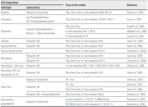
Table 1. composition and focus of the studies on solid lipid nanoparticles for paclitaxel delivery
SLN Composition
Focus of the studies Reference Solid lipid Surfactant(s)
Trilaurine Polyoxyl 20-stearyl ether Phys. Chem. Char.; In-vitro evaluation; MDA-MB-231 (Dong et al., 2009)
Trimyristine Egg Phosphatidylcholine +
PEG-Phosphatidylethanolamine Phys.Chem.Char.; In-vitro evaluation ; OVCAR-3; MCF-7 (Lee et al., 2007)
Tripalmitine Soybean Phosphatidylcholine + Butanol + Sodium taurocholate
Phys.Chem.Char (Cavalli et al., 2000) In-vitro evaluation; MCF-7; HC60 (Miglietta et al., 2000) In-vitro evaluation HT-29 (Serpe et al., 2004) Tristearin Poloxamer 188 Phys.Chem.Char.; In-vitro evaluation A549 (Yuan et al., 2008) Glyceryl Behenate Poloxamer 188 Phys.Chem.Char.; In-vitro evaluation A549 (Yuan et al. 2008) Glyceryl Palmito stearate Phys.Chem.Char.; In-vitro evaluation B16F10 (Shenoy et al., 2009a)
Monostearin Poloxamer 188 Phys.Chem.Char.; In-vitro evaluation A549 (Yuan et al. 2008) Poloxamer 407 Phys.Chem.Char.; In-vitro evaluation B16F10 (Shenoy et al., 2009b) Monostearin + Oleic acid Poloxamer 188 In-vitro evaluation; MCF-7; MCF-7/ADR; SKOV3; SKOV3-TR30 (Zhang et al., 2008) Monostearin-PEG-SA,
Monostearin-FA-SA Poloxamer 188 Phys.Chem.Char.; In-vitro evaluation A549 (Yuan et al. 2008)
Stearic Acid
Polyoxyl 20-stearyl ether PK - mice (Chen et al., 2001)
Poloxamer 188 PK - mice (Chen et al. 2001) Phys.Chem.Char.; In-vitro evaluation A549 (Yuan et al. 2008) Poloxamer 188 + Phosphatidylcholine Phys.Chem.Char.; In-vitro evaluation HepG2 (Pandita et al., 2009) Cetyl alcohol +
polysorbate 60 Polyoxyl 20-stearyl ether
In-vitro evaluation; U-118; HCT-15; BBB perfusion (Koziara et al., 2004) BD (BALB/c mice); In-vitro evaluation; U-118; HCT-15 (Koziara et al., 2006)
Abbreviations: phys.chem.char. = physicochemical characterization (zeta-potencial, scaning electron microscopy, transmission electron microscopy, Differential scanning calorimetry, Wide-angle Xray scattering, in-vitro release); pK = pharmacokinetic studies, bD = body distribution studies; bbb = blood-brain barrier
spe-day 12 of the study. the body distribution of the paclitaxel also did not differ significantly from free drug (Koziara et al., 2006). the explanation suggested by this research team was the relatively fast release of the drug already in in-vitro experiments and its low loading in sln, which stress the need to design a formulation which would entrap higher amount of drug and assure its controlled release. moreover, this research group suggested that pacli-taxel encapsulated in sln could overcome the membrane transporter-mediated resistance to taxenes.
more in-vitro studies confirmed that sln and nlc formulations could be effective even in taxenes-resistant cell lines. Zhang and colleagues reported similar inhibition concentrations (ic50) of paclitaxel-loaded nlc required to cease the growth of both sensitive and resistant cancer cell lines, while 30 times higher concentration of taxol was required to kill the resis-tant cells (Zhang et al., 2008). the authors also reported that folic acid-surfaced nlc were required in even lower concentrations to inhibit cancer cell growth.
Yuan and colleagues observed cellular uptake of paclitaxel-loaded sln (here loaded also with fluorescein isothiocyanate) by confocal microscopy (Yuan et al., 2008). this study also gives interesting results of very low cytotoxicity of blank sln formulation composed of va-rious lipids (see table 1), illustrated by ic50 of 308.72 to 471.48 μg ml−1 in contrast to ic
50 of 0.21 to 1.86 μg ml−1 of paclitaxel-loaded sln (which is still less than ic
50 of taxol).
3.4.
LIPID NANOCAPSULES
paclitaxel solubility in medium chain triglycerides or short-chain phospholipids makes it a perfect candidate for encapsulation in lipid nanocapsules (lnc). indeed, stable lipid nano-capsules were developed by some research groups. lnc composed of labrasol, soy phos-phatidylcholine and polyethylene glycol-660 hydroxystearate with size below 100 nm were prepared and characterized by lacoeuille and colleagues. these carriers showed a sustained release of paclitaxel over two weeks. During the release studies, lnc were reported as stable; nevertheless, long-term stability over this period is not reported (lacoeuille et al., 2007a). in an in-vivo study conducted in Wistar rats, pharmacokinetic parameters and survival rates similar to taxol were obtained (lacoeuille et al., 2007b).
lnc composed of combination of miglyol 812 with brij 78 and D-alpha-tocopheryl polyethyle-ne glycol 1000 succinate were developed by the same statistical analysis as trilauripolyethyle-ne-compo- trilaurine-compo-sed sln and proved efficient in inhibiting the growth of mDA-mb-231 cells (Dong et al., 2009).
91
4.
FURTHER PERSPECTIVES
All up-to-date published in-vitro studies compare effectiveness of lipid nanoparticles to that of taxol. As a nanoparticle-bound formulation is already marketed, it would be of great in-terest to compare a lipid nanoparticle formulation to this protein-based marketed formu-lation. As one of the reasons of development of nanoparticulate formulations is to provide controlled release and protect the drug until it reaches its site of action – and this is doubtful in case of Abraxane (Henderson and bhatia, 2007), the future nanoparticle-based formula-tion will need to fulfill these expectaformula-tions.
lipid nanoparticles are explored for a shorter period of time than liposomes, polymeric na-noparticles or micelles, yet some of their features make them more advantageous, namely in terms of biocompatibility and toxicity (degradation products including), and scaling up the manufacture process. if the lipid nanoparticle-based formulations are intended for in-travenous administration, size of the particles needs to be maintained in suitable size range. some of the authors proposed their nanoparticle formulations with larger diameters for oral administration, but the suitability for this administration route also needs to be proven by preclinical studies.
5.
CONCLUSIONS
As the efforts to develop a safer and more efficient formulation for paclitaxel still continue, it is expected that more nanoparticle-based formulations will enter clinical trials soon. based on the pre-clinical data obtained for sln, nlc and lnc, these can be considered as suitable carriers for paclitaxel. in addition to advantages of lipid-based colloidal carriers in the gene-ral, these colloidal carriers might be promising for the future therapy of cancer.
REFERENCES
ARICA YEGIN, B., BENOIT, J. P. AND LAMPRECHT, A. (2006). paclitaxel-loaded lipid nano-particles prepared by solvent injection or ultrasound emulsification. in: Drug Development and industrial pharmacy, 32, nº9, pp. 1089-1094.
BABU DHANIKULA, A., MOHAMED KHALID, N., LEE, S. D., YEUNG, R., RISOVIC, V., WA-SAN, K. M. AND LEROUX, J.-C. (2007). long circulating lipid nanocapsules for drug detoxifi-cation. in: biomaterials, 28, nº 6, pp. 1248-1257.
CAVALLI, R., CAPUTO, O. AND GASCO, M. R. (2000). preparation and characterization of solid lipid nanospheres containing paclitaxel. in: european Journal of pharmaceutical sciences, 10, nº 4, pp. 305-309
DONG, X., MATTINGLY, C. A., TSENG, M., CHO, M., ADAMS, V. R. AND MUMPER, R. J.
(2009). Development of new lipid-based paclitaxel nanoparticles using sequential simplex optimization. in: european Journal of pharmaceutics and biopharmaceutics, 72, nº 1, pp. 9-17.
FENG, S.-S. AND HUANG, G. (2001). effects of emulsifiers on the controlled release of pacli-taxel (taxol®) from nanospheres of biodegradable polymers. in: Journal of controlled release, 71, nº 1, pp. 53-69.
pharmaceutical bulletin (tokyo), 49, nº 11, pp. 1444-1447.
KOZIARA, J., OH, J., AKERS, W., FERRARIS, S. AND MUMPER, R. (2005). blood compatibility of cetyl Alcohol/polysorbate-based nanoparticles. in: pharmaceutical research, 22, nº 11, pp. 1821-1828.
KOZIARA, J. M., LOCKMAN, P. R., ALLEN, D. D. AND MUMPER, R. J. (2004). paclitaxel nano-particles for the potential treatment of brain tumors. in: Journal of controlled release, 99, nº 2, pp. 259-69.
KOZIARA, J. M., WHISMAN, T. R., TSENG, M. T. AND MUMPER, R. J. (2006). in-vivo efficacy of novel paclitaxel nanoparticles in paclitaxel-resistant human colorectal tumors. in: Journal of controlled release, 112, nº 3, pp. 312-319.
LACOEUILLE, F., GARCION, E., BENOIT, J. P. AND LAMPRECHT, A. (2007a). lipid nanocap-sules for intracellular drug delivery of anticancer drugs. in: Journal of nanoscience and nano-technology, 7, nº 12, pp. 4612-4617.
LACOEUILLE, F., HINDRE, F., MOAL, F., ROUX, J., PASSIRANI, C., COUTURIER, O., CALES, P., LE JEUNE, J. J., LAMPRECHT, A. AND BENOIT, J. P. (2007b). in vivo evaluation of lipid nanocapsules as a promising colloidal carrier for paclitaxel. in: international Journal of phar-maceutics, 344, nº 1-2, pp. 143-149.
MIGLIETTA, A., CAVALLI, R., BOCCA, C., GABRIEL, L. AND ROSA GASCO, M. (2000). cellular uptake and cytotoxicity of solid lipid nanospheres (sln) incorporating doxorubicin or pacli-taxel. in: international Journal of pharmaceutics, 210, nº 1-2, pp. 61-67.
MICHA, J. P., GOLDSTEIN, B. H., BIRK, C. L., RETTENMAIER, M. A. AND BROWN III, J. V.
(2006). Abraxane in the treatment of ovarian cancer: the absence of hypersensitivity reac-tions. in: Gynecologic oncology, 100, nº 2, pp. 437-438.
MüLLER, R. H., RADTKE, M. AND WISSING, S. A. (2002). nanostructured lipid matrices for improved microencapsulation of drugs. in: international Journal of pharmaceutics, 242, sup-plement 1, pp. 121-128.
SERPE, L., CATALANO, M. G., CAVALLI, R., UGAZIO, E., BOSCO, O., CANAPARO, R., MUN-TONI, E., FRAIRIA, R., GASCO, M. R., EANDI, M. AND ZARA, G. P. (2004). cytotoxicity of anti-cancer drugs incorporated in solid lipid nanoparticles on Ht-29 colorectal anti-cancer cell line. in: european Journal of pharmaceutics and biopharmaceutics, 58, nº 3, pp. 673-680.
SINGLA, A. K., GARG, A. AND AGGARWAL, D. (2002). paclitaxel and its formulations. in: international Journal of pharmaceutics, 235, nº 1-2, pp. 179-192.
VAN TELLINGEN, O., HUIZING, M. T., PANDAY, V. R., SCHELLENS, J. H., NOOIJEN, W. J. AND BEIJNEN, J. H. (1999). cremophor el causes (pseudo-) non-linear pharmacokinetics of paclitaxel in patients. in: british Journal of cancer, 81, nº 2, pp. 330-335.
VIDEIRA, M., FLORINDO, H. F., GOUVEIA, L. F., LOBATO, M. R. AND ALMEIDA, A. J. (2005). triglyceride nanoparticles as potencial carriers for paclitaxel. in: 15th international symposium on Microencapsulation, parma,. italy,, 18-21 september 2005, pp. 225-226
93
YUAN, H., MIAO, J., DU, Y. Z., YOU, J., HU, F. Q. AND ZENG, S. (2008). cellular uptake of solid lipid nanoparticles and cytotoxicity of encapsulated paclitaxel in A549 cancer cells. in: international Journal of pharmaceutics, 348, nº 1-2, pp. 137-145.
ZHANG, X.-G., MIAO, J., DAI, Y.-Q., DU, Y.-Z., YUAN, H. AND HU, F.-Q. (2008). reversal activi-ty of nanostructured lipid carriers loading cytotoxic drug in multi-drug resistant cancer cells.