The Efect of Processing Parameters and Solid Concentration on the Microstructure and
Pore Architecture of Gelatin-Chitosan Scafolds Produced by Freeze-Drying
Daniel Enrique López Anguloa*, Paulo José do Amaral Sobrala
aDepto de Eng. de Alimentos – FZEA, University of São Paulo – USP, Av. Duque de Caxias Norte, 225, CEP 13635-900, Pirassununga, SP, Brazil
Received: December 28, 2015; Revised: April 18, 2016; Accepted: June 5, 2016
One of the main components for being successful in tissue engineering is developing a scafold with an appropriate architecture for allow migration, cell proliferation, and diferentiation. A gelatin– chitosan scafold by vacuum freeze-drying has been developed for tissue engineering applications. The efects of solid concentration and freezing processing on the scafold morphology and porous size were investigated. As the chitosan content was increased the viscoelastic properties of pigskin gelatin was modiied, the maximum G’ values were lower than the values for pure gelatin solution, and the thermal transition points also occurred at lower temperatures, as well as a decrease of pore size tendency was observed and the scafold visibly increased porosity, the structure scafold was observed with an interconnected and more homogeneous pore matrix. The pore sizes become smaller and pore walls thinner, while interconnectivity increases along with declining pre-freezing temperature. The chitosan–gelatin scafold will be a promising candidate in tissue engineering.
Keywords: tissue engineering, cell proliferation, interconnected pore matrix
1. Introduction
Tissue engineering is an emerging science enclosing
such diverse ields as molecular biology and materials engineering. It attempts to develop biological substitutes
for damaging organs and tissues1. Porous scafolds have
been extensively utilized in tissue engineering for playing
an important role in manipulating cell function and guidance of new tissue formation as well as transporting nutrients and removing wastes2,3.
An ideal tissue engineered scafold must have overall
constructions, internal structures, surface properties, mechanical properties, and material properties to meet the requirements of host tissue4,5. Ideally, the porosity of the scafolds, mean
pore size and the pore structure should be appropriate for cell adhesion, proliferation, migration, diferentiation and extracellular matrix regeneration. High porosity (generally greater than 90%) and a large pore size (about 100-200 μm)
as well as highly interconnected pore structure are necessary
for the transport of cells and metabolites6,7. The materials
should be biocompatible without inlammation or toxicity
in vivo and processed into 3D structure1,8.
Moreover, an ideal wound dressing scafold should present several speciic properties for the intended application, including capacity to absorb wound exudates, maintenance of the moisture, protection from secondary infection, provision of adequate gaseous exchange, provision of thermal insulation, free from particulate or toxic contaminants, mechanical durability, lexibility, and non toxicity9,10.
Many polymers and biopolymers have been extensively
investigated as potential materials for wound covering
scafold production. The use of natural biopolymers such
as chitosan, collagen and gelatin in the construction of
wound healing devices is quite attractive, mostly because of their biocompatibility, possibility of various chemical modiications, and relatively low cost of production (in many cases directly from renewable sources)11-15. Particularly,
gelatin and chitosan are non-toxic, biodegradable, and
biocompatible14. This biopolymer has the capacity to form
thermoreversible gel. Gelatin gel formation, at a molecular level, implicate a structural re-arrangement of the protein, involving the change from a messy stage to a more ordered one, formed by triple helix structures. The knowledge of the gel forming suspensions rheological properties by stationary and dynamic tests, is important for the design and processing of freezing gels.
Among several approach to produce biopolymers-based scafolds, the freeze-drying processing is interesting because do not demand special equipment. With this processing, material pore characteristics such as size, homogeneity and orientation are dependent on ice crystal nucleation and growth, and can be controlled by modulating the freezing
process9-11. While pore size is largely governed by freezing
temperature, pore homogeneity is achieved by controlling freezing rate and providing a uniform contact surface10,11.
In these contexts, aiming to develop a low cost scafold, we are trying to develop a material based on blends of gelatin and chitosan by freeze-drying processing. We hypothesized that these biopolymers can have opposite charge depending of pH and this will contribute to a better material. Moreover, they can be modiied and/or crosslinked by using a reactant as the glutaraldehyde. Thus, the objective of this work was focused on the study of the inluence of gelatin:chitosan ratio, and on the efect of two freezing processing on scafold microstructure (pore size range, average pore size). The efect of gelatin and chitosan concentration on the low behavior
and viscoelastic properties of gel forming suspensions was
2. Materials and methods
2.1. Materials
A pig skin gelatin type-A was used (bloom 260; molecular weight ~5.2x10⁴ Da; moisture content = 9.98 %), supplied by
Gelnex South America (Itá Santa Catarina, Brazil). Chitosan (derived from crab shell with minimum deacetylation degree of 85%, MW: 2x10⁵) was purchased from Sigma (St. Louis,
MO, USA). Acetic acid (Sigma) and glycerol (Synth) were all chemical reagents of analytical grade.
2.2. Fabrication of Porous Scafolds.
Blends of gelatin (GE) and chitosan (CH) were prepared by thorough mixing of gelatin (2%) and chitosan (1%) solutions in 0.05 M acetic acid, with GE:CH ratios of 3:7, 5:5, 7:3, 9:1, under stirring with a magnetic bar, at 50°C, for 2 h., then the glycerol (gly) was added in 0,3%, the pH of solutions was adjusted to 5. The solution blends were then poured into a glass petri dish and then: a) freezing in refrigerator at -27°C (for evaluating the efect of the solid
concentration), and b) the glass petri was left 2 hours in liquid
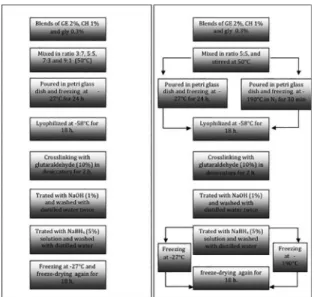
nitrogen system chamber -190°C, and then was kept in a refrigerator -27°C until the lyophilization (for evaluating the efect of freezing rate it was utilized two temperatures). After that, these products were submitted to the irst freeze-drying at -55°C for 18 h. reaching a sponge-like material (Figure 1).
2.3. Rheological measurements
The scafolds forming solutions were analyzed to determine it rheological and visco-elastic properties. Rheological tests were carried out in a rheometer (AR2000 Advanced Rheometer; TA Instruments, New Castle, DE, USA) using a cone and plate sensor geometry (cone angle 4°, 60 mm diameter). This equipment controlled the temperature with a peltier system. The results were analyzed using the software Rheology Advantage Data Analysis V.5.3.1 (TA Instruments).
2.3.1. Dynamic tests
For the dynamic tests, the extent of the linear viscoelastic region was determined by performing a strain (0–20%) sweep in gel forming solutions both as a gel (5°C) and as a sol (30°C), in all cases at 1 Hz of frequency. Thus, according
to the results of these tests, the strain value used for all
following dynamic tests was ixed at 4%, within the linear viscoelastic domain. The viscoelastic parameters determined in these trials were the storage or elastic modulus (G’) and the loss or viscous modulus (G’’)16.
2.3.2. Temperature sweep tests
Early, the scafold forming solutions were heated up to 50°C without a predestined heating rate program, starting
the tests when that measurement temperature was attained16.
The measures were separated in two parts, irst, the sample was cooled down from 50 to 5°C, remaining at this latter temperature for 5 min., and second heating up the sample to 50°C, at heating rate of 2°C/min. G’ and G’’ values as a
function of temperature were used to establish the following
parameters: gelling (Tsol–gel) and melting (Tgel–sol) temperatures,
calculated as the temperature where G’ changed drastically as an inlexion point; and the viscoelastic moduli (G’ and G’’) of the gel, calculated at 5°C17.
2.4. Scanning Electron Microscopy
The surface morphology, structural integrity and interconnectivity of the pores in the scafold were observed using Scanning Electron Microscopy (Hitachi TM 3000, Japan) at a magniication of 100x. The sample was placed
on double sided carbon tape in a vacuum chamber prior to
measurement. The surface of the samples was scanned at 15 kV, without previous treatment18. The pore sizes of the
scafolds were measured using image visualization software (Image J 1.45s, NIH Image, USA). The values were expressed as the mean ± standard error.
2.5. Statistical analysis
Statistical analysis and interpretation of all results was done by using GraphPad Prism version 6.00 for Windows (GraphPad Software, San Diego, California, USA). One way ANOVA with Bonferroni posttest was performed, to obtain the variance between and within all treatments. P-values less
than 0.05 denoted statistical signiicance.
Figure 1: Diagram of the steps of obtaining scafolds, left (efect of the solid concentration) and right (efect of freezing rate).
To crosslinking the GE:CH, the obtained sponge was placed inside desiccators containing glutaraldehyde (10%) in 200 ml ethanol (90%) during 2 h. Then, the sponge was immersed in NaOH solution (1%) and washed with distilled water twice, and treated with a NaBH4 (5% w/v) solution to
3. Results and discussions
3.1. Scanning temperature in dynamic rheological tests.
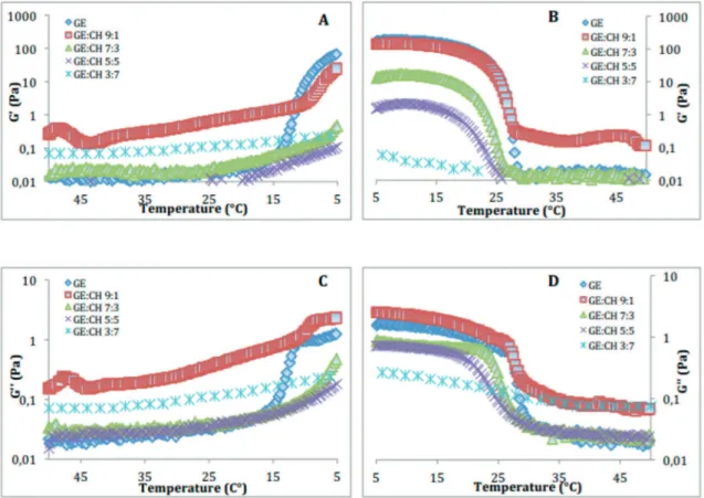
Rheology provides information about the polymer low properties, including the changes in the behaviour of the polymer when it subjected to a stress with variation in temperature. The storage and loss modulus in case of viscoelastic solids is the measure of stored energy, which represents their elastic portion, and the energy dissipated in the form of heat, representing the viscous portion, respectively19.The preliminary tests allowed to determine the conditions of tests inside the linear viscoelastic domain: frequency = 1 Hz, and strain amplitude = 0-20%. Thus, scanning temperature in dynamic rheological tests was carried out (Figure 2), allowing the determination of both moduli (G’and G’’), which had higher values at low than at higher temperatures. Between these two domains, an inlexion was observed in both moduli. These behaviors are typical of physical gels presenting a sol–gel (Tsol–gel) and a gel–sol (Tgel–sol) transition. The result shown, when the systems showed a gel character, it was also observed that the stifness (elastic character) increased with gelatin concentration in the blend. Similar proiles have been observed by several authors in gelatin based systems17,20,21.
When the chitosan concentration was increased, the viscoelastic properties of pig gelatin was modiied, the maximum G’ values
were lower than the values for pure gelatin solution, and the
thermal transition points also occurred at lower temperatures.
A decrease in percentage renaturation of food grade gelatin
by incorporation of chitosan has been previously reported22.
The temperatures of the sol–gel and gel–sol transitions, itted as the temperature where the peak of the curve of the irst derivative of G’ occurred, were afected by the chitosan concentration. The increase in the chitosan concentration in
the blend, as it was noted above, caused an intense reduction
(46.4%) in the Tsol–gel, which decreased from around 15.3°C to 8.2°C, whereas the Tgel–sol decreased (24.3%) from around 28.8°C to about 21.8°C (Table 1). These phenomena may well be related with the processes involved in the coil-helix/ helix-coil transition. The gelatin triple helix formation (coil-helix transition) is a slow process, which continues almost indeinitely, while the dissociation (helix-coil transition) of the triple helix is an equilibrium process linked with the
transition temperature23. Therefore, this is indicative of a
pronounced loss of the gelatin’s ability to refold into triple-helix chains in the presence of chitosan. Adding chitosan
at the higher proportion led to a substantial increase in the
viscous modulus (G’’) during cooling.
It is interesting to analyze the isoelectric point of the gelatin in terms of molecular interactions with chitosan. The gelatin used in this study is type-A, therefore the isoelectric point should be around pH 8-9, further solutions were adjusted to pH 5, therefore gelatin below its isoelectric point it is positively charged. Also, chitosan is a cationic polysaccharide, where the interactions between gelatin and chitosan are produced by both electrostatic and hydrogen bonding24. The latter occur extensively
between the -COOH, -NH2, and -OH groups on the amino acids in the gelatin and the -OH and -NH2 groups on the
Table 1: Transition temperatures Tsol–gel and Tgel–sol of gel forming
solutions based on diferent rate of gelatin and chitosan.
GE-CH ratio Tsol-gel (°C) Tgel-sol (°C)
10:0 15.3a ± 0.07 28.8
a ± 0.14
9:1 11.9b ± 0.04 28.3
a ± 0.09
7:3 9.1c ± 0.01 25.8b ± 0.14
5:5 8.8d ± 0.06 25.4
b ± 0.14
3:7 8.2e ± 0.06 21.8
c ± 0.21
Diferent letters indicate signiicant diferences (p<0.05).
chitosan, and are particularly notable at low temperatures. The chitosan structure is formed by units of glucosamine and N-acetyl D-glucosamine linked by β-(1→4) where the deacetylation degree is the percentage of amino
groups that remaining free in the chitosan molecule, in fact, a high amount of options is presented to generate electrostatic interactions with pig gelatin which is more
polar. Due to the acidic pH of the suspensions, these amine residues of chitosan are protonated (NH3
+) and
could form electrostatic interactions preferably with acid residues of gelatin (COO- from Glutamine and Aspartic acid), they will also form bridges with polar residues above. However, the amount of formed interactions will depend on the degree of exposure of residues of the protein
to interact with the chitosan and that will be determined
by the degree of unfolding of the protein, as well as by
the relative abundance of acid residues in protein versus
polar residues and the stoichiometry of the interaction.
3.2. Efect of biopolymers ratios on morphology
The average thickness of GE:CH scafold was between 1.4-1.7 mm (Table 2). As pig gelatin being a macromolecules and forms cross-linkage with chitosan molecules through hydrogen bonding and electrostatic interaction, the highest proportion of gelatin it had increased the thickness of scafold. The porous microstructure of the scafolds has a remarkable inluence on the cell intrusion, proliferation and function,when tissue engineering uses are envisioned14. Because
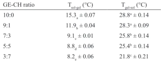
of that, this structure must be well controlled. Then, the GE:CH ratio was evaluated and optimized through the their intrinsic microstructure and porous size observation. It can be observed in Figure 3 that the chitosan have an important efect on material microstructure allowing the formation of sponge type structure.
Table 2: Efect of GE:CH ratio on the porous size and porous size range of the scafolds.
GE-CH Pore size (μm) Pore size range (μm) Thickness Scafolds (mm)
3:7 178.5a ± 69.1 132-311 1.46a ± 0.03
5:5 198.3ab ± 47.9 162-296 1.40a ± 0.02
7:3 202.8b ± 64.1 163-338 1.70b ± 0.04
9:1 192.5ab ± 49.8 160-393 1.66b ± 0.03
Diferent letters indicate signiicant diferences (p<0.05)
Then, scafold fabricated with higher proportion of gelatin (GE:CH= 9:1, Figure 3a) have irregular pore structure without a well visible porous networking structure. The pore walls were thicker with very few holes in the pore walls, which may indicate that the interconnectivity of the scafolds could be poor. This efect could diicult luid exchange and growing of native tissue. Similar structure was observed for GE:CH= 7:3 (Figure 3b) ratio material. As the chitosan
content was increased, a more homogeneous structure can
be observed, with some tendency for lower porous size formation. Moreover, these scafolds visibly presented increased porosity, and with matrix with an interconnected and more homogeneous pore structure (GE:CH=5:5, Figure 3c; GE:CH=3:7, Figure 3d). Very thin walls can be observed for equal proportion of biopolymers, but with less homogeneous porous size distribution (GE:CH= 5:5, Figure 3c). This increasing in homogeneity of porous size allow more hollow space, meaning that more luid and liquid component could be introduced to the scafolds.
The porous size was calculated after image treatments
allowing calculation of data presented at Table 2, where it
can be observed that, in overall, the pore size varied between
132 and 393 μm, for all formulations. The smallest and the
largest (P < 0.05) average pore size was 179 μm, for scafolds
produced with GE:CH= 3:7 ratio, and 203 μm, for GE:CH= 7:3, respectively (Table 2). These results suggested that the proportion of GE and CH was directly related to the pore size. Previous research25 reported that the mean pore size of
collagen based scafolds was afected by the viscosity of the material solution. Increasing the chitosan content raises the viscosity of the polymeric solution, reducing the density of the complex solution and leads to an increased porosity26 after
freeze-drying. The viscosity of the solution is large (>2%) ,
which is unfavorable to the water and the molecular chains
moving in the solution. Therefore, the number of ice nuclei
in the solution with high concentration is smaller and more
diicult to grow larger than that of the low concentration
one26. So, the pore size is inversely proportional to the
concentration of the scafolds27. All scafolds obtained in
this work were freeze-dried at the same time and under the same conditions; thus, the diference in the pore structure is associated only with the GE:CH ratios.
Thus, considering these quali-quantitative results of SEM, it could be concluded that GE:CH 5:5 formulation were with good pore size and porosity, which indicated that the sponge scafold meet the basic requirements as tissue engineering material. However, the structure scafold can even be improved by altering the conditions of freezing before freeze-drying; the efect of freezing rate is an important factor to be evaluated in terms of its interconnected porous structure.
Figure 3: Supericial SEM Images of scafolds obtained with diferent GE:CH ratios: 9:1 (A), 7:3 (B), 5:5 (C) and 3:7 (D).
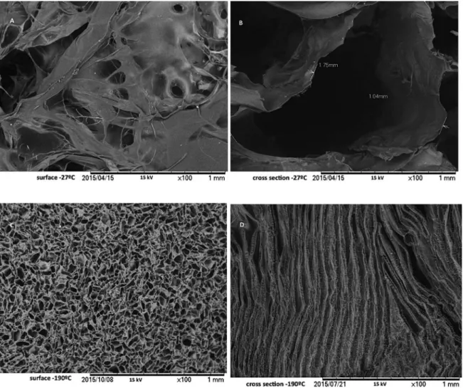
results indicated that freezing processing had an evident efect on the pore size and distribution in these scafolds, due to the diference in the growth velocity of ice crystals formed during freezing of materials. Scafolds prepared by quenching samples in liquid nitrogen (-190°C, Figure 4 c,d) exhibited smaller pores with a uniform distribution and with pore walls thinner, whereas the scafolds frozen at higher temperatures (-27°C, Figure 4 a,b) exhibited a poor porous microstructure with heterogeneous and big size pores. These results agree with those reported by28. After
ice crystals sublimation during freeze-drying, the scafold structure contains pores that correspond to the size and shape of the ice crystals29.
Analysis of the images of surface structure allowed calculation of the average pores size: 119.2±38.1 μm, for
material frozen at -190°C, and 196.8±36.9, for the specimen frozen at -27°C. Both pore size ranges are within the ideally expected for the transport of cells and metabolites in this kind of material6,7, however, the cross-sectional image shows
a much more homogeneous structure and arranged in layers to the material obtained after freezing at lower temperature. It is important to consider how variations in pore architecture afect cell-material interactions. Experimental
studies have led to the determination of a characteristic
interaction parameter, by careful analysis, it can be shown
that two matrices with identical chemistries but difering pore diameters have vastly diferent abilities to provide for cellular attachment. For example, scafolds containing pores
of 300 mm have a much lower internal surface area than matrices with pore diameters of less than 50 mm and, as such,
bind a much lower number of cells in the matrix. Because of the low cellular iniltration, tissue formation in these constructs is slow and in certain cases inhibited due to lack
of interactions between cells30. In general, research supports
that pores larger than 10 mm (the average cell diameter) will permit cell iniltration31. In terms of successful histogenesis,
both pore size and the degree of porosity are tissue and scafold speciic. For example, in vivo osteogenesis occurs in biomaterials of high porosity (>70%) with average pore sizes >300 mm32. However, in skin regeneration, successful
scafolds need only to exhibit pore sizes of 20-125 mm30.
Therefore, in this study the proposed material according to its porosity and average pore diameter, could be a good candidate for use in tissue regeneration as skin and cartilage.
4. Conclusions
The increase in the chitosan concentration in the blend caused an intense reduction in the Tsol–gel, and Tgel–sol this is
Figure 4: Surface (left) and cross-section (right) images of scafolds frozen at -27°C (a, b) and -190°C (c,d) measured by SEM.
triple-helix chains in the presence of chitosan. Morphology of the pigskin gelatin and chitosan scafolds prepared by freeze-drying technique and diferent solid proportion and freezing rate was investigated. The incorporation of chitosan improved the pore structure and size uniformity in supericial area of the scafolds. The pore sizes becomes smaller and pore walls thinner, while an improved of interconnectivity was observed with declining pre-freezing temperature. It has the porous structure, surface morphology, could be favorable to penetrate nutrients, export metabolic substances and leave new spaces for cells to ill in and produce new intercellular matrix. Finally, the study underscores gelatin–chitosan composite as a potential and promissory scafold material for skin regeneration.
5. Acknowledgments
The authors would like to thank at the Brazilian National Council for Scientiic and Technological Development (CNPq) and TWAS, the academy of sciences for the developing world, for the research grants (190232/2014-5 CNPq/ TWAS-FR: 3240279900). And to the Food´s Laboratory of Sao Paulo University.
6. References
1. Teimouri A, Azadi M, Emadi R, Lari J, Chermahini AN. Preparation, characterization, degradation and biocompatibility of diferent silk ibroin based composite scafolds prepared by freeze-drying method for tissue engineering application. Polymer Degradation and Stability. 2015;121:18-29.
2. Chen G, Ushida T, Tateishi T. Scafold design for tissue engineering. Macromolecular Bioscience. 2002;2(2):67-77.
3. Zmora S, Glicklis R, Cohen S. Tailoring the pore architecture in 3-D alginate scafolds by controlling the freezing regime during fabrication. Biomaterials. 2002;23(20):4087-4094.
4. Sengers BG, Taylor M, Please CP, Orefo ROC. Computational
modelling of cell spreading and tissue regeneration in porous
scafolds. Biomaterials. 2007;28(10):1926-1940.
5. Chen M, Zuo B. Summarization of cartilage scafolds material
for tissue engineer. Modern Silk Science and Technology.
2011;26(3):110-113.
6. Freyman TM, Yannas IV, Gibson LJ. Cellular materials as porous scafolds for tissue engineering. Progress in Materials Science. 2001;46(3-4):273-282.
8. Yang S, Leong KF, Du Z, Chua CK. The design of scafolds for use in tissue engineering. Part I. Traditional factors. Tissue Engineering. 2001;7(6):678-689.
9. Purna et . Collagen based dressings–a review. Burns. 2000;26(1):54-62. 10. Haugh MG, Murphy CM, O’Brien FJ. Novel freeze-drying
methods to produce a range of collagen–glycosaminoglycan
scafolds with tailored mean pore sizes. Tissue Engineering:
Part C: Methods. 2009;16(5):887-894.
11. Jayakumar R, Prabaharan M, Sudheesh Kumar PT, Nair SV, Tamura H. Biomaterials based on chitin and chitosan in wound dressing
applications. Biotechnology Advances. 2011;29(3):322-337.
12. Busilacchi A, Gigante A, Mattioli-Belmonte M, Manzotti S, Muzzarelli RA. Chitosan stabilizes platelet growth factors and modulates stem cell diferentiation toward tissue regeneration. Carbohydrate Polymers. 2013;98(1):665-676.
13. Hoyer B, Bernhardt A, Heinemann S, Stachel I, Meyer M, Gelinsky M. Biomimetically mineralized salmon collagen scafolds for
application in bone tissue engineering. Biomacromolecules.
2012;13(4):1059-1066.
14. Renó CO, Lima BFAS, Sousa E, Bertran CA, Motisuke M. Scafolds of calcium phosphate cement containing chitosan and gelatin. Materials Research. 2013;16(6):1362-1365. 15. Yang C, Fang C. Microporous nano-hydroxyapatite/collagen/
phosphatidylserine scafolds embedding collagen microparticles for controlled drug delivery in bone tissue engineering. Materials Research. 2015;18(5):1077-1081.
16. Moraes ICF, Carvalho RA, Bittante AMQB, Solorza-Feria J, Sobral PJA. Film forming solutions based on gelatin and poly(vinyl alcohol) blends: Thermal and rheological characterizations. Journal of Food Engineering. 2009;95(4):588-596.
17. Gómez-Guillén MC, Ihl M, Bifani V, Silva A, Montero P. Edible ilms made from tuna-ish gelatin with antioxidant extracts of
two diferent murta ecotypes leaves (Ugni molinae Turcz).
Food Hydrocolloids. 2007;21(7):1133-1143.
18. Jorge MFC, Alexandre EMC, Flaker CHC, Bittante AMQB, Sobral PJA. Biodegradable Films Based on Gelatin and Montmorillonite Produced by Spreading. International Journal of Polymer Science. 2015, ID 806791, 9p.
19. Meyers MA, Chawla KK. Mechanical behavior of materials.
New York: Cambridge University Press; 1999. 882p. 20. Van den Bulcke AI, Bogdanov B, De Rooze N, Schacht EH,
Cornelissen M, Berghmans H. Structural and rheological properties of methacrylamide modiied gelatin hydrogels. Biomacromolecules. 2000;1(1):31-38.
21. Cho SM, Gu YS, Kim SB. Extracting optimization and physical properties of yellowin tuna (Thunnus albacares) skin gelatin
compared to mammalian gelatins. Food Hydrocolloids.
2005;19(2):221-229.
22. Arvanitoyannis IS. Formation and properties of collagen and gelatin ilms and coatings. In: Gennadios A, ed. Protein-based Films and Coatings. Boca Raton: CRC Press; 2002. p.275-304. 23. Fitzsimons SM, Mulvihill DM, Morris ER. Segregative
interactions between gelatin and polymerized whey protein. Food Hydrocolloids. 2008;22(3):485-492.
24. Taravel MN, Domard A. Collagen and its interaction with chitosan. II. Inluence of the physicochemical characteristics of collagen. Biomaterials. 1995;16(11):865-871.
25. Doillon CJ, Whyne CF, Brandwein S, Silver FH. Collagen-based wound dressings: Control of the pore structure and morphology. Journal of Biomedical Materials Research. 1986;20(8):1219-1228. 26. O’Brien FJ, Harley BA, Yannas IV, Gibson L. Inluence of
freezing rate on pore structure in freeze-dried collagen-GAG
scafolds. Biomaterials. 2004;25(6):1077-1086.
27. Alizadeha M, Abbasi F, Khoshfetrat AB, Ghaleh H. Microstructure and characteristic properties of gelatin/chitosan scafold prepared by a combined freeze-drying/leaching method. Materials Science & Engineering: C. 2013;33(7):3958-3967.
28. Mao JS, Zhao LG, Yin YJ, Yao KD. Structure and properties of bilayer chitosan–gelatin scafolds. Biomaterials. 2003;24(6):1067-1074.
29. Hu L, Wang CA, Huang Y, Sun C, Lu S, Hu Z. Control of pore channel size during freeze casting of porous YSZ ceramics with unidirectionally aligned channels using diferent freezing
temperatures. Journal of the European Ceramic Society.
2010;30(16):3389-3396.
30. Yannas IV. Facts and theories of induced organ regeneration. In: Yannas IV, ed. Regenerative Medicine I: Theories, Models and Methods. Berlin: Springer; 2005. p.1-38.
31. Agrawal CM, Ray BR. Biodegradable polymeric scafolds for
musculoskeletal tissue engineering. Journal of Biomedical
Materials Research. 2001;55(2):141-150.
32. Karageorgiou V, Kaplan D. Porosity of 3D biomaterial scafolds