Short Report
*e-mail: [email protected]
Thin Film of ZnAl
2O
4:Eu
3+Synthesized by a non-Alkoxide Precursor Sol-Gel Method
Renata F. Martins and Osvaldo A. Serra*
Laboratório de Terras Raras, Departamento de Química, FFCLRP, Universidade de São Paulo, Av. Bandeirantes 3900, 14040-901 Ribeirão Preto-SP, Brazil
Filmes de aluminato de zinco dopados com európio (ZnAl2O4:Eu3+) foram preparados pelo método sol-gel usando precursores não-alcóxidos em meio etanólico através da técnica de
dip-coating. Os espectros de fotoluminescência do ilme aquecido a 500 ºC apresentaram bandas
de excitação em 270 e 393 nm e bandas de emissão em 578, 590, 612, 650 e 698 nm, atribuídas às transições 5D
0→ 7FJ (J = 0, 1, 2, 3, 4). Os ilmes mostraram-se transparentes acima de 320 nm.
Europium-doped zinc aluminate (ZnAl2O4:Eu3+) ilms were prepared by the dip-coating technique using a non-alkoxide precursor sol-gel method in ethanolic medium. Photoluminescence spectra displayed the main excitation bands at 270 and 393 nm. The emission spectra of the ilm heated at 500 ºC displayed bands at 578, 590, 612, 650 and 698 nm, assigned to the 5D
0→7FJ (J = 0, 1, 2, 3, 4) transitions. The ilms are transparent above 320 nm.
Keywords: zinc aluminate, europium, non-alkoxide, sol-gel process, UV-Vis transparent and luminescent ilms
Introduction
In recent years, spinel-type zinc aluminate (ZnAl2O4)
has become technologically important due to its peculiar optical properties. With an optical bandgap of 3.8 eV, which results in effective transparency at wavelengths above 320 nm, it is a useful component of photoelectronic
devices operating in the ultraviolet region.1-3 Also, its high
chemical and mechanical stabilities make it suitable for a variety of applications such as lat panel display electrodes,
ceramics, and electronic and catalytic materials.4,5 Bulk
non-doped ZnAl2O4 has been prepared by the sol-gel
process employing alkoxides as precursors, as well as
by coprecipitation and wet mixing.6,7 Phani et al.8 have
obtained ZnAl2O4 thin ilms by utilizing an alkoxide
sol-gel route and deposition by spin-coating.Davolos et al.9
have successfully prepared bulk zinc aluminate by a non-alkoxide sol-gel method. Bulk rare earth (RE)-doped zinc
aluminate (ZnAl2O4:RE3+) has been produced by spray
drying, combustion and hydrothermal methods.10-14 The
sol-gel process has also been employed in the preparation of luminescent rare earth-based materials incorporated into
inorganic hosts in very mild conditions.15
The non-alkoxide sol-gel method has the advantage of employing stable and less expensive precursors; alkoxides are very reactive and need special care during manipulation and storage (especially aluminum alkoxides). So far, there has been no study devoted to the preparation of rare
earth-doped ZnAl2O4 using a non-alkoxide sol-gel process. In the
present work, thin ilms of photoluminescent ZnAl2O4:Eu3+
have been synthesized by a non-alkoxide precursor sol-gel process using the dip-coating technique.
Experimental
Zinc acetate dihydrate (0.329 g, 0.0015 mol) and aluminium nitrate nonahydrate (1.125 g, 0.0030 mol), selected as starting materials, were dissolved in 13.4 mL ethanol. An ethanolic solution of europium chloride
(1.6 mL, 0.095 mol L-1, 0.00015 mol) was then added;
the resulting solution, containing Al/Zn 2:1 and Eu/Al 5:100 (molar ratios), was reluxed until formation of a homogeneous and stable sol (4 h).
Thin Film of ZnAl2O4:Eu3+ Synthesized by a non-Alkoxide Precursor Sol-Gel Method J. Braz. Chem. Soc. 1396
at 500 ºC for 15 min, and this procedure was repeated ten times. Bulk samples were fabricated by evaporating the initial solution and heating the solid at 500 and 700 ºC for 1 h.
The ilm and bulk material were characterized at room temperature by photoluminescence (PL), using a SPEX TRIAX 550 FLUOROLOG III spectroluorometer. UV-Vis absorbance spectra of the ilm on the glass substrate were obtained on an HP 8353 diode array spectrophotometer. The X-ray diffraction (XRD) experiment for the bulk materials (500 and 700 ºC) was performed at room temperature with
a Siemens/Bruker D.5005 instrument using CuKα radiation
(λ = 1.542 Å). In order to determine the thickness of the
ilm, the substrate was fractured and imaged on a Zeiss EVO 50 scanning electron microscope (SEM).
Results and Discussion
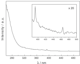
Figure 1 shows the PL excitation spectrum of the 10-layered ilm with emission wavelength at 612 nm. The spectrum presents a wide band below 260 nm, which
corresponds to a charge-transfer transition from O2− to
Eu3+ ions, as well as sharp lines of lower intensity, which
correspond to f–f transitions 7F
0 → 5L6 (393 nm) and
7F
0→5D2 (462 nm).11 The emission spectrum of the ilm,
obtained with excitation at 270 and 393 nm, displays bands
at 578, 590, 612, 650 and 698 nm assigned to the 5D
0→7FJ
(J = 0, 1, 2, 3, 4) transitions, with the hypersensitive
5D
0 → 7F2 red emission centered at 612 nm being the
most prominent (Figure 2). Due to the resolution (spectral bandwidth: 1 nm), the only possible consideration about the
emission spectrum is that the environment of the Eu3+ ion
leads to a low symmetry system, as indicated by the high
intensity of the 5D
0→7F2 electric dipole (ED) transition
as compared with the 5D
0 → 7F1 magnetic-dipole (MD)
transition.11
The chromaticity coordinates (CIE 1931) were
calculated using the Spectra Lux Software.16 The obtained
results demonstrated high color purities: x = 0.68 and
y = 0.32, which are eligible for optical applications.11,17,18
The spectra are similar to that obtained by Barros et al.,11
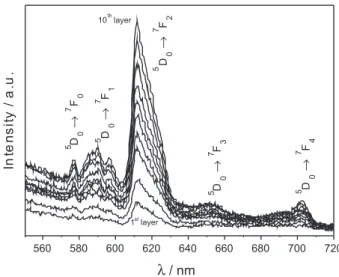
who also considered the europium-doped ZnAl2O4 as
having properties required for display applications. Figure 3 exhibits the emission spectra of the ilm after each layer transfer from the irst until the tenth, and this same figure shows that the luminescence intensity is approximately linear with respect to the number of layers. For comparison purposes, Figure 4 depicts the emission spectra of the powder (700 ºC) presenting the same features as the ilm spectrum. The bulk material obtained at 500 ºC remains dark due to carbonaceous impurities,
and it is not appropriate for optical measurements, especially with respect to transparency studies. The close similarity of the emission spectra of the ilm and bulk
samples with that presented by Barros et al.11 led us to
conclude that Eu3+ is embedded predominantly in a Gahnite
spinel structure.
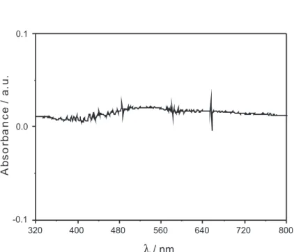
The UV-Vis spectrum of the ZnAl2O4:Eu3+ film
displays low absorbance from 320 nm to 800 nm (Figure S1, Supplementary Information), resulting in good transparency in a large spectral window. It was not possible to obtain the absorption spectrum of the bulk material obtained at 500 ºC. On the other hand, the spectrum of the powder obtained at 700 ºC (without carbonaceous material) presented low absorbance above 320 nm, which is very similar to what was observed in the case of the ilm spectra.
Figure 5 presents the X-ray diffraction patterns of the powder obtained at 500 and 700 ºC. The main peaks are
Figure 1. Excitation spectrum of the ZnAl2O4:Eu3+ ilm (λ
em = 612 nm).
Figure 2. Emission spectra of the ZnAl2O4:Eu3+ ilm: (a) λ
in agreement with the Gahnite spinel crystalline phase
(JCPDS 01-082-1043). Analysis of the X-ray pattern depicts the formation of the main crystalline planes
(220), (311), (511) and (440) at 2θ = 31, 37, 59 and 65,
respectively, and it is evident that (311) is the preferential
orientation, as previously reported by Barros et al.11 Some
less intense peaks due to ZnO are also present in the sample patterns, as described in Barros’ paper.
In order to estimate the thickness of the 10-layered ilm, the fractured cross-section of the substrate was imaged by SEM (Figure 6). A 300 nm thickness was observed, which led us to infer that each deposited layer has a thickness of approximately 30 nm. From this value, we can estimate that the size of the particles forming the ilm is less than 30 nm.
Recently, Kapilashrami et al.19 used the same technique
(cross-section) to determine the thickness of ZnO ilms
measuring between ca. 150 and 1000 nm.
Conclusions
We have successfully fabricated ZnAl2O4:Eu3+ ilms
with nanosized thickness by the dip-coating technique using a non-alkoxide sol-gel method. The films are transparent above 320 nm, and the close similarities between the emission spectra of the ilm and those of the
bulk samples (known to have a Gahnite spinel structure as
evidenced by XRD) have led us to conclude that it consists
mainly of the Gahnite structure, where Eu3+ is embedded.
Also, the determined CIE 1931 chromaticity coordinates
x ≅ 0.68 and y ≅ 0.32 make this very thin ilm a possible
component for photoelectronic devices transparent in the UV-Vis region.
Figure 3. Emission spectra (λexc = 393 nm) of the ZnAl2O4:Eu3+ ilm from one to ten layers.
Figure 4. Emission spectra of the ZnAl2O4:Eu3+ powder annealed at 500 ºC (a) and 700 ºC (b).
Figure 5. X-ray diffraction pattern of the ZnAl2O4:Eu3+ powder annealed at 500 and 700 ºC, together with the reference Gahnite ZnAl2O4 spinel (JCPDS 01-082-1043).
Thin Film of ZnAl2O4:Eu3+ Synthesized by a non-Alkoxide Precursor Sol-Gel Method J. Braz. Chem. Soc. 1398
Supplementary Information
The UV-Vis absorbance spectrum of the ZnAl2O4:Eu3+
film (Figure S1) is available free of charge at http://jbcs.sbq.org.br, as a PDF ile.
Acknowledgments
The authors are grateful to R. F. Silva, J. F. de Lima and C. M. P. Manso for helpful discussions, and to the Brazilian research funding agencies Coordenação de Aperfeiçoamento de Pessoal de Nível Superior (CAPES), Conselho Nacional de Desenvolvimento Científico e Tecnológico (CNPq) and Fundação de Amparo à Pesquisa do Estado de São Paulo (FAPESP) for inancial support and grants.
References
1. Sampath, S. K.; Cordor, J. F.; J. Am. Ceram. Soc.1998, 81, 649. 2. Khenata, R.; Sahnoun, M.; Baltachea, H.; Rérat, M.; Reshak,
Ali H., Al-Douri, Y.; Bouhafs, B.; Phys. Lett. A2005, 344, 271. 3. Mathur, S.; Veith, M.; Hass, M.; Shem, H.; Lecerf, N.; Huch,
V.; Huier, S.; Haberkorn, R.; Beck, H. P.; Jilavi, M.; J. Am. Ceram. Soc.2001, 84, 1921.
4. El-Nabarawy, T.; Attia, A. A.; Alaya, M. N.; Mater. Lett.1995, 24, 105.
5. Girgis, B. S.; Youssef, A. M.; Alaya, M. N.; Surf. Technol.1980, 10, 105.
6. Duan, X.; Yuan, D.; Wang, X.; XU, H.; J. Sol-Gel Sci. Technol. 2005, 35, 221.
7. Valenzuela, M. A.; Bosch, P.; Aguilar-Rios, G.; Montoya, A.; Schifter, I.; J. Sol-Gel Sci. Technol.1997, 8, 107.
8. Phani, A. R.; Passacantando, M.; Santucci, S.; Mater. Chem. Phys.2001, 68, 66.
9. da Silva, A. A.; Gonçalves, A. S.; Davolos, M. R.; J. Sol-Gel Sci. Technol.2009, 49, 101.
10. García-Hipólito, M.; Hernández-Pérez, C. D.; Alvarez-Fregoso, O.; Martinez, E.; Guzmán-Mendoza, J.; Falcony, C.; Opt. Mater. 2003, 22, 345.
11. Barros, B. S.; Melo, P. S.; Kiminami, R. H. G. A.; Costa, A. C. F. M.; de Sá, G. F.; Alves Jr., S.; J. Mater. Sci.2006, 41, 4744. 12. Strek, W.; Deren, P.; Bednarkiewicz, A.; Zawadzki, M.;
Wrzyszcz, J.; J. Alloys Compd.2000, 300, 456.
13. Yanga, C.-C.; Chena, S.-Y.; Cheng, S.-Y.; Powder Technol. 2004, 148, 3.
14. Singh,V.; Natarajan, V.; Zhu J-J.; Opt. Mater.2007,29, 1447. 15. Chea, P.; Meng, J.; Guoa, L.; J. Lumin.2007, 122, 168. 16. Santa-Cruz, P. A.; Teles, F. S.; Spectra Lux Software v.2.0, Ponto
Quântico Nanodispositivos/RENAMI (Brazil), 2003. 17. Sousa Filho, P. C.; Serra, O. A.; J. Lumin.2009, 129, 1664. 18. Wang, Z.; Liang, H.; Gong, M.; Su, Q.; J. Alloys Compd.2007,
432, 308.
19. Kapilashrami, M.; Xu, J.; Ström, V.; Rao, K.V.; Belova, L.; Appl. Phys. Lett. 2009, 95, 033104.
Received: November 16, 2009
Web Release Date: May 17, 2010
Supplementary Information
*e-mail: [email protected]
Thin Film of ZnAl
2O
4:Eu
3+Synthesized by a non-Alkoxide Precursor Sol-Gel Method
Renata F. Martins and Osvaldo A. Serra*
Laboratório de Terras Raras, Departamento de Química, FFCLRP, Universidade de São Paulo, Av. Bandeirantes 3900, 14040-901 Ribeirão Preto-SP, Brazil