From the Division of Gynecologic Oncology of the Departments of Gynecology and Obstetrics, and the Department of Radiology, Hospital das Clínicas, Faculty of Medicine, University of São Paulo - São Paulo/SP, Brazil.
Received for publication on July 03, 2002.
ORIGINAL ARTICLE
COLOR DOPPLER SONOGRAPHY WITH CONTRAST
IN THE DIFFERENTIATION OF OVARIAN TUMORS
Eduardo Cardoso Blanco, Ayrton Roberto Pastore, Angela Maggio da Fonseca, Filomena Marino Carvalho, Jesus Paula Carvalho and José Aristodemo Pinotti
BLANCO EC et al. - Color Doppler sonography with contrast in the differentiation of ovarian tumors. Rev. Hosp. Clín. Fac. Med. S. Paulo 58(4):185-192, 2003.
The objective of this study was to differentiate benign ovarian tumors from malignant ones before surgery using color and pulsed Doppler sonography, and to compare results obtained before and after use of contrast medium, thereby verifying whether contrast results in an improvement in the diagnostic sensitivity.
METHODS: Sixty two women (mean age 49.9 years) with ovarian tumors were studied, 45 with benign and 17 with malignant tumors. All women underwent a transvaginal color Doppler ultrasonographic exam. A study of the arterial vascular flow was made in all tumor areas, as well as an impedance evaluation of arterial vascular flow using the resistance index.
RESULT: Localization of the vessels in the tumor revealed a greater proportion of malignant tumors with detectable internal vascular flows (64%) than benign tumors with such flows (22%). There was a considerable overlap of these findings. The use of contrast identified a greater number of vessels with confirmation in the totality of tumors, but did not improve the Doppler capacity in tumoral differentiation. Malignant tumors presented lower values of resistance index than the benign ones, whether or not contrast was used. The cutoff value for resistance index that better maximized the Doppler sensitivity and specificity was 0.55. Through this value, an increase of the sensitivity after contrast use was obtained, varying from 47% to 82%, while specificity remained statistically unchanged.
CONCLUSION: Although the injection of a microbubble agent improved the sensitivity of the method detecting vascularization of tumors, a positive finding for vascularization by this method was not clinically useful in the differentiation of benign and malignant ovarian tumors.
DESCRIPTORS:Transvaginal ultrasound. Color Doppler sonography. Adnexal mass. Ovarian neoplasms. Contrast medium.
Considerable evidence indicates that neovascularization is responsible for the tumoral growth and invasion1.
Ultrasonography with color and pulsed Doppler provides dynamic information concerning the distribution of tumor vessels and their impedance value through the determination of resist-ance (RI) and pulsatility (PI) indices. Preliminary data suggests that it is possible to detect vascular changes as-sociated with angiogenesis in early ovarian cancer through this tech-nique2. A possible explanation for
these changes is that the new blood vessels present in the carcinomas have limited muscular tonus due to the ab-sence of the medial layer and that to-gether with numerous existing
arterio-venous shunts, they would be respon-sible for the low impedance registered by the RI and PI indices. However, this method presents some restrictions as-sociated with the inaccessibility to the small caliber vessels, thus demonstrat-ing low sensitivity for the intended purpose3.The advent of
echo-enhanc-ing substances for endovenous use, according to experiences in other or-gans4, would allow better visibility of
The objective of this study was to differentiate benign from malignant tumors prior to surgery by using color and pulsed Doppler and to compare the results before and after use of con-trast so that any improvements in re-sulting diagnostic sensitivity and specificity could be verified.
MATERIAL AND METHODS
This study comprised 62 women with ages varying from 13 to 84 years (average 49.9 years) with histologically confirmed ovarian tumors, 45 benign and 17 malignant ones. All patients underwent surgery, and the classification of histologic findings was made according to the World Health Organization criteria. The patients signed a consent form, as approved by the Research Ethics Com-mittee of our University.
Contrast medium
All patients received the ultra-sound contrast agent Levovist® 400
mg/mL (8 mL) intravenously via pe-ripheral vein. Levovist® (Schering AG,
Berlin FRG) is a suspension of espe-cially manufactured galactose contain-ing 99.9% galactose microparticles and a very low concentration of pal-mitic acid. Each vial contained 4 g of microparticles, and after suspension in water for injection was administered as a bolus via an 18-gauge plastic can-nula inserted into the arm vein (injec-tion speed: 1-2 mL/second continu-ously). This was followed by an addi-tional 10 mL of physiological saline solution to flush the cannula using the same injection rate. A comparison was made of the Doppler sonographic find-ings before and after injection of Levovist®. Although repetitive
injec-tions of the product were permitted (up to the maximum of 6 units) none of the cases required an additional injection.
Equipment and analysis of color Doppler signals
The ultrasonographic transvaginal exam with Doppler was performed with a commercially available scanner, GE LOGICTM 500 , version 2.0, with
ab-dominal and transvaginal transducers of 3.5 MHz and 5.0 MHz respectively. A Doppler filter of 50 Hz was used to eliminate low-frequency signals re-sulting from vessel wall motion. The color Doppler gain was increased un-til noise appeared and then was re-duced until the noise was totally sup-pressed (usually this was a 50% reduc-tion). Whenever the tumors exceeded 10 cm diameter, the study was com-pleted via transabdominal ultrasonog-raphy. In premenopausal patients, the examination was performed between the 3rd and 10th days of the menstrual
cycle in order to avoid the effects on the ovarian vascularization provoked by ovulation and the luteal body. The settings of the scanner controls were not altered after the injection of con-trast. Vascular arterial flows were stud-ied in all tumor areas, as was the im-pedance of vascular flows using RI de-termination. The vessels were classi-fied as internal or peripheral according to their location. When the flow was measured in more than one vessel, the vessel with the lowest RI was chosen for study.
The video output of the scanner was tape recorded starting at 5 seconds prior to injection and continuing un-til the intensity of the color Doppler signals was subjectively judged to re-turn to the base-line level. The number of vessels was evaluated within 10 mm of the tumors in the peripheral and central location.
The same examiner performed all ultrasonographic examinations.
The chi-square test was used to sta-tistically evaluate the quantitative variables. A canonical correlation was applied to the sets of variables
ob-served in both methods (with and with-out contrast) aiming at evaluating their relationship. The classification power regarding malignancy of the method used, with or without contrast, was analyzed with focus on the specificity and sensitivity of measure-ments that were based on Youden’s in-dex and the receiver operating charac-teristics (ROC). A p value below 0.05 was considered to be a statistically sig-nificant difference.
RESULTS
When determining the RI by Dop-pler technique without the use of con-trast, it was possible to reproduce ar-terial flow in only 45 cases, which were therefore used for the compara-tive analysis between the 2 examina-tion modalities, without and with con-trast (Table 1).
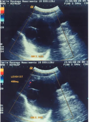
After injection of the contrast dur-ing the color Doppler exam, enhance-ment of vascular signals presented by the adnexal masses was found at a mean time of 25 seconds ±14 (SD), this effect remaining for a variable period of time of 185 seconds ±45 (SD). All patients presented an increase of Dop-pler sign intensity, with the appearance of new vessels in 77% of the tumors (Figs. 1, 2).
Regarding the RI, the measures of central tendency (mean) and of vari-ability (standard deviation) observed in the benign and malignant tumor groups are presented in table 2.
Statistically significant differences (p < 0.001) were observed between the average values for both RI1 and RI2 in the benign vs. malignant compari-sons. The average values of RI1 and RI2 were not significantly different in either the benign or the malignant tumor group comparisons (Table 2).
Figure 1 - Serous cystadenoma of the ovary. (a) Cystic formation of anechoic contents with presence of heterogeneous septa and lack of blood vessels at transvaginal color Doppler ultrasonography. (b) After administration of echographic contrast, very few vessels of peripheral location were identified.
Figure 2 - Mucinous cystadenoma of the ovary. (a) Cystic formation of irregular walls, predominant anechoic of contents with discreet echogenic points (debris) in its interior; presence of numerous heterogeneous and confluent septations. Color Doppler shows rare blood vessels of peripheral location. (b) After use of contrast, an increase of the vascular signal with the appearance of multiple vessels in the interior septa and in the tumor periphery was noted.
within a solid component or septation (64%) than did benign tumors with in-ternal flows (22%). A significantly greater number (c2 = 10.06, p = 0.0065) of benign compared to malignant tumors had peripheral flow without evi-dence of internal flow (Table 1). The use of contrast allowed the identification of a greater number of centrally located vessels in both groups of tumors, with this increase being more evident in be-nign tumors, in contrast with the results obtained without the use of contrast (Fig. 2).
Whether or not contrast was used, the malignant tumors had smaller RI values than the benign ones did (Ta-ble 2).
Table 1 - Histologic diagnosis and results of blood flow distribution in 62 ovarian tumors without use of contrast.
Pathologic Diagnosis Number Vessels
Absent Peripheral Central Benign
Cystadenoma 25 1 1 1 2 2
Endometrioma 3 3
Dermoid cyst 9 3 2 4
Thecoma 1 1
Fibroma 1 1
Cystadenofibroma 4 1 2 1
Others 2 1 1
Malignant
Cystadenocarcinoma 1 0 1 9
Borderline 5 2 2 1
Clear cell carcinoma 1 1
Table 4 - Specificity, sensibility and predictive positive (PPV), and negative (PNV) values of the classification based on vessel localization by the Doppler technique without and with contrast
Color Doppler Sensibility Specificity PPV PNV Youden
Without contrast 6 5 7 8 5 2 8 5 4 2
With contrast 8 8 4 7 3 8 9 1 3 5
Note: Values are shown as percentages.
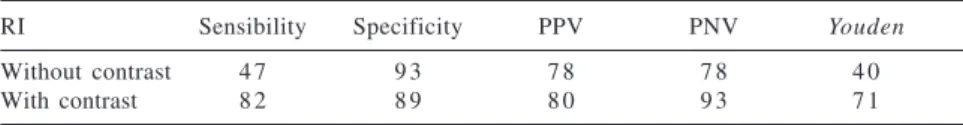
Table 3 - Sensibility, specificity, predictive positive (PPV), and negative (PNV) values of Doppler without and with contrast for RI = 0.55
RI Sensibility Specificity PPV PNV Youden
Without contrast 4 7 9 3 7 8 7 8 4 0
With contrast 8 2 8 9 8 0 9 3 7 1
Note: Values are shown as percentages.
Table 2 - Averages and confidence intervals for RI values obtained as a result of using the Doppler technique without contrast (RI1) and with contrast (RI2)
Method p Diagnosis Average Range
RI1 <0.001 Malignant 0.565 0.38 – 0.81
Benign 0.773 0.42 – 1
RI2 <0.001 Malignant 0.461 0.33 – 0.57
Benign 0.712 0.31 – 0.89
Note: The p value (descriptive level) is related to the Fisher’s exact test for comparison of averages.
The cutoff RI value that better maximized the Doppler sensitivity and specificity was 0.55, as demonstrated by the use of receiver operating char-acteristics (ROC) methodology.
It was noted that the Youden’s in-dex for all cutoff RI values was higher when the contrast was used. The com-parison of sensitivity values for both methods, when considering the 0.55 cutoff value, showed a significant dif-ference at the 8% level (the chi-square test was applied with the Yates’ correc-tion factor), thereby confirming the su-perior Doppler results when using con-trast, based on the Rl determination (Table 3).
When the internal flow was used as an indicator of malignancy, the sensi-tivity and specificity for internal flow versus either peripheral or no flow through color Doppler were 65% and 78%, respectively. After use of the con-trast, the sensitivity and specificity
were 88% and 47% respectively (Ta-ble 4).
As it can be seen, the sensitivity and specificity values of the color Doppler are low, independent of the use or not of the contrast for the clas-sification of the tumors. There is no evidence that the increase of sensitiv-ity observed with the use of contrast is significant (χ2=1.47, p = 0.2252,
with Yates’ correction).
DISCUSSION
The analysis of the epidemiologi-cal aspects of ovarian cancer has gen-erated a great deal of interest, espe-cially regarding the detection and characterization of the ovarian masses through radiological imaging. Since 1958, when for the first time Donald identified a voluminous pelvic-ab-dominal ovarian cyst
ultrasonogra-phically, this technique has been sys-tematically used in differentiating be-tween benign and malignant tumors5.
Although the transvaginal route is presently considered to produce re-sults that are sensitive for the detection of malignancy, the diagnostic confi-dence is not sufficient to obviate in-vasive procedures like laparoscopy and laparotomy. Thus, notwithstand-ing the efforts for improvnotwithstand-ing the diag-nostic accuracy, many studies have demonstrated that morphological evaluation of transvaginal ultrasound has found 80% to 90% sensitivity and from 65% to 95% specificity6-8.
Color and pulsed Doppler have been widely investigated and proposed as possible means to augment ultra-sonography in differentiating adnexal masses. Unfortunately, there is much controversy in the literature regarding the actual utility of the Doppler tech-niques for this application.
The presence and localization of the blood flow demonstrated through ultrasonography with color Doppler were initially used in an attempt to
characterize ovarian masses9-12.
Fleischer et al.10 found statistically
significant differences in the location of blood vessels in benign lesions, where they tend to be peripheral, com-pared to malignant lesions where they tend to be central. Our data also showed a significantly higher percent-age of malignant compared with be-nign tumors with internal flow. Simi-larly, the presence of a peripheral lo-cation of vessels or their absence is better related to benignity (PNV=85%). It is interesting to point out that 88% of the tumors in our study in which no blood flow was observed were benign (n = 15). Previous studies using Dop-pler technology also reported the rar-ity of identifying flows in predomi-nantly benign ovarian masses12-15.
malignant nature, do not present blood vessels at ultrasonographic evaluation with Doppler technology. The im-proper concept of “lack of blood flow” in benign tumors presented by some authors8,16-18 could be resulting from
technical limitations associated with Doppler ultrasound rather than from phenomena actually related to angio-genesis. Thus, care must be taken when considering whether the absence of vascularization is to be used as a cri-terion of assessment, since information revealed through Doppler ultrasonog-raphy is highly dependent on varia-tions in technology and could easily change as technology improves.
Currently, the use of some new tech-nological resources has been funda-mental for the increase in the detection of blood vessels, and consequently for the diagnostic accuracy of Doppler use in the differentiation of pelvic masses. The first resources that were of technological origin did not always have satisfactory results with the use
of amplitude Doppler (Angio®,
Power®, Energy®, Power-Angio, etc.). Subsequently, based on experiences with other organs19-26, results have been
obtained by applying echo-enhancer agents via intravenous administration. In this connection, the use of LevovistÒ permitted the identification of new blood vessels in tumors that had undetectable vascular flow ini-tially and in other tumors that already had revealed vascular flow of periph-eral distribution. In 12 cases of the lat-ter, the new localization (central) was chosen for diagnostic purposes. Al-though more vessels were detected, the use of contrast did not improve the method’s capacity for predicting ma-lignancy. Whether or not contrast is used, color Doppler showed a limited utility in the differentiation of ovarian tumors.
When determining RI, signifi-cantly lower values were registered in malignant tumors as compared with the benign ones, independent of the use or not of contrast (Table 2). Con-siderable overlap of RI values was ob-served between the 2 groups of tumors, thus confirming the findings in other publications27-33. The lower RI values
found in both groups of tumors after use of contrast would result from the detection of vascular signs of low am-plitude and obviously lower resistance that were not detected by the conven-tional Doppler technique.
In contrast with the first studies performed with pulsed Doppler in ovarian tumors16,34, where high
sensi-tivity (96%) and specificity (95%) in the identification of malignant tumors were obtained with RI <0.40 values, later works questioned the veracity of these findings. Thus, authors such as TEKAY & JOUPILLA14 obtained, for a RI cutoff value of 0.50, 46% sensitivity and false positive rates of 11% in the detection of malignancy. The results of SCHNEIDER et al.35, for RI <0.40 values were remarkably similar. Subsequent studies have also showed low rates of detection for ovarian cancer when us-ing different cutoff values36-39. The
false positive rates in these investiga-tions varied from 2% to 48%.
In the population covered by this study, the discriminatory power range of RI in the differentiation of tumors was between the 0.50 and 0.60 values, from which 0.55 was the one that best maximized the sensitivity and specificity simultaneously. For this cutoff value, the pulsed Doppler with-out and with contrast showed specificity rates statistically similar to 93% and 89%, respectively.
A variety of benign lesions might lead to an erroneous diagnosis, includ-ing teratomas34, endometriomas40, and
thecomas41. In these studies, the
pres-ence of these types of tumors was con-firmed following initial detection us-ing pulsed Doppler.
A significant increase in sensitiv-ity (from 47% to 82%) was observed after administration of the contrast agent, leading to a significant differ-ence at the level of 8% (chi-square test with Yates’ correcting factor). The pri-mary clinical usefulness of this tech-nique is associated with reliability of the method for predicting benignity (PNV for malignity = 93%). However, the problem still persists regarding the identification of malignity.
CONCLUSION
The use of echo-enhancing agents such as Levovist considerably
im-proved the performance of the Doppler ultrasonographic technique through the increase of the vascular signal, thereby permitting better identification of the blood vessels and analysis of the im-pedance standard of the blood flow originating from them. Although our data show that statistical statements can be made about benign and malignant tumors following the use of contrast agents, they are of little clinical use, es-pecially in the context of evaluating an individual patient, because of the low sensitivity that is still present.
ACKNOWLEDGMENTS
This study was supported by the
generous grants from the FAPESP
(“Fundação de Amparo à Pesquisa do Estado de São Paulo”).
RESUMO
pesquisa de fluxo vascular foi realiza-do em todas as áreas tumorais, assim como a avaliação da impedância atra-vés do índice de resistência.
RESULTADOS: A localização dos vasos no tumor revelaram uma maior proporção de fluxo vascular interno detectável nos tumores malignos (64%) do que nos tumores benignos (22%). Houve entretanto uma conside-rável sobreposição destes achados. O uso de constrate foi capaz de identifi-car um grande número de vasos na to-talidade dos tumores, mas não melho-rou a capacidade do Doppler na dife-renciação entre tumores benignos e malignos. Os tumores malignos apre-sentaram valores mais baixos de índi-ce de resistência.IR do que os benig-nos, independentemente do uso de BLANCO EC e col. - Ultra-sonografia
com Doppler colorido e uso de contraste na diferenciação dos
tumores ovarianos. Rev. Hosp.
Clín. Fac. Med. S. Paulo 58(4): 185-192, 2003.
O objetivo deste estudo foi dife-renciar tumores ovarianos benignos e malignos antes da cirurgia através da ultra-sonografia com uso de Doppler colorido pulsátil e comparar os resul-tados obtidos antes e após o uso de contraste.
MÉTODO: Foram estudadas ses-senta e duas mulheres (idade média de 49,9 anos) com tumores ovarianos, sen-do 45 benigos e 17 malignos. Todas foram submetidas a ultra-sonografia transvaginal com Doppler colorido. A
contraste. O valor de corte do índice de resistência. que maximizou a sen-sibilidade e a especificidade do Doppler foi de 0,55. Com este valor foi obtido um aumento na sensibilidade após o uso de contraste, variando de 47% a 82 %, enquanto a especifici-dade se manteve equivalente.
CONCLUSÃO: Embora a injeção de agentes produtores de microbolhas aumentem a sensibilidade do método na diferenciação da natureza do tumor, este valor não se mostrou clinicamen-te util na avaliação de tumores ovari-anos.
DESCRITORES: Ultra-sonografia
transvaginal. Doppler sonografia. Massas anexiais. Neoplasia ovaria-na. Meios de contraste.
REFERENCES
1 . FOLKMAN J, WATSON KJ, INGBER D et al. - Induction of angiogenesis during transition from hyperplasia to neoplasia. Nature 1989; 339; 58-61.
2 . HATA K, HATA T - Intratumoral blood flow analysis in ovarian cancer. J Ultrasound Med 1996; 15: 571-5.
3 . LEVINE D- Color Doppler imaging for the detection of ovarian malignancy is unreliable (letter). Ultrasound Obstet Gynecol 1995; 6: 451-4.
4 . ERNST H, HAHN EG, BALZER T et al. - Color Doppler ultrasound of liver lesions: signal enhancement after intravenous injection of the ultrasound contrast agent Levovist. J Clin Ultrasound 1996; 24: 31.
5 . BONILLA-MUSOLES F - Posibilidades de diagnóstico precoz en el cáncer de ovario. In: BONILLA-MUSOLES F. Tratado de endosonografia en obstetricia y ginecologia. 2nd ed.
6 . BENACERRAF BR, FINKLER NJ, WOJCIECHOWSKI C et al. KNAPP RC - Sonographic accuracy in the diagnosis of ovarian masses. J Reprod Med 1990; 35: 491-5.
7 . SASSONE AM, TIMOR-TRITSCH IE, ARTNER A et al. Transvaginal sonographic characterization of ovarian disease: evaluation of a new scoring system to predict ovarian malignancy. Obstet Gynecol 1991; 78: 70-6.
8 . SENGOKU K, SAITOH T, SITOH S et al. - Evaluation of transvaginal color Doppler sonography, transvaginal sonography and CA 125 for prediction of ovarian malignancy. Int J Obstet Gynecol 1994; 46: 39-43.
9 . KAWAI M, KANO T, KIKKAWA F et al. - Transvaginal Doppler ultrasound with color flow imaging in the diagnosis of ovarian cancer. Obstet Gynecol 1992; 79: 163-7.
10. FLEISCHER AC, RODGERS WH, KEPPLE DM et al. - Color Doppler sonography of ovarian masses: a multiparameter analysis. J Ultrasound Med 1993; 12: 41-8.
11. KURJAK A, PREDANIC M, KUPESICUREK S et al. -Transvaginal color and pulsed Doppler assessment of adnexal tumor vascularity. Gynecol Oncol 1993; 50: 3-9.
12. CARTER J, LAU M, FOWLER J et al. - Blood flow characteristics of ovarian tumors: implications for ovarian cancer screening. Am J Obstet Gynecol 1995; 172: 901-7.
13. KURJAK A, SCHULMAN H, SOCIC A et al. - Transvaginal ultrasound, color flow, and Doppler waveform of the postmenopausal adnexal mass.Obstet Gynecol 1992; 80: 917-21.
14. TEKAY A, JOUPPILA P - Validity of pulsatility and resistance indices in classification of adnexal tumors with transvaginal color Doppler ultrasound. Ultrasound Obstet Gynecol 1992; 2: 338-44.
15. TIMORTRITSCH IE, LERNER JP, MONTEAGUDO A et al. -Transvaginal ultrasonographic characterization of ovarian masses by means of color flow-directed Doppler measurements and a morphologic scoring system. Am J Obstet Gynecol 1993; 168: 909-13.
16. KURJAK A, ZALUD I, JURCOVIC D et al. - Transvaginal color Doppler for the assessment of pelvic circulation. Acta Obstet Gynecol Scand 1989; 68: 131-5.
17. KURJAK A, JURCOVIC D, ALFIVERIC Z et al. - Transvaginal color Doppler imaging. J Clin Ultrasound 1990; 18: 227-34. 18. KURJAK A, ZALUD I - Transvaginal colour flow imaging and
ovarian cancer. Br Med J 1990; 300: 330-5.
19. BURNS PN, LIU JB, HILPERT P - Intravenous US contrast agent for tumor diagnosis: quantitative studies. Radiology 1990; 177: 140.
20. FRITZSCH T, HAUFF P, HELDMANN F et al. - Preliminary results with a new liver specific ultrasound contrast agent. Ultrasound Med Biol 1994; 20: 137.
21. LLICETO S, CAIATI C, ARAGONA P et al. -Improved Doppler signal intensity in coronary arteries after intravenous peripheral injection of a lung-crossing contrast agent (Levovist). J Am Coll Cardiol 1994; 23: 184-190.
22. COSGROVE D, MCCREADY VR, BAMBER JC et al. - Microbubble contrast agent for color Doppler effect on breast masses. Work in progress. Radiology 1996; 198: 679-86.
23. RIES F - Clinical experience with echo-enhanced transcranial Doppler and duplex imaging. J Neuroimaging 1997; 7: 15-21. 24. BOGERS HA, SEDELAAR JP, BEERLAGE HP et al. -
Contrast-enhanced three-dimensional power Doppler angiography of the human prostate: correlation with biopsy outcome. Urology 1999; 54: 97-104.
25. HOSTEN N, PULS R, LEMKE AJ et al. - Contrast-enhanced power Doppler sonography: improved detection of characteristic flow patterns in focal liver lesions. Rofo Fortschr Geb Rontgenstr Neuen Bildgeb Verfahr 1999; 27: 107-15.
26. HARVEY CJ, BLOMLEY MJ, ECKERSLEY RJ et al. - Pulse-inversion mode imaging of liver specific microbubbles: improved detection of subcentimetre metastases. Lancet 2000; 355: 807-8. 27. FLEISCHER AC, RODGERS WH, RAO BK et al. - Assessment of
ovarian tumor vascularity with transvaginal color Doppler sonography. JUltrasound Med1991; 10: 563-8.
28. HATA K, HATA T, MANABE A, SUGIMURA K et al. - A critical evaluation of transvaginal Doppler studies, transvaginal sonography, magnetic resonance imaging and CA 125 in detecting ovarian cancer. Obstet Gynecol 1992; 80: 922-6. 29. HAMPER UM, SHETH S, ABBAS FM et al. - Transvaginal color
Doppler sonography of adnexal masses: differences in blood flow impedance in benign and malignant lesions. Am J Radiol 1993; 160: 1225-8.
30. PRÖMPELER HJ, MADJAR H, SAUERBREI W et al. - Quantitative flow measurements for classification of ovarian tumors by transvaginal color Doppler sonography in postmenopausal patients. Ultrasound Obstet Gynecol 1994; 4: 406-13. 31. WU CC, LEE CN, CHEN TM et al. -Incremental angiogenesis
assessed by color Doppler ultrasound in the tumorigenesis of ovarian neoplasms. Cancer 1994; 73: 1251-6.
32. BROWN DL, FRATES MC, LAING FC et al. - Ovarian masses: can benign and malignant lesions be differentiated with color and pulsed Doppler US? Radiology 1994; 190: 333-6.
33. BLANCO EC - O valor da Dopplervelocimetria com mapa de cores no estudo dos tumores ovarianos [Dissertação]. São Paulo, 1996. (Faculdade de Medicina da Universidade de São Paulo). 34. BOURNE T, CAMPBELL S, STEER C et al. - Transvaginal color flow imaging: a possible new screening technique for ovarian cancer. Br Med J 1989; 299: 1367-70.
35. SCHNEIDER VL, SCHNEIDER A, REED KL et al. - Comparison of Doppler with two-dimensional sonography and CA 125 for prediction of malignancy of pelvic masses. Obstet Gynecol 1993; 81: 983-8.
36. WU CC, LEE CN, CHEN TM, LAI JI et al. - Factors contributing to the accuracy in diagnosing ovarian malignancy by color Doppler ultrasound. Obstet Gynecol 1994b; 84: 605-8. 37. BROMLEY B, GOODMAN H, BENACERRAF BR - Comparison
38. LEVINE D, FELDSTEIN V, BABCOOK C et al. - Sonography of ovarian masses: poor sensitivity of resistive index for identifying malignant lesion. Am J Roentgenol 1994; 162: 1355-9.
39. BLANCO EC, CARVALHO JP, CARVALHO FM et al. - Valor del Doppler colorido transvaginal en la diferenciación de los tumores ovarianos benignos de malignos. Rev Imag2000; 4: 37-9.
40. KIRCHLER H, SCHWEGEL P, ENTNER C - Physiologische durchblutung des ovars: endosonographische farb Doppler-blutflussanalyse. Gynakol Geburtshilfe Rundsch 1992; 1: 90-100.