Key words:
Prostate cancer; Prostate; Gleason score; Diagnosis
Int Braz J Urol. 2012; 38: 760-8
__________________ Submitted for publication: February 14, 2012
__________________ Accepted after revision: August 14, 2012
Introduction: The widespread screening programs prompted a decrease in prostate can-cer stage at diagnosis, and active surveillance is an option for patients who may harbor clinically insignifi cant prostate cancer (IPC). Pathologists include the possibility of an IPC in their reports based on the Gleason score and tumor volume. This study determi-ned the accuracy of pathological data in the identifi cation of IPC in radical prostatec-tomy (RP) specimens.
Materials and Methods: Of 592 radical prostatectomy specimens examined in our la-boratory from 2001 to 2010, 20 patients harbored IPC and exhibited biopsy fi ndings suggestive of IPC. These biopsy features served as the criteria to defi ne patients with potentially insignifi cant tumor in this population. The results of the prostate biopsies and surgical specimens of the 592 patients were compared.
Results: The twenty patients who had IPC in both biopsy and RP were considered real positive cases. All patients were divided into groups based on their diagnoses following RP: true positives (n = 20), false positives (n = 149), true negatives (n = 421), false ne-gatives (n = 2). The accuracy of the pathological data alone for the prediction of IPC was 91.4%, the sensitivity was 91% and the specifi city was 74%.
Conclusion: The identifi cation of IPC using pathological data exclusively is accurate, and pathologists should suggest this in their reports to aid surgeons, urologists and radiotherapists to decide the best treatment for their patients.
INTRODUCTION
The detection of patients with nonpalpable prostate cancer has increased since the advent of prostate-specifi c antigen (PSA) screening. Modi-fi ed prostatic biopsy schemes have also contribu-ted to higher detection rates (1). Many of these earlier, smaller cancers are low volume (< 0.5 cm3),
low grade and clinically insignifi cant tumors. Ap-proximately one third of patients with stage T1c
cancer have potentially clinically insignifi cant tumors, and approximately 5% of radical prosta-tectomy (RP) patients have small cancers that are diffi cult to identify histologically (2).
Epstein et al. initially created a set of four criteria to predict insignifi cant prostate cancer prior to defi nitive therapy: PSA density 0.1-0.15, low or intermediate cancer grade, core involve-ment of less than 3 mm, and involveinvolve-ment of only one needle biopsy core. These criteria identifi ed
The accuracy of pathological data for the prediction of
insignifi cant prostate cancer
___
_______________________________________________
____________________________________________
Betina Katz, Miguel Srougi, Luiz H. Camara-Lopes, Alberto A. Antunes, Luciano Nesrallah, Adriano
Nesrallah, Marcos Dall’Oglio, Katia R. M. Leite
Laboratory of Medical Research - LIM55, Department of Urology, University of São Paulo Medical School (BK, MS, AAA, LN, AN, MD, KRML) and Laboratory of Surgical and Molecular Pathology - Sirio Libanes Hospital (BK, CL, KRML), São Paulo, Brazil
ABSTRACT
ARTICLE
INFO
761
IBJU |PATHOLOGICAL DATA FOR THE PREDICTION OF INSIGNIFICANT PROSTATE CANCER
79% of tumors with a volume < 0.5 cm3 that were
organ confi ned and did not qualify as high-grade lesions at the time of RP (3).
However, the Epstein criteria, which were later updated (4), are insuffi cient, and 20% of pa-tients who fulfi ll these criteria may have unfavo-rable pathological cancer characteristics at RP (5). The validity of these criteria has been questioned. Jeldres et al. demonstrated that the Epstein criteria may not be applicable to European men becau-se prostate cancer was underestimated in 24% of these patients (1).
The aim of this study was to assess the prediction of insignifi cant prostate cancer based on the biopsy fi ndings of patients who underwent RP and exhibited insignifi cant cancer. The biopsy features from these subjects were evaluated, and patients whose biopsies had similar parameters were selected. The biopsies were correlated with the respective RP specimens to identify the lesion characteristics and the clinical signifi cance of the tumor.
MATERIALS AND METHODS
A total of 592 patients underwent trans-rectal ultrasound-guided (TRUS) prostate biopsy followed by radical prostatectomy for prostate cancer from January 2001 to December 2010. A single surgeon (MS) treated all patients and a sin-gle uropathologist (KRML) examined all biopsies and surgical specimens. The surgical specimens were fi xed in 10% buffered formalin, the entire surgical margin was stained with India ink, the left and right lobes were separated, 3 mm trans-verse serial sections were taken from each lobe, and the entire gland was submitted for histolo-gic examination. Sections of the bladder neck, prostatic apex, seminal vesicles, and pelvic lym-ph nodes were also submitted to exam. The Gle-ason score (GS) was used for histologic grading (6). The tumor volume was evaluated as described by Humphrey et al. (7). Briefl y, a grid was placed below the slides, on which the area involved by the tumor had been previously sketched out. The percentage of tumor on a slide was determined by dividing the number of squares involved by tumor by the number of squares occupied by the whole
section on the slide. Tumor volume was defi ned as the mean percentage of tumor in the prostate gland (the percentage of tumor on each slide di-vided by the number of slides from the prostate gland). Extraprostatic involvement was defi ned as tumor infi ltration of the adipose tissue, the neuro-vascular plexus, or the parenchyma of the seminal vesicles. The TNM 2010 system was used for tumor staging and patients were classifi ed as pT2 when tumor was confi ned to the organ and pT3 when EPE or seminal vesicles were infi ltrated by tumor. PM was considered when tumor glands were inked with India ink.
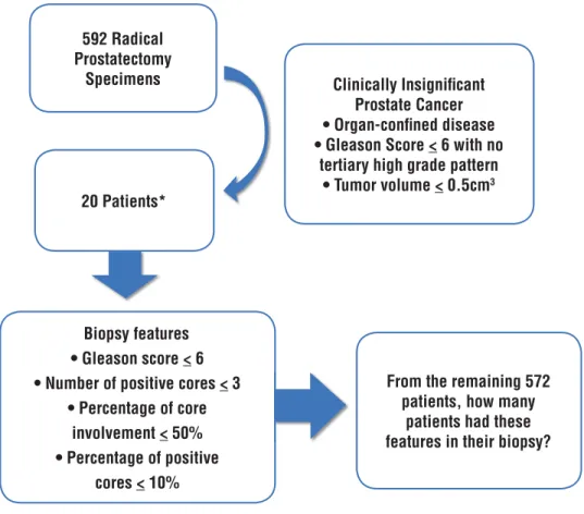
Twenty-two (3.7%) of the 592 patients exhibited insignifi cant tumor in their RP, which was defi ned as an organ-confi ned adenocarcino-ma, Gleason score (GS) < 6 with no tertiary high grade Gleason pattern, and a tumor volume 0.5 cm3 (Figure-1). The biopsies from these patients
were analyzed, and twenty cases (91%) presented the following features: adenocarcinoma (GS) < 6, one to three positive cores that consisted of less than 50% tumor and a total percentage of positive fragments < 10%. These features, which are simi-lar to the Epstein criteria regarding pathological fi ndings and are referred during the paper as “our criteria”, served as the parameters for the selection of patients with potentially insignifi cant prostate cancer. In order to evaluate the importance of pa-thological fi ndings in defi ning these tumors, our criteria were purposely created based solely on the biopsy features and did not include PSA or any imaging method. These criteria were used to re-view the biopsy and radical prostatectomy results and defi ne the sensitivity, specifi city, and positive and negative predictive value of these parameters for the identifi cation of insignifi cant prostate car-cinoma in our population.
RESULTS
our criteria mentioned above, exhibited a mean GS of 5.9 and mean positive cores of 6%. This group was considered real positive (Table-1). The remaining 2 patients (9%) had GS of 7 in the biop-sy, 3 cores positive for tumor, a higher percentage of a single core (40% and 70%) and 4% and 6% positive cores (in 13 and 14 cores, respectively). We consider this group as false negative, since we would not expect them to have insignifi cant tu-mor in the RP. The RPs of additional patients who exhibited similar prostate biopsy characteristics of the real positive group were re-examined. A total of 149 patients (26%) had biopsies that met our criteria and were characterized as probable IPC.
The mean GS was also 5.9, the mean percentage of tumor in a single core was 23.3%, and the mean percentage of cores that were positive for tumor was 5.4% (range 1.5 to 10%), which represented one to two positive cores in a mean of 15 biop-sies. However, their RPs revealed clinically sig-nifi cant carcinomas, including patients with pT3 disease (4.7%) as well as patients with interme-diate and high grade tumor (56 patients with GS 7 and 16 with GS > 7). This group was considered false positive, which means they could have been considered as having IPC based on their biopsy features, despite having tumor with adverse pa-thological characteristics. The real negative group * 22 patients had clinically insignifi cant prostate cancer; however, 2 patients had unfavorable features in their biopsy.
Figure 1 - Selection of patients who had clinically insignifi cant prostate cancer in radical prostatectomy as well as features of insignifi cant tumor in biopsy.
592 Radical Prostatectomy
Specimens
20 Patients*
Clinically Insignifi cant Prostate Cancer • Organ-confi ned disease • Gleason Score < 6 with no
tertiary high grade pattern • Tumor volume < 0.5cm3
From the remaining 572 patients, how many
patients had these features in their biopsy? Biopsy features
• Gleason score < 6 • Number of positive cores < 3
• Percentage of core involvement < 50% • Percentage of positive
763
IBJU |PATHOLOGICAL DATA FOR THE PREDICTION OF INSIGNIFICANT PROSTATE CANCER
Table 1 - Comparison between biopsy and RP of the patients regarding clinical signifi cance of the tumor.
Number of patients (%) Biopsy Radical Prostatectomy 1
20 (3.4%) insignifi cant insignifi cant Real Positive
149 (25.2%) insignifi cant signifi cant False Positive
2 (0.34%) signifi cant insignifi cant False Negative
421 (71.11%) signifi cant signifi cant Real Negative
1 Gold Standard
was composed of 421 men whose biopsy and RP revealed signifi cant prostate cancer. All patient data are detailed in Table-2.
The sensitivity of our criteria for the iden-tifi cation of clinically insignifi cant prostate cancer in RP was 91% with 74% specifi city. The positive predictive value was only 12%, and the negative predictive value was 99.5%. Therefore, if our criteria were used to predict signifi cant cancer, the probabi-lity of a patient exhibiting signifi cant cancer would be almost certain at level of accuracy of 91.4%.
DISCUSSION
Several studies have questioned the effi cacy of diagnosing limited cancer by needle biopsy, and the possibility of predicting tumor extent at RP ba-sed on biopsies. The applicability and validity of the criteria and nomograms that are commonly used to predict insignifi cant prostate cancer have also been discussed. The current study designed a set of novel criteria that were based on our own data and restric-ted to morphological aspects without the conside-ration of clinical stage or tumor markers. Although clinical fi ndings, such as PSA level and PSA density, were not used, the features of the biopsies from pa-tients with insignifi cant tumors at RP were similar to the biopsy criteria in the literature, such as a GS
< 6 and limited tumor extent on biopsy. The current study does not propose criteria or models for the prediction of insignifi cant tumor. Conversely, this study clarifi ed the use of precise morphological fi n-dings of prostate biopsy in the identifi cation of in-signifi cant prostate cancer.
Our data for the prediction of organ-con-fi ned tumors are consistent with the literature. Bastian et al. updated the Epstein criteria, which are the most widely used criteria for the prediction of clinically insignifi cant prostate cancer, and de-monstrated concordance with pathologically or-gan-confi ned disease and a favorable grade (GS 6) in 83.9% of patients (4). Although 91.6% of these patients had organ-confi ned disease, 7.6% had a GS of 7 or higher.
The validity of the Epstein criteria has been questioned in European men. In a study that evaluated 366 patients who fulfi lled the contem-porary Epstein criteria demonstrated a similar rate of organ-confi ned disease (91.7%). But the percent of patients with a GS of 7 was substantially hi-gher; 24% of patients had a GS of 7 at RP, which yielded lower overall accuracy (76% vs. 84%) (1). Unfortunately, these authors did not mention the number of patients with clinically insignifi cant tumor at RP. According to our criteria, 95.3% of the tumors were organ-confi ned, but 48.3% were GS 7 (37.6% patients were GS 7 and 10.7% were GS 8 or 9), which is substantially higher than the reported rate in previous studies.
IBJU
|
P
A
THOLOGICAL D
A
T
A FOR THE PREDICTION OF INSIGNIFICANT PROST
A
TE CANCER
764
Insignifi cant biopsy and insignifi cant RP (N = 20)
Biopsy Radical Prostatectomy
Age #cores GS #positive
cores
%cores Greatest% PNI% GS Tumor volume (cm3) pT2(%)
Mean 60.4 16.6 5.9 1.3 6 20.5 0 5.9 0.43 100
Median 59.5 16.5 6 1 6 20 6 0.40
Min 39 7 5 1 3 10 5 0.40
Max 74 24 6 3 10 50 6 0.50
Insignifi cant biopsy and signifi cant RP (N = 149)
Age #cores GS #positive
cores
%cores Greatest% PNI% GS Tumor volume (cm3) pT2(%)
Mean 59.5 15 5.9 1.6 5.4 23 0 6.6 3.10 95.3
Median 60 14 6 1 5.1 20 6 2.70
Min 39 6 4 1 1.5 5 5 0.40
765
IBJU
|
P
A
THOLOGICAL D
A
T
A FOR THE PREDICTION OF INSIGNIFICANT PROST
A
TE CANCER
Signifi cant biopsy and insignifi cant RP (N = 2)
Age #cores GS #positive
cores
%cores Greatest% PNI% GS Tumor volume (cm3) pT2(%)
Mean 57 13.5 7 3 5.4 55 0 6 0.4 100
Signifi cant biopsy and signifi cant RP (N = 421)
Age #cores GS #positive
cores
%cores Greatest% PNI GS Tumor volume (cm3) pT2(%)
Mean 61.3 14.3 7.1 5.1 8.5 66.3 22.6% 7.2 6.08 76.7
Median 61 14 7 4 7.5 70 7 5.00
Min 41 6 5 1 1.2 2 4 0.33
even when the tumor is organ confi ned, and it is the most informative predictor of biochemical recurrence (11).
Epstein et al. created their criteria to pre-dict insignifi cant tumor in patients with stage T1c, and it accurately predicted 73% of insignifi cant tumor (3). The current study predicted only 12% of the clinically insignifi cant tumors, but our sam-ples were not limited to stage T1c patients. Despi-te this low positive predictive value, the negative predictive value was 99%, which indicated that ninety-nine percent of the tumors that were iden-tifi ed as signifi cant and required active therapy were indeed signifi cant tumors. This result addres-ses the concern of overdiagnosis, which occurs at a rate of approximately 56% (12,13).
The retrospective assessment of biopsies from patients with insignifi cant or signifi cant tu-mors at RP revealed that all of the parameters that are usually used to estimate tumor extent, such as the number of positive cores, maximal involve-ment of a single core and the percentage of posi-tive cores, were similar in both groups. These re-sults suggest an absence of rules for insignifi cant tumor behavior on needle biopsy.
Numerous diffi culties exist in the use of prostatic needle biopsies to predict limited can-cer at RP, and even the smallest cancan-cer focus on a needle biopsy does not guarantee a clinically insignifi cant tumor. Small amounts of carcinoma (total linear extent less than 3 mm) do not pre-dict insignifi cant tumors (14,15). Patients who are diagnosed based on a single focus less than 3 mm (GS 6) have only a 30% chance of harboring an insignifi cant tumor (16). Samaratunga et al. exa-mined 58 patients with a single minute focus of a GS 6 on biopsy, and only 10 patients (17%) had insignifi cant tumor at RP (17). Forty-eight patients had signifi cant tumor, 8 patients had extraprosta-tic extension and 32% of the patients had a GS > 6. These authors concluded that a minute focus of prostate cancer on a needle biopsy is not indi-cative of insignifi cant carcinoma in most cases. Interestingly, this study was performed without PSA screening. However, these authors demons-trated that a larger prostate size was signifi cantly correlated with potentially insignifi cant cancer. These patients likely presented earlier for PSA
tes-ting because of symptoms, and an elevated PSA level due to the benign enlargement might lead to biopsies at an earlier stage. Cupp et al. demonstra-ted only an 8% risk of insignifi cant cancer using tumor volume at biopsy (14), which is similar to our results. The percentage of tumor extension in millimeters relative to the total extension of all of the cores was 5% and a GS less than 7.
In contrast, Allan et al. evaluated 54 PSA--screened patients with limited adenocarcinoma (< 0.5 mm) on biopsy; the majority of these pa-tients exhibited potentially insignifi cant cancer, but only one-third warranted defi nitive thera-py (2). Potentially clinically insignifi cant tumors were present in 67% of the patients in this study, and 44% of these patients had small tumors at RP (less than 0.1 cc). These authors also repor-ted that a PSA density cutoff of 0.15 or less was correlated with clinically insignifi cant tumor, and the association of these criteria with limited can-cer on biopsy predicted patients with insignifi cant tumors with a greater than 80% accuracy.
The Epstein criteria are not perfectly ac-curate, but no alternatives for prediction of cli-nically insignifi cant prostate cancer are available (1). Kattan et al. derived several nomograms for the prediction of pathologically confi rmed insig-nifi cant prostate cancer with an accuracy of 64 to 79% (18). However, this series included 13 to 20% of Gleason patterns of 2 as part of the GS, which is much lower than the GS consensus from 2005. Nakanishi et al. improved the accuracy of the existing tools to 73%, especially in patients with a single positive core at biopsy (19).
767
IBJU |PATHOLOGICAL DATA FOR THE PREDICTION OF INSIGNIFICANT PROSTATE CANCER
or a 76 to 79% accuracy when pathologically con-fi rmed insignicon-fi cant prostate cancer is predicted.
Do these data suggest that a signifi cant number of patients might be left undertreated? Is active surveillance a dangerous choice that could jeopardize the curability of prostate cancer in some men? The answer to these questions appears to be no.
A review of active surveillance revealed that the majority of patients stay on active sur-veillance, and once a patient requires active treat-ment, that patient presents with curable prostate cancer (12). The detection of prostate cancer pro-gression in a patient who is selected for active sur-veillance remains a continuing challenge, and the PSA level remains important during the decision process. The progression of Gleason grade and the increased percentage of cancers per core are also indicators for the cessation of active surveillance.
Duffi eld et al. have also studied the RP fi ndings of patients in whom active surveillance has failed (13). These authors relied solely on sub-sequent biopsy pathology to determine progres-sion, and more extensive disease was observed in surveillance biopsies during the fi rst 2 years of follow-up in the majority of cases.
Despite their high accuracy rates, curren-tly available models for the prediction of insigni-fi cant prostate cancer are incorrect in 10 to 20% of cases. The addition of novel markers is required, and current imaging techniques, such as multipa-rametric magnetic resonance, may have a poten-tial role.
Tumor location is problematic for sampling adequately. Anterior tumors are diffi cult to assess and sample clinically, and the amount of these tu-mors in biopsies is lower than the amount of tumor from equivalently sized posterior tumors (20,21). These tumors appeared smaller on biopsy, but they were also undersampled. Takashima et al. analyzed the anatomical patterns of disease distribution in nonpalpable tumors and demonstrated that these tumors were localized predominantly in the ante-rior half of the prostate at the apical to midprostate level (21). These authors suggested that additional cores from the anterior apical site could enhance the detection rate of prostate cancer. Miyake et al. evaluated the signifi cance of additional cores in the
dorsal apex and demonstrated a signifi cant incre-ase (9.3%) in the cancer detection rate, particularly for early stage disease (22).
In conclusion, we have shown that the biopsy data exclusively are accurate to diagnose IPC, and pathologist should suggest this possibili-ty in their reports helping urologists, oncologists and radiotherapists to choose the better treatment for each patient.
CONFLICT OF INTEREST
None declared.
REFERENCES
1. Jeldres C, Suardi N, Walz J, Hutterer GC, Ahyai S, Lattouf JB, et al.: Validation of the contemporary epstein criteria for insignifi cant prostate cancer in European men. Eur Urol. 2008; 54: 1306-13.
2. Allan RW, Sanderson H, Epstein JI: Correlation of minute (0.5 MM or less) focus of prostate adenocarcinoma on needle biopsy with radical prostatectomy specimen: role of prostate specifi c antigen density. J Urol. 2003; 170: 370-2. 3. Epstein JI, Walsh PC, Carmichael M, Brendler CB: Patho-logic and clinical fi ndings to predict tumor extent of non-palpable (stage T1c) prostate cancer. JAMA. 1994; 271: 368-74.
4. Bastian PJ, Mangold LA, Epstein JI, Partin AW: Character-istics of insignifi cant clinical T1c prostate tumors. A con-temporary analysis. Cancer. 2004; 101: 2001-5.
5. Chun FK, Haese A, Ahyai SA, Walz J, Suardi N, Capitanio U, et al.: Critical assessment of tools to predict clinically insignifi cant prostate cancer at radical prostatectomy in contemporary men. Cancer. 2008; 113: 701-9.
6. Epstein JI, Allsbrook WC Jr, Amin MB, Egevad LL; ISUP Grading Committee. The 2005 International Society of Uro-logical Pathology (ISUP) Consensus Conference on Glea-son Grading of Prostatic Carcinoma. Am J Surg Pathol. 2005; 29: 1228-42.
7. Humphrey PA, Vollmer RT: Intraglandular tumor extent and prognosis in prostatic carcinoma: application of a grid method to prostatectomy specimens. Hum Pathol. 1990; 21: 799-804.
9. Moreira Leite KR, Camara-Lopes LH, Dall’Oglio MF, Cury J, Antunes AA, Sañudo A, Srougi M: Upgrading the Gleason score in extended prostate biopsy: implications for treat-ment choice. Int J Radiat Oncol Biol Phys. 2009; 73: 353-6. 10. Stamey TA, McNeal JE, Yemoto CM, Sigal BM, Johnstone
IM: Biological determinants of cancer progression in men with prostate cancer. JAMA. 1999; 281: 1395-400. 11. Chun FK, Briganti A, Jeldres C, Gallina A, Erbersdobler A,
Schlomm T, et al.: Tumour volume and high grade tumour volume are the best predictors of pathologic stage and biochemical recurrence after radical prostatectomy. Eur J Cancer. 2007; 43: 536-43.
12. Bastian PJ, Carter BH, Bjartell A, Seitz M, Stanislaus P, Montorsi F, et al.: Insignifi cant prostate cancer and active surveillance: from defi nition to clinical implications. Eur Urol. 2009; 55: 1321-30.
13. Duffi eld AS, Lee TK, Miyamoto H, Carter HB, Epstein JI: Radi-cal prostatectomy fi ndings in patients in whom active surveil-lance of prostate cancer fails. J Urol. 2009; 182: 2274-8. 14. Cupp MR, Bostwick DG, Myers RP, Oesterling JE: The
volume of prostate cancer in the biopsy specimen cannot reliably predict the quantity of cancer in the radical pros-tatectomy specimen on an individual basis. J Urol. 1995; 153: 1543-8.
15. Bruce RG, Rankin WR, Cibull ML, Rayens MK, Banks ER, Wood DP Jr: Single focus of adenocarcinoma in the pros-tate biopsy specimen is not predictive of the pathologic stage of disease. Urology. 1996; 48: 75-9.
16. Boccon-Gibod LM, Dumonceau O, Toublanc M, Ravery V, Boccon-Gibod LA: Micro-focal prostate cancer: a com-parison of biopsy and radical prostatectomy specimen fea-tures. Eur Urol. 2005; 48: 895-9.
17. Samaratunga H, Yaxley J, Kerr K, McClymont K, Duffy D: Signifi cance of minute focus of adenocarcinoma on pros-tate needle biopsy. Urology. 2007; 70: 299-302.
18. Kattan MW, Eastham JA, Wheeler TM, Maru N, Scardino PT, Erbersdobler A, et al.: Counseling men with prostate cancer: a nomogram for predicting the presence of small, moderately differentiated, confi ned tumors. J Urol. 2003; 170: 1792-7.
19. Nakanishi H, Wang X, Ochiai A, Trpkov K, Yilmaz A, Don-nelly JB, et al.: A nomogram for predicting low-volume/ low-grade prostate cancer: a tool in selecting patients for active surveillance. Cancer. 2007; 110: 2441-7.
20. Bott SR, Young MP, Kellett MJ, Parkinson MC; Contribu-tors to the UCL Hospitals’ Trust Radical Prostatectomy Database: Anterior prostate cancer: is it more diffi cult to diagnose? BJU Int. 2002; 89: 886-9.
21. Takashima R, Egawa S, Kuwao S, Baba S: Anterior distribu-tion of Stage T1c nonpalpable tumors in radical prostatec-tomy specimens. Urology. 2002; 59: 692-7.
22. Miyake H, Harada K, Inoue TA, Takenaka A, Hara I, Fujisawa M: Additional sampling of dorsal apex on systematic pros-tate biopsy: impact on early detection of prospros-tate cancer. Urology. 2007; 69: 738-42.