ORIGINAL ARTICLE
739
Key words:
Penis; Penis Cancer; Carcinoma, Squamous Cell; Plasma; Proteomics; Complement C3b Inactivator Proteins
Int Braz J Urol. 2012; 38: 739-49
__________________
Submitted for publication: August 02, 2012
__________________
Accepted after revision: October 16, 2012 Purpose: To investigate the use of ClinProt technique to identify cancer markers in
plasma of patients suffering from squamous cell carcinoma of the penis (SCCP).
Materials and Methods: Plasma of 36 healthy subjects and 25 patients with penile carcinoma who underwent surgical treatment between June 2010 and June 2011 was collected and analyzed by the ClinProt/MALDI/ToF technique. Then the peptides were identifi ed from the C8 MB eluted fraction of patients’ and control subjects’ plasma by LIFT MS/MS.
Results: A cluster of 2 peptides (A=m/z 1897.22 ± 9 Da and B=m/z 2021.99 ± 9 Da) was able to discriminate patients from control subjects. Cross validation analysis using the whole casuistic showed 62.5% and 86.76% sensitivity and specifi city, respectively. The cluster also showed very high sensitivity (100%) and specifi city (97%) for SCCP pa-tients that died due to the disease. Furthermore, papa-tients with lymph node involvement presented sensitivity and specifi city of 80% and 97%, respectively. These two peptides were identifi ed by the proteomic approach based on a MALDI-TOF/TOF as fragments of C3 (m/z 1896.17) and C4a/b (m/z 2021.26) complement proteins.
Conclusions: The results showed that as the disease progresses, the fragments C3 and C4 A/B are less expressed in comparison with healthy subjects. These results may be useful as prognostic tools.
INTRODUCTION
Squamous cell carcinoma of the penis (SCCP) is a rare disease in developed countries, but in emergent countries it can account for 10% of male neoplasms (1,2). Its etiology is not fully understood, but there is a strong association with poor hygienic conditions, phimosis and human
papillomavirus (HPV) infection (2,3). Metastases from penile carcinoma usually spread through penile lymphatic vessels to regional nodes, espe-cially the superfi cial and deep inguinal nodes, and subsequently to the iliac nodes within the pelvis. Tumor involvement of inguinal lymph nodes is the best indicator of long-term survival in patients with invasive SCCP (1). Twenty to fi fty percent of
Vol. 38 (6): 739-749, November - December, 2012
Downregulation of C3 and C4A/B complement factor
fragments in plasma from patients with squamous cell
carcinoma of the penis
___
_______________________________________________
____________________________________________
Paulo Ornellas, Antonio Augusto Ornellas, Clizia Chinello, Erica Gianazza, Veronica Mainini, Marta
Cazzaniga, Denise Abreu Pereira, Vanessa Sandim, Ana Sheila Cypriano, Leandro Koifman, Paulo
Cesar Barbosa da Silva, Gilda Alves, Fulvio Magni
Laboratory of Applied Genetics, Hematology Service (PO, DAP, VS, ASC, GA) and Department of Urology, Brazilian National Cancer Institute (AAO,PCBS); Urology Service, Mário Kröeff Hospital (AAO,LK), Rio de Janeiro, Brazil and Department of Experimental Medicine, University of Milano-Bicocca (CC,EG,VM,MC,FM), Milan, Italy
ABSTRACT
ARTICLE
INFO
IBJU |DOWNREGULATION OF COMPLEMENT FACTOR FRAGMENTS IN PLASMA FROM PATIENTS WITH CARCINOMA OF THE PENIS
740
patients with penile carcinoma present inguinal in-volvement at diagnosis and physical examination is a not a reliable predictor or lymph nodes sta-tus (2). Therefore, reliable staging information can only be acquired through surgical procedures with subsequent histologic examination of the inguinal lymph nodes. The pathologic factors with known prognostic value, other than the presence of lym-ph node metastasis, include tumor thickness, gra-de, histologic type, lymphovascular embolization, presence of koilocytosis and stage (4-6). On the clinical nodal status there is a dilemma: a signifi -cant number of patients with palpable lymph no-des do not have metastasis, in the other hand, 20% of patients with clinically non-suspicious nodes present micrometastasis at pathological examina-tion. Thus, prophylactic bilateral inguinal lymph node dissection is considered unnecessary in up to 80% of penile carcinoma patients with clinically negative regional lymph nodes (7).
While DNA is the information archive, proteins do the work for the cell. It has been sho-wn that direct gene expression is only responsible for a small part of the complexity of a living or-ganism (8). Proteomics is the large-scale study of proteins, and is associated traditionally with dis-playing a large number of proteins from a given cell line or organism. Curiously, there is no strict linear relationship between genes and the protein complement or ‘proteome’ of a cell. Proteomics is complementary to genomics, because it focuses on the gene products, which are the active agents in cells. There are two strategies for fi nding protein biomarkers in tissues or in biological fl uids. Seve-ral possible biomarkers have been identifi ed using a gel-based approach in bi-dimensional electro-phoresis (with and without stable isotopic labe-ling) and mass spectrometry (9,10). However, since this approach is very laborious and time-consu-ming, it is not practicable in clinical chemistry la-boratories. Other methods based on the biological fl uid proteome prefractionation have been recen-tly made available (11,12). The successful disco-very of a proteomic profi le correlated to an altered state by the ClinProt method has been reported in various human diseases, such as oral, bladder, na-sopharyngeal and neck cancer (13-15). Recently, we have used this approach to fi nd possible ccRCC
biomarkers in urine and serum (12,16). In view of these results, we investigated the possibility of using the ClinProt technique to fi nd possible plas-ma diagnostic plas-markers that can better distinguish healthy subjects from patients affected by SCCP.
MATERIALD AND METHODS
Chemicals
The C8-Hydrophobic kit, α-cyano-4-hydroxycinnamic acid (HCCA) and Protmix1 were purchased from Bruker Daltonics, GmbH (Bremen, Germany). acetonitrile from Merck KGaA (Darms-tadt, Germany) and methanol from Sigma-Aldri-ch, Inc. (St. Louis, MO).
Patients and Blood Sample Collection
Twenty-fi ve patients with penile cancer being treated by the National Cancer Institute and Mário Kröeff Hospital were enrolled in this study after permission of the hospital’s ethic committee. In addition, we used blood samples from thirty-six healthy subjects (blood donor volunteers) who un-derwent circumcision in Santa Veronica Hospital. Informed consent was obtained from all patients. After diagnosis of penile cancer, 5 ml of blood was collected before surgery and stored in a BD vacutainer with EDTA. About 2.5 ml of plasma was obtained after centrifugation at 1500 g for 10 min. within 4 hours of collection. The samples were frozen on dry ice and sent to Milano Bicocca University in Italy. All the samples were stored at -80 ºC until being sent to Italy.
The mean age of the patients and controls was 63.56 (range, 38-90) and 60 years (range, 23-83), respectively. Follow-up was evaluated in all patients. Median follow-up was 9 months (range 1 to 36). The data collected from the patients’ medi-cal records are shown in Table-1. Due to the small number of patients, no stratifi cation according to risk factors for penile cancer was performed.
Study Design
IBJU |DOWNREGULATION OF COMPLEMENT FACTOR FRAGMENTS IN PLASMA FROM PATIENTS WITH CARCINOMA OF THE PENIS
741
used for the identifi cation of signals related to peptides expressed differentially in penile cancer patients compared with controls (pattern recogni-tion). The second group (test data set: 8 controls and 8 penile cancer patients) was used for prelimi-nary pattern validation of the cluster.
Sample Purifi cation
ClintProt was applied to analyze the plas-ma samples collected from SCCP patients. Heal-thy subjects and SCCP patients were randomly split into two groups for the training and the test experiments. Because of the high complexity of spectrum profi les, two algorithms were tested for biomarker discovery. After the basic statis-tical analysis, the two algorithms were used for the selection of a signal cluster that was able to differentiate patients from controls. Peptides were extracted from the plasma by ClintProt C8 magnetic beads according to the kit instructions. Plasma aliquots (40 µL each) were mixed with 5 µL of magnetic beads and 40 µL of the kit buffer, allowing peptides and proteins to bind to hydro-phobic C8 surface of the magnetic micro parti-cles. The supernatant was removed after 1 minute of incubation and the beads were washed twice with 45 µL and once with 15 µL of the recommend washing solution. Peptides were then eluted with 10 µL of 50% acetonitrile. The procedure was au-tomatically carried out using the ClinProt robot (Bruker Daltonics, GmbH, Bremen, Germany), thus reducing variability.
Mass Spectrometry Analysis
Aliquots of eluted peptides (5 µL) were mi-xed with 10 µL of HCCA matrix solution (6.2 g/L HCCA in methanol/acetonitrile/water 50/40/10). About 1 µL of this mixture was spotted four ti-mes onto a MALDI-ToF MTP 384 target plate ground steel F (Bruker Daltonics, GmbH, Bremen, Germany). All preparation steps were performed automatically by a robot (ClinProt robot, Bruker Daltonics, GmbH, Bremen, Germany). Mass spec-tra were acquired by an Ulspec-trafl eXtreme MALDI TOF/TOF MS instrument (Bruker Daltonics, GmbH, Bremen, Germany) operated in positive-ion line-ar or refl ectron mode, recording m/z values from 1000-10000 Da. External calibration was
per-formed using a set of peptide/protein standards (ProtMix1). MALDI-TOF acquisition parameters were as follows: total of 1200 shots (200 x 6), laser power about 70% (linear mode) and 40% (refl ec-tron mode), laser movement hexagonal.
Data Analysis
Data analysis was performed using the ClinProtTools 2.1 software package (17) (Bruker Daltonics, GmbH, Bremen, Germany). ClinPro-Tools was used for multiple spectra comparison and protein pattern identifi cation with the follo-wing workfl ow: (i) spectra normalization to their total ion current; (ii) spectra recalibration using the prominent peaks; (iii) baseline subtraction and peak detection; and (iv) calculation of peak areas for each spectrum. Peak detection was done with signal-to-noise ratio of 5 and peak areas were calculated using endpoint level integration type. Spectra were also “top hat” baseline sub-tracted with a minimum baseline width of 10% and processed in a range of 1-10KDa. Only 1 of the 8 spectra obtained from the plasma of each subject by MALDI-TOF was used for the next sta-tistical analysis, with the help of “Support Spec-tra Grouping” and “Enable Similarity Selection” options. The program, after calculation of the mean spectrum of each subject’s data set, selects the spectrum that is most similar to the average for subsequent statistical analysis. Basic statistical analysis, genetic algorithm (GA) (18), and Support Vector Machine (SVM) (19) were then used for the selection of signal clusters that were able to di-fferentiate controls from penile cancer patients. These signals were preliminarily tested for their diagnostic capability using the two different data sets separately. The receiver operating characteris-tic curve analysis and area under curve (AUC) cal-culation were performed by the ClinProTools 2.1 software to determine the diagnostic effi cacy of each marker. The cut-off value (P < 0.001) corres-ponding to the highest accuracy (the lowest false negative and false positive results) was also calcu-lated, along with specifi city and sensitivity.
Peptide Identifi cation by MALDI-TOF/TOF
sub-IBJU
|
DOWNREGULA
TION OF COMPLEMENT F
A
CT
OR FRA
GMENTS IN PLASMA
FROM P
A
TIENTS WITH CARCINOMA OF THE PENIS
742
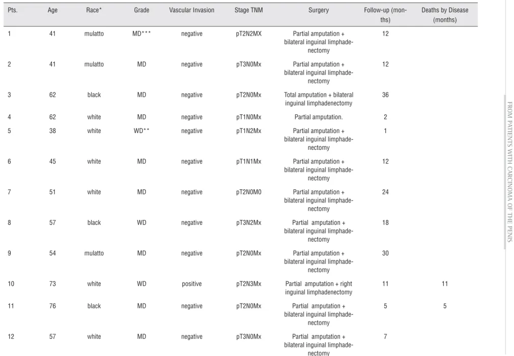
Table 1 - Patient characteristics, histopathologic fi ndings, pathologic staging, type of surgery and follow-up.
Pts. Age Race* Grade Vascular Invasion Stage TNM Surgery Follow-up (mon-ths)
Deaths by Disease (months)
1 41 mulatto MD*** negative pT2N2MX Partial amputation + bilateral inguinal
limphade-nectomy
12
2 41 mulatto MD negative pT3N0Mx Partial amputation + bilateral inguinal
limphade-nectomy
12
3 62 black MD negative pT2N0Mx Total amputation + bilateral inguinal limphadenectomy
36
4 62 white MD negative pT1N0Mx Partial amputation. 2
5 38 white WD** negative pT1N2Mx Partial amputation + bilateral inguinal
limphade-nectomy
1
6 45 white MD negative pT1N1Mx Partial amputation + bilateral inguinal
limphade-nectomy
12
7 51 white MD negative pT2N0M0 Partial amputation + bilateral inguinal
limphade-nectomy
24
8 57 black WD negative pT3N2Mx Partial amputation + bilateral inguinal
limphade-nectomy
18
9 54 mulatto MD negative pT2N0Mx Partial amputation + bilateral inguinal
limphade-nectomy
30
10 73 white WD positive pT2N3Mx Partial amputation + right inguinal limphadenectomy
11 11
11 76 black MD negative pT2N0Mx Partial amputation + bilateral inguinal
limphade-nectomy
5 5
12 57 white MD negative pT3N0Mx Partial amputation + bilateral inguinal
limphade-nectomy
IBJU
|
DOWNREGULA
TION OF COMPLEMENT F
A
CT
OR FRA
GMENTS IN PLASMA
FROM P
A
TIENTS WITH CARCINOMA OF THE PENIS
743
13 80 white WD negative pT2N0Mx Partial amputation + bilateral inguinal
limphade-nectomy
24
14 84 white MD negative pT2NxMx Partial amputation. 3 3
15 86 white MD negative pT3NxMx Total amputation. 8
16 64 white MD negative pT3N3Mx Partial amputation + bilateral inguinal
limphade-nectomy
5 5
17 80 white MD positive pT4NxMx Emasculation 8 8
18 90 white PD**** negative pT1NxMx Partial amputation. 1
19 83 white WD negative pT2N3Mx Partial amputation + bilateral inguinal
limphade-nectomy
8 8
20 55 mulatto MD negative pT3N2Mx Emasculation + bilateral inguinal limphadenectomy
4
21 61 mulatto MD negative pT2N0Mx Total amputation + bilateral inguinal limphadenectomy
9
22 71 white MD positive pT2N0Mx Partial amputation + bilateral inguinal
limphade-nectomy
1
23 56 mulatto WD negative pT2N0Mx Partial amputation + bilateral inguinal limphade-nectomy
4
24 60 black WD negative pT2NxMx Total amputation. 1
25 62 white WD negative pT2N3Mx Partial amputation + bilateral inguinal limphade-nectomy
24
* In Brazil, race defi nition is not accurate due to miscegenation. The column represents the self defi ned skin color of the patients ** WD: well differentiated
IBJU |DOWNREGULATION OF COMPLEMENT FACTOR FRAGMENTS IN PLASMA FROM PATIENTS WITH CARCINOMA OF THE PENIS
744
jects’ plasma by LIFT MS/MS. For peptide identi-fi cation, the LIFT-TOF/TOF spectra were recorded in the Ultrafl eXtremeTM MALDI-TOF/TOF MS ins-trument. The fragment masses were analyzed after their ion refl ector detection. Analyses were per-formed using the following acquisition settings: ion source 1, 7.5 kV; ion source 2, 6.7 kV; lens 3.6 kV; refl ector, 29.5 kV; refl ector 2, 13.95 kV; lift 1, 19 kV; lift 2, 3.15 kV; pulsed ion extraction 80 ns.
Raw MS/MS data were processed with the FlexAnalysisTM 3.3 software (Bruker Daltonics, Germany). Database searching was performed by an in-house Mascot search engine (Version: 2.3.02) using the following parameters: human Swissprot (accessed Feb. 2012- 20,317 sequences) database, no enzyme and fi xed and variable modifi cations. MS and MS/MS tolerances were generally set at 1 Da. Only identifi cations with a score higher than Mascot identity thresholds were accepted.
RESULTS
The pathological fi ndings of the SCCP are shown in Table-1. Most of the patients (n = 14) were at pT2 level, whereas the remaining were at pT1 (n = 4), pT3 (n = 6) and pT4 (n = 1) le-vels. Histopathology confi rmed invasive SCCP in all patients. Of the 25 cases, 8 (32%) were well differentiated, 16 (64%) moderately differentia-ted and 1 (4%) poorly differentiadifferentia-ted. Amputation and inguinal lymphadenectomy was performed in 20 patients. Of these patients, 11 (55%) were N0, 1 (5%) was N1, 4 (20%) were N2 and 4 (20%) were N3.
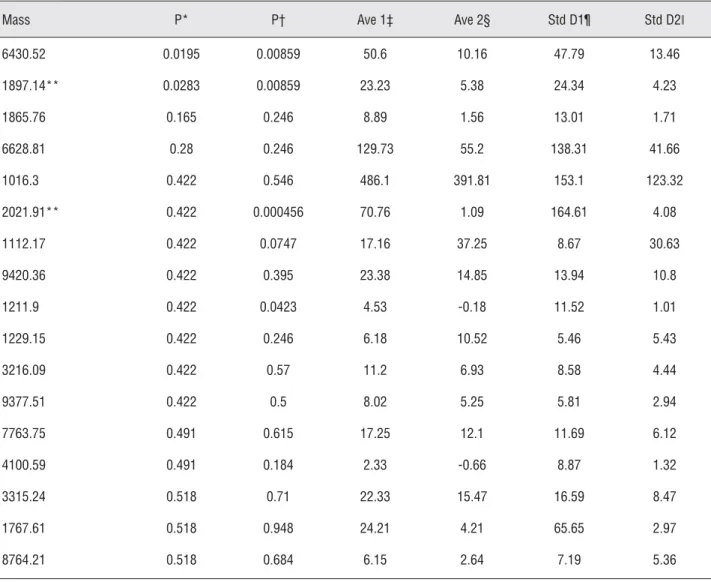
ClintProt was applied to analyze the plas-ma samples collected from SCCP patients and a cluster of two peaks was identifi ed. Comparison of spectrum profi les obtained from the data set used in the training phase showed several ions diffe-rentially expressed in the two studied populations (Table-2). On the basis of the GA and SVM results, a cluster of two statistically different signals (P < 0.05), at m/z 2021.99 ± 9, 1897.22 ± 9, were identifi ed as able to differentiate the populations. Preliminary statistical analysis was carried out for each marker and for the cluster of signals by the receiver operating characteristic curve analysis. The AUC of peak A at m/z 1897.22 (P < 0.0001)
was 0.85, which corresponds to a moderately ac-curate test, according to the criteria suggested by Swets (20). The AUC of peak B at m/z 2021.99 (P < 0.0001) was 0.91, which corresponds to a highly accurate test. The combination of the two peaks indicated an improvement in the performance compared to the single signals, with specifi city and sensitivity of 100% and 80%, respectively. This pattern was subsequently tested for its abili-ty to differentiate SCCP patients from controls by external validation using data obtained from a se-cond group of normal subjects and patients. Sen-sitivity and specifi city were at 63.6% and 100%, respectively. Cross-validation analysis using the whole casuistic showed 62.5% and 86.76% sen-sitivity and specifi city, respectively. The cluster of signals was also evaluated using the entire set of patient data grouped according to deaths due to disease and lymph node involvement. The results showed 100% sensitivity and 97% specifi city for patients who had died of disease. Among patients with lymph node involvement, the sensitivity and specifi city was 80% and 97%, respectively. Pa-tients without lymph node involvement showed 54% sensitivity and 97% specifi city (Table-3). Both peeks A and B, included in the diagnostic cluster, were under-expressed (p < 0.05) in pa-tients compared with healthy subjects (Figure-1). Some of the peaks observed in the plasma protein profi le could be identifi ed by LIFT MS/MS (Ta-ble-4). In particular, signals at m/z 1897.22 (m/z 1896.17 in refl ector mode) and at m/z 2021.99 (m/z 2021.26 in refl ector mode) were identifi ed by the proteomic approach based on MALDI-TOF/ TOF as fragments of C3 (m/z 1896.17) and C4a/b (m/z 2021.26) complement proteins (Table-4).
Preliminary evaluation of the diagnostic effi cacy was determined with an internal valida-tion. The cluster showed a very high specifi city value in an external validation test with plasma samples collected from different patients and con-trols (Table-3).
DISCUSSION
IBJU |DOWNREGULATION OF COMPLEMENT FACTOR FRAGMENTS IN PLASMA FROM PATIENTS WITH CARCINOMA OF THE PENIS
745
Table 2 - Selection of peaks differently expressed (P < 0.05) between controls (n = 28) and penile cancer patients (n = 17) used for the training phase.
Mass P* P† Ave 1‡ Ave 2§ Std D1¶ Std D2ǁ
6430.52 0.0195 0.00859 50.6 10.16 47.79 13.46
1897.14** 0.0283 0.00859 23.23 5.38 24.34 4.23
1865.76 0.165 0.246 8.89 1.56 13.01 1.71
6628.81 0.28 0.246 129.73 55.2 138.31 41.66
1016.3 0.422 0.546 486.1 391.81 153.1 123.32
2021.91** 0.422 0.000456 70.76 1.09 164.61 4.08
1112.17 0.422 0.0747 17.16 37.25 8.67 30.63
9420.36 0.422 0.395 23.38 14.85 13.94 10.8
1211.9 0.422 0.0423 4.53 -0.18 11.52 1.01
1229.15 0.422 0.246 6.18 10.52 5.46 5.43
3216.09 0.422 0.57 11.2 6.93 8.58 4.44
9377.51 0.422 0.5 8.02 5.25 5.81 2.94
7763.75 0.491 0.615 17.25 12.1 11.69 6.12
4100.59 0.491 0.184 2.33 -0.66 8.87 1.32
3315.24 0.518 0.71 22.33 15.47 16.59 8.47
1767.61 0.518 0.948 24.21 4.21 65.65 2.97
8764.21 0.518 0.684 6.15 2.64 7.19 5.36
* P value by t test and ANOVA; values lower than .0.05 indicate statistical relevance.
† P value calculated with the Wilcoxon/Kruskal–Wallis test; values lower than 0.05 suggest statistical relevance. ‡ Average area of peaks for control subjects.
§ Average area of peaks for SCCP patients. ¶ Standard deviation of peaks for control subjects.
ǁS tandard deviation of peaks for SCCP patients.
** Marked peaks represent signals selected for the diagnostic model
Table 3 - Cluster evaluation: diagnostic effi cacy of pattern based on deaths from disease and lymph node involvement.
Group Sensitivity Specifi city
6 patients dead by disease 100% 97%
9 patients with positive lymph nodes 80% 97%
IBJU |DOWNREGULATION OF COMPLEMENT FACTOR FRAGMENTS IN PLASMA FROM PATIENTS WITH CARCINOMA OF THE PENIS
746
role that proteins plays in health and disease. By applying this knowledge, innovative discoveries in early cancer detection have been made, inclu-ding kidney, prostate, ovarian, and bladder cancer. To our knowledge, this is the fi rst study of penile cancer proteomics.
Although several markers have been eva-luated, currently the clinical application of these markers is limited. HPV positive tumors show a variable prognostic outcome. In a previous article we reported the analysis of 80 consecutive cases of patients with penile cancers and HPV status was not signifi cantly associated with the presence of
re-Figure 1 - Signal intensity of 2 peaks (1897.22 and 2021.99 m/z) discriminating control (class 1) and tumor (class 2).
A B
Table 4 - Identifi ed peptides in the C8 MB eluted fraction of patients’ and control subjects’ plasma by LIFT MS/MS. All peptide identifi cation scores were above Mascot identity threshold. Table reports the m/z values of the ions observed in MALDI linear and refl ectron mode.
Refl ectron mode m/z
Linear mode m/z
Calc. mass (Da) in all pre-fractionated
plasma samples
Peptide sequence Accession
(UNIPROT)
Description Peptide
score
1896.17 1897.22 1895.024
NGFKSHALQLNNRQIR
CO4A_HUMAN
CO4B_HUMAN
Complement C4-A OS=Homo sapiens GN=C4A PE=1 SV=1 Complement C4-B OS=Homo sapiens GN=C4B PE=1 SV=1
61
2021.26 2021.99 2020.097 SSKITHRIHWESASLLR CO3_HUMAN Complement C3 OS=Homo
sapiens GN=C3 PE=1 SV=2
72
gional metastases (21). Up to now, P53 status may correlate with survival in T1 disease but further studies are required to establish the link to lymph node spread (22) Therefore, it is still necessary to fi nd biomarkers for early detection of metastases, prognosis and follow-up of this disease (22).
Before the use of proteomics in penile carcinoma, a critical assessment of its diagnos-tic and prognosdiagnos-tic value will be required. There is a controversy about whether and when patients should undergo lymphadenectomy. The presence and the extent of metastasis to the inguinal region is the most powerful prognostic factor for
survi-Relative Intensity
IBJU |DOWNREGULATION OF COMPLEMENT FACTOR FRAGMENTS IN PLASMA FROM PATIENTS WITH CARCINOMA OF THE PENIS
747
val in patients with SCCP. The principal aim in the management of this disease is correct staging of patients without invasive procedures, since a presence of metastatic lymph nodes is an indepen-dent prognostic factor for survival (6). Therefore, research into potential biomarkers and biological targets is very important.
The C3, C4a and C4b complement frag-ments are involved in the classic and the alterna-tive complement cascade pathway. These peptides were under-expressed (p < 0.05) in SCCP patients compared with healthy subjects. During disea-se progress, under-expression of C3, C4a/b frag-ments become more evident. More interestingly, we have found very high sensitivity (100%) and specifi city (97%) for under-expression of these fragments in SCCP patients that have succumbed to the disease. Besides this, patients with lymph node involvement present sensitivity and speci-fi city of 80% and 97%, respectively. In the case of patients without lymph node involvement, we obtained a sensitivity rate of 57% and specifi city of 97%.
Degradation of complement system con-vertases was observed for patients with breast cancer (BCP). For C3, the degradation was solely on the C3d, C3g, C3ɑ´1 and C3β. Convertases C3, which occupies a central position in the system, displays differential degradation in the BCP (5-fold more peptides than in the healthy subjects). For C4, the degradation was solely on the C4b and C4β. The degradation of the front portion of the C4b fragment was observed solely for the tested BCP (23).
The contribution of the complement sys-tem to the control of tumor growth has been ne-glected for a long time, since the main emphasis has been put on cell-mediated immune response against cancer (24).The innate immune system is the fi rst line of defense, comprised of cells and mechanisms that defend the host from infection in a non-specifi c manner. Taneja et al. (25) studied the plasma protein profi le in patients with hepati-tis E. In that study, the levels of complement pro-teins C3, C3f, C4, and bradykinin and kininogen were found to be lower in the plasma of hepatitis E patients compared to healthy controls. Any per-turbation, such as a viral infection in the liver,
should trigger a strong innate response as the fi rst line of host defense, but the opposite was found to occur, with these proteins being in downregula-tion. The exact mechanism of this reduction is not understood at this time.
In our patients, the downregulation of C3 and C4 could be caused by HPV and/or EBV in-fection, viruses that are highly prevalent in SCCP lesions (26). Viral proteins counteract the immune response (27). This could explain the progression of the disease along with C3 and C4a/b under--expression. It is a hypothesis to be tested in the future.
The over-expression of C regulatory pro-teins (CRPs) in tumor cells is one way these cells protect themselves from C attack. CD46, CD55 and CD59 are thought to be the most important mem-brane C regulatory proteins (mCRPs), expressed both on normal and tumor cells. Tumor cells can also evade C attack by binding soluble C inhibitors from serum such as factor H (fH). It is also interes-ting to note that fH or a related protein is a marker for bladder cancer, suggesting a link between C resistance and escape from immune surveillance (28). CD55 has been identifi ed as a tumor-associa-ted antigen and a high expression level of CD55 in colorectal cancer tissue is correlated with a signi-fi cant decrease in survival (29). Also, lower CD46 has been found to be inversely related with high levels of C3 deposited in renal and cervical cancer tissue (30).
In addition, in a previous study (31) we de-tected lower activity of natural killer cells (NK) in patients with cancer of the penis. NK cells are also part of the innate immune response and were fi rst identifi ed for their ability to kill tumor cells without deliberate immunization or activation. Subsequen-tly, they were also found to be able to kill cells that are infected with certain viruses and to attack preferentially cells that lack expression of major histocompatibility complex (MHC) class I antigens. The innate immune response of patients to cancer therefore may be involved, since natural killer (NK) cell activity can be signifi cantly decreased in SCCP patients compared to control groups (31).
IBJU |DOWNREGULATION OF COMPLEMENT FACTOR FRAGMENTS IN PLASMA FROM PATIENTS WITH CARCINOMA OF THE PENIS
748
known about the timeline and reciprocal connec-tion between these processes. The widely accep-ted theory of immuno-editing describes a rational, time-structured evolution of the relationship that occurs between tumor and host immune system (32,33). Tumor escape in cancer patients appears to be a cumulative process that involves tumor--derived soluble factors (TDSFs), induction of regulatory elements of various cell lineages and different anatomical environments (34). In a sim-plifi ed view, tumor-induced immune subversion results from two main activities, namely the in-duction of tolerance toward tumor antigens and the functional suppression of the effector lym-phocytes that normally counteract tumor growth (32-36).
CONCLUSIONS
Our results suggest that a proteomic ap-proach based on magnetic beads is a useful me-thod to discover possible clinical biomarkers. We demonstrated the capability of selected signals to differentiate SCCP patients from normal subjects. The peptides identifi ed from the C8 MB eluted fraction of patients’ and control subjects’ plasma correspond to fragments of the C3 (m/z 1896.17) and C4 (m/z 2021.26) complement protein. The results showed that when the disease progresses, they are more under-expressed. These fragments are mainly downregulated in patients with metas-tatic involvement. The innate immune response of patients could be suppressed. Downregulation of C3 and C4A/B represents a promising prognostic tool for SCCP.
ACKNOWLEDGEMENTS
This work was supported by grants from the Italian Ministry of Universities and Research: PRIN 2006 (no. 69373), FIRB 2007 (Rete nazionale per lo studio del proteoma umano, no.RBRN07BMCT_11), FAR 2006-2011 (ex 60%), from the Italian Institu-te of Technology (IIT), Project SEED: “IPG-CHIP”, by “FONDO PER LA PROMOZIONE DI ACCOR-DI ISTITUZIONALI” Regione Lombardia DGR N. 5200/2007, project no. 14546: “Network Enabled Drug Design (NEDD)” The work was also
suppor-ted by grants from Programa de Oncobiologia and FAPERJ (APQ1-E26/110.812/2009), Brazil.
CONFLICT OF INTEREST
None declared.
REFERENCES
1. Misra S, Chaturvedi A, Misra NC: Penile carcinoma: a chal-lenge for the developing world. Lancet Oncol. 2004; 5: 240-7. 2. Ornellas AA, Kinchin EW, Nóbrega BL, Wisnescky A, Koif-man N, Quirino R: Surgical treatment of invasive squamous cell carcinoma of the penis: Brazilian National Cancer Insti-tute long-term experience. J Surg Oncol. 2008; 97: 487-95. 3. Ravi R: Correlation between the extent of nodal involve-ment and survival following groin dissection for carcinoma of the penis. Br J Urol. 1993; 72: 817-9.
4. Soria JC, Fizazi K, Piron D, Kramar A, Gerbaulet A, Haie-Meder C, et al.: Squamous cell carcinoma of the penis: multivariate analysis of prognostic factors and natural his-tory in monocentric study with a conservative policy. Ann Oncol. 1997; 8: 1089-98.
5. Lopes A, Bezerra AL, Pinto CA, Serrano SV, de MellO CA, Villa LL: p53 as a new prognostic factor for lymph node metastasis in penile carcinoma: analysis of 82 patients treated with amputation and bilateral lymphadenectomy. J Urol. 2002; 168: 81-6.
6. Ornellas AA, Nóbrega BL, Wei Kin Chin E, Wisnescky A, da Silva PC, de Santos Schwindt AB: Prognostic factors in invasive squamous cell carcinoma of the penis: analysis of 196 patients treated at the Brazilian National Cancer Insti-tute. J Urol. 2008; 180: 1354-9.
7. Abi-Aad AS, deKernion JB: Controversies in ilioinguinal lymphadenectomy for cancer of the penis. Urol Clin North Am. 1992; 19: 319-24.
8. Alpantaki K, Tsiridis E, Pape HC, Giannoudis PV: Applica-tion of clinical proteomics in diagnosis and management of trauma patients. Injury. 2007; 38: 263-71.
9. Theodorescu D, Wittke S, Ross MM, Walden M, Conaway M, Just I, et al.: Discovery and validation of new protein biomarkers for urothelial cancer: a prospective analysis. Lancet Oncol. 2006; 7: 230-40.
10. Sarto C, Proserpio V, Magni F: Insight on renal cell carci-noma proteome D.S. Sayed (ed.), Cancer Proteomics From Bench to Bedside, Humana Press, Totowa, NJ. 2008; pp. 121-37.
IBJU |DOWNREGULATION OF COMPLEMENT FACTOR FRAGMENTS IN PLASMA FROM PATIENTS WITH CARCINOMA OF THE PENIS
749
12. Bosso N, Chinello C, Picozzi SC, Gianazza E, Mainini V,Gal-busera C, et al.: Human urine biomarkers of renal cell car-cinoma evaluated by ClinProt. Proteomics Clin Appl. 2008; 2: 1036-46.
13. Tolson JP, Flad T, Gnau V, Dihazi H, Hennenlotter J, Beck A, et al.: Differential detection of S100A8 in transitional cell car-cinoma of the bladder by pair wise tissue proteomic and im-munohistochemical analysis. Proteomics. 2006; 6: 697-708. 14. Chang JT, Chen LC, Wei SY, Chen YJ, Wang HM, Liao CT, et
al.: Increase diagnostic effi cacy by combined use of fi nger-print markers in mass spectrometry--plasma peptidomes from nasopharyngeal cancer patients for example. Clin Bio-chem. 2006; 39: 1144-51.
15. Freed GL, Cazares LH, Fichandler CE, Fuller TW, Sawyer CA, Stack BC Jr, et al.: Differential capture of serum proteins for expression profi ling and biomarker discovery in pre- and posttreatment head and neck cancer samples. Laryngo-scope. 2008; 118: 61-8.
16. Chinello C, Gianazza E, Zoppis I, Mainini V, Galbusera C, Picozzi S, et al.: Serum biomarkers of renal cell carcinoma assessed using a protein profi ling approach based on Clin-Prot technique. Urology. 2010; 75: 842-7.
17. Ketterlinus R, Hsieh SY, Teng SH, Lee H, Pusch W: Fishing for biomarkers: analyzing mass spectrometry data with the new ClinProTools software. Biotechniques. 2005; (Suppl): 37-40.
18. Holland JH: Adaptation in Natural and Artifi cial Systems. Ann Arbor, MI: University of Michigan Press. 1975. 19. Vapnik V: Statistical Learning Theory. New York, NY, Wiley.
1998; pp. 1-736.
20. Swets JA: Measuring the accuracy of diagnostic systems. Science. 1988; 240: 1285-93.
21. Scheiner MA, Campos MM, Ornellas AA, Chin EW, Ornellas MH, Andrada-Serpa MJ: Human papillomavirus and penile cancers in Rio de Janeiro, Brazil: HPV typing and clinical fea-tures. Int Braz J Urol. 2008; 34: 467-74; discussion 475-6. 22. Muneer A, Kayes O, Ahmed HU, Arya M, Minhas S:
Mo-lecular prognostic factors in penile cancer. World J Urol. 2009; 27: 161-7.
23. Shen Y, Tolić N, Liu T, Zhao R, Petritis BO, Gritsenko MA, et al.: Blood peptidome-degradome profi le of breast cancer. PLoS One. 2010; 5: e13133.
24. Macor P, Tedesco F: Complement as effector system in can-cer immunotherapy. Immunol Lett. 2007; 111: 6-13. 25. Taneja S, Ahmad I, Sen S, Kumar S, Arora R, Gupta VK, et
al.: Plasma peptidome profi ling of acute hepatitis E patients by MALDI-TOF/TOF. Proteome Sci. 2011; 9: 5.
26. Afonso LA, Moyses N, Alves G, Ornellas AA, Passos MR, Oliveira Ldo H, et al.: Prevalence of human papillomavirus and Epstein-Barr virus DNA in penile cancer cases from
Brazil. Mem Inst Oswaldo Cruz. 2012; 107: 18-23. 27. Campo MS, Graham SV, Cortese MS, Ashrafi GH, Araibi EH,
Dornan ES, et al.: HPV-16 E5 down-regulates expression of surface HLA class I and reduces recognition by CD8 T cells. Virology. 2010; 407: 137-42.
28. Fedarko NS, Fohr B, Robey PG, Young MF, Fisher LW: Fac-tor H binding to bone sialoprotein and osteopontin enables tumor cell evasion of complement-mediated attack. J Biol Chem. 2000; 275: 16666-72.
29. Durrant LG, Chapman MA, Buckley DJ, Spendlove I, Rob-ins RA, Armitage NC: Enhanced expression of the comple-ment regulatory protein CD55 predicts a poor prognosis in colorectal cancer patients. Cancer Immunol Immunother. 2003; 52: 638-42.
30. Blok VT, Daha MR, Tijsma OM, Weissglas MG, van den Broek LJ, Gorter A: A possible role of CD46 for the protec-tion in vivo of human renal tumor cells from complement-mediated damage. Lab Invest. 2000; 80: 335-44.
31. Campos MM, de Souza MH, Pires V, Scheiner MA, Esteves EB, Ornellas AA: Clinical implications of natural killer cy-totoxicity in patients with squamous cell carcinoma of the penis. Nat Immun. 1998; 16: 256-62.
32. Dunn GP, Bruce AT, Ikeda H, Old LJ, Schreiber RD: Cancer immunoediting: from immunosurveillance to tumor es-cape. Nat Immunol. 2002; 3: 991-8.
33. Smyth MJ, Dunn GP, Schreiber RD: Cancer immunosurveil-lance and immunoediting: the roles of immunity in sup-pressing tumor development and shaping tumor immuno-genicity. Adv Immunol. 2006; 90: 1-50.
34. Bronte V, Cingarlini S, Marigo I, De Santo C, Gallina G, Dol-cetti L, et al.: Leukocyte infi ltration in cancer creates an unfavorable environment for antitumor immune responses: a novel target for therapeutic intervention. Immunol Invest. 2006; 35: 327-57.
35. Rabinovich GA, Gabrilovich D, Sotomayor EM: Immuno-suppressive strategies that are mediated by tumor cells. Annu Rev Immunol. 2007; 25: 267-96.
36. Zitvogel L, Tesniere A, Kroemer G: Cancer despite immu-nosurveillance: immunoselection and immunosubversion. Nat Rev Immunol. 2006; 6: 715-27.
_____________________
Correspondence address: