Key words:
Phimosis; Methods; Anatomy and histology; Histology; Circumcision
Int Braz J Urol. 2012; 38: 802-8
__________________
Submitted for publication: January 19, 2012
__________________
Accepted after revision: May 22, 2012
Objectives: To evaluate histological alterations in prepuce of patients with phimosis submitted to topic treatment with betamethasone in association with hyaluronidase.
Materials and Methods: We studied sixty patients (mean age 4.5), presenting true phi-mosis and treated with a topical treatment with betamethasone cream (0.2%) + hyalu-ronidase. The parents of seven of these patients opted for circumcision (control group). The other fi fty-three patients were submitted to clinical treatment. The samples were stained with Weigert’s resorcin-fuchsin (analysis of the elastic fi bers) and Picro-Sirius Red, for analysis of the collagen. The volumetric density of the elastic fi bers was deter-mined by stereological methods.
Results: Only eight (15%) of the fi fty-three patients submitted to topical treatment presented failure, being indicated for circumcision (histological analysis). We observed an increase of the collagen type III of the patients submitted to topical treatment. The quantifi cation showed a reduction of the volumetric density of the prepuce’s elastic fi bers of the patients submitted to the cream treatment, when compared to the con-trol group (p = 0.056). The volumetric density of the elastic fi bers of the prepuce at the group not submitted to topical treatment showed an average of 14.60% (11.06 to 21.64%); in the group submitted to the cream treatment, the volumetric density of the elastic fi bers of the prepuce showed an average of 10.34% (3.45 to 17.9%).
Conclusion: The topical treatment of phimosis with betamethasone 0.2% + hyaluro-nidase had a success rate of 85%. Patients with failure of the topical treatment with steroid had histological alterations in the prepuce.
INTRODUCTION
The foreskin is a specialized and inner-vated mucous-cutaneous tissue that covers and protects the glans. After 3 years of age, cysts of keratin are formed below the foreskin adherences and together with intermittent erections are able to enlarge the phimotic ring, exposing the glans. Around 80 to 90% of the boys that were not
circu-mcised became capable to expose the glans after 3 years of age (1).
Pathological phimosis is characterized by a foreskin fi brotic ring with adherences that does not allow the exposition of the glans (1). This alte-ration hinders adequate penile hygiene that favors foreskin infections, repeated urinary infections, sexually transmitted diseases and, at adult age, penile carcinoma (2).
Structural analysis of the phimotic prepuce in patients
with failed topical treatment compared with untreated
phimosis
___
_______________________________________________
____________________________________________
Luciano Alves Favorito, Carlos M. Balassiano, João Pedro Rosado, Luiz Eduardo M. Cardoso,
Waldemar Silva Costa, Francisco José Barcellos Sampaio
Urogenital Research Unit, State University from Rio de Janeiro, Brazil
ABSTRACT
ARTICLE
INFO
803
Circumcision at childhood must be perfor-med under general anesthesia. Moreover, the pro-cedure itself is not free of risks, presenting com-plication rates up to 34% (3,4). The most common complications of circumcision are hemorrhage, urethral meatus and preputial ring stenosis, and even glans amputation (5). Besides these proble-ms, circumcision presents considerable costs, whi-ch could approawhi-ch a mean of US$ 1,920.00 per procedure, according to recent reports (6,7).
The clinical treatment of phimosis with topical corticosteroids has been proposed as an alternative to surgery in the beginning of 1990, demonstrating acceptable results (8-10) and low cost (11). Since then, several authors have presen-ted results varying from 67% to 95% of sucess, independently of the patients’ age. The most com-mon used topical treatments were betamethasone, clobetasol, diclofenac sodium, mometasone furo-ate 0.05% and triamcinolone acetfuro-ate (10,12,13). Recent randomized prospective studies have con-fi rmed that topical use of corticosteroids as treat-ment for phimosis is superior to placebo (14,15).
Studies on the foreskin structure in pa-tients with phimosis are scarce. Also, to our kno-wledge, there are no studies on histological alte-rations of the foreskin after topical corticosteroids treatment. Therefore, the objective of the present work was to evaluate the possible histological al-terations in the foreskin of patients with phimosis submitted to topical application of an association of betamethasone 0.2% and hyaluronidase cream.
MATERIALS AND METHODS
The present work received institutional committee review and parental approval. This work was carried out in accordance with the ethi-cal standards of the institutional committee res-ponsible for human experimentation.
We studied 60 patients ranging from 3 to 10 years (mean 4.5), during the period from Janu-ary 2006 to October 2010. All patients presented true phimosis with foreskin stenosis and attended the pediatric urology ambulatory care to be sub-mitted to circumcision. All 60 patients presented total impossibility of foreskin retraction, which characterizes the type A foreskin anatomy
accor-ding to Marques (16). The exclusion criteria were patients with less than 3 years of age, patients without true phimosis and patients with clinical suspicion of balanitis xerotica obliterans.
To the parents of the boys referred for cir-cumcision we offered an alternative clinical ma-nagement of the phimosis. The regimen proposed was a topical application of an association of be-tamethasone 0.2% and hyaluronidase (150 units of turbid retention) cream on the phimotic ring. The parents were oriented to perform gentle trac-tion of the foreskin until the stenotic ring could be seen. The cream was applied twice a day, for 6 consecutive weeks, associated with appropriate hygiene of the penis. The children had a follow up every 3 weeks in our ambulatory.
The parents of 7 children did not agree with the clinical treatment and opted for circu-mcision directly. These 7 patients served as the control group and the fi nal treated sample was composed of 53 patients.
The topical treatment was considered suc-cessful if the patient was able to expose the glans completely. We considered failure if the glans could not be exposed after treatment or if occur-red foreskin infection during treatment. In these cases, circumcision was performed. The patients submitted to topical treatment were followed up for 6 months.
Histological Analysis
In controls and in those cases on which the patients failed treatment and were submitted to circumcision, the foreskin was fi xed in a 10% buffered formalin solution, routinely processed for embedding in paraffi n and 5-µm thick sec-tions were obtained. The secsec-tions were stained with Picro-Sirius Red, for qualitative analysis of the collagen under microscope of polarization and Weigert’s Resorcin Fuscin with previous oxidation by oxona, for characterization and quantifi cation of the elastic system fi bers.
Quantitative Analysis
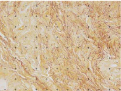
elastic system fi bers were expressed as volume-tric density (Vv - %). The sections were observed with X400 magnifi cation using an Olympus li-ght microscope coupled to a video camera Sony CCD, and the images transferred to a Sony mo-nitor KX14-CP1. The selected histological areas were then quantifi ed using M42 test-grid system on the digitized fi elds (17,18). (Figure-1). All nu-merical results are presented as mean ± standard deviation.
The data were analyzed with the Graphpad software. To compare the quantitative data in both groups (controls and treated) and the outcomes, the Student’s t-test was used (p < 0.05 was consi-dered signifi cant) (19).
RESULTS
From the 53 patients submitted to topical treatment with the association of betamethasone and hyaluronidase cream, 8 (15%) presented fai-lure (6 could not expose the glans and 2 remai-ned with foreskin stenosis) and were referred to circumcision. The foreskin of these patients was submitted to histological analysis and composed our treated group. The others 45 patients presen-ted signifi cant improvement of the phimosis, with
total exposure of the glans without foreskin ste-nosis. After 6 months of follow-up, these patients considered as success did not present recurrence of the phimosis.
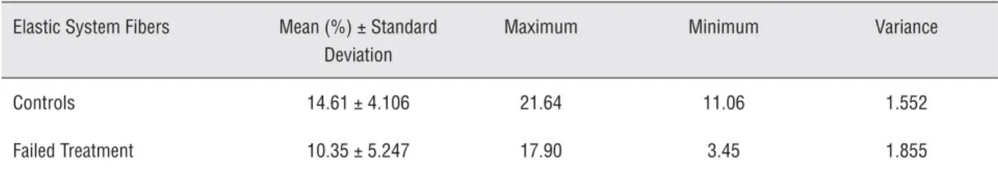
We analyzed the alterations on the elastic fi bers and collagen of the foreskin. Elastic system fi bers were analyzed in the foreskin of the control (n = 7) and treatment failed (n = 8) groups. Figu-re-2 shows the elastic system fi bers analysis in the foreskin. One may note in Figure-2B, in a patient who failed the topical treatment, an apparent lo-wer amount of elastic fi bers when compared to controls (Figure-2A). The quantifi cation demons-trated that the mean volumetric density of elastic fi bers in the control group was 14.60% (11.06 to 21.64%) and in the group who failed treatment was 10.34% (3.45 to 17.9%); nevertheless, the di-fference was not signifi cant (p = 0.056). Data on the stereological quantifi cation of elastic system fi bers in the foreskin are presented in details on Table-1.
The Figure-3 evidences the alterations on the concentration of the collagen of the two groups studied, through the PicroSirius Red stain (with polarization). We can see the alteration on the pattern of the collagen of the patients submit-ted to the treatment with the cream. In Figure-3B,
805
Table 1 - Quantifi cation of elastic system fi bers in the foreskin of controls (n = 7) and of patients who failed topical treatment (n = 8) with the association of betamethasone and hyaluronidase cream.
Elastic System Fibers Mean (%) ± Standard Deviation
Maximum Minimum Variance
Controls 14.61 ± 4.106 21.64 11.06 1.552
Failed Treatment 10.35 ± 5.247 17.90 3.45 1.855
p = 0.056; not signifi cant.
Figure 2 - Analysis of the elastic fi bers: A) Photomicrography of the prepuce of a 3-year old patient not treated with topical steroid. It is possible to observe that the elastic fi bers (purple) are distributed over the whole fi eld. Weigert stain reduced X400. B) Photomicrography of the prepuce of a 3-year old patient submitted to topical steroid treatment. It is possible to oberserve the elastic fi bers (purple). Weigert stain reduced X400.
A B
Figure 3 - Qualitative analysis of the collagen: A) Photomicrography of the prepuce of a 4 year old patient without treatment with topical steroid. Collagen type I - red; Collagen type III - green. Picro-Sirius Red stain reduced X 400. B) Photomicro-graphy of the prepuce of a 4 year old patient of the group submitted to topical steroid treatment. It is possible to observe an enlargement of green color (collagen type III), in this group. Picro-Sirius Red stain reduced X 400.
one may observe the increase of the collagen type III (colored in green), on patients submitted to tre-atment with topical steroid.
DISCUSSION
The physiological phimosis affects 96% of the newborns and its incidence reduces with age. At 3 years of age, 10% of the patients present phi-mosis and at 14 years of age, this incidence affects only 1% (20). The natural process of enlargement of the prepuce can suffer alterations when facing episodes of balanoposthitis and lesion of the pre-puce by traction of the ring that leads to the for-mation of a healing fi brosis and the impossibility to expose the glans.
The clinical treatment of the phimosis with topic corticosteroids is well accepted by the pa-rents, since it is a simple procedure, presents low costs and risks, no side effects and a good com-pliance to the treatment when the guardians of the patients are well oriented (14,15). In our studies we did not have reports of signifi cant side effects and all fi fty-three patients submitted to clinical treatment completed the six weeks of treatment.
The success rate of the phimosis topic tre-atment with corticosteroids is signifi cant, with satisfactory results (67%-95%) (8-10,14,15). The success rate of the topical treatment of the phi-mosis is lower when smaller concentrations of be-tamethasone are used (21). We opted for the use of a larger concentration of betamethasone (0.2%) associated with hyluronidase. This larger concen-tration of corticosteroids does not present a larger rate of side effects and could enhance the success rate of the treatment. After six weeks of treatment, among the fi fty-three patients treated, 85% sho-wed signifi cant improvement and were not sub-mitted to surgery, demonstrating the effi ciency of the treatment.
Betamethasone is one of the substances that present better rates of effi ciency as a topical treatment of phimosis (20,22), therefore the cho-sen medication in this study. Corticosteroids re-duce the arachidonic and hydroxyecosatetranoic acids on the reproductive infl ammatory skin di-sease, inhibiting the liberation of prostaglandins and enlarging the level of dismutase
super-oxida-tive activity, being able to liberate anti-oxidants (20). Side effects, such as the suppression of the hypothalamo-pituitary-adrenal axis or cutaneous atrophy, may ocurr; nevertheless, the doses used in the topical treatment of the phimosis are not suffi cient to lead to this type of complications (1).
Hyaluronidase is an enzyme obtained from cattle testicles, which depolymerize hyalu-ronic acid, a mucopolysaccharide that is present in the interstitial tissue spaces (conjunctive tissue of the preputial adherences). The depolimerization of the hyaluronic acid reduces the inter-cellular viscosity, allowing the tissue to remain availa-ble to the dispersion of substances. Besides that, hyaluronidase presents elastic cohesion functions, being applied with the objective of cleaning the adherences and also because it presents the effects of a local anesthesia.
Collagen and elastic fi bers are the fi brotic components of the cellular matrix and are related to pathological alterations in different tissues. In our studies, we noticed a reduction of volumetric density of the elastic fi bers of the patient´s prepu-ces submitted to topical treatment, with corticos-teroids + hyaluronidase.
The reduction of elastic fi bers of the pre-puce in patients submitted to topical treatment with the cream was not signifi cant but is typical of the healing process of this treatment (23). The increasing concentration of elastic fi bers is related to a larger widening of the tissues. For an easy exposure of the glans, the prepuce needs a larger concentration of elastic fi bers (24). The reduction of the elastic fi bers in the prepuce of the patients presenting phimosis and submitted to a betame-thasone + hyaluronidase treatment is similar to what occurs in the healing process and could be related to a larger diffi culty of the exposure of the glans; these patients did not report infections of the prepuce.
failu-807
re of the cream applied; nevertheless, to confi rm this hypothesis, it would be necessary to analyze the prepuces of the patients that received the tre-atment which obviously, for ethical reasons, is not possible.
The use of the betamethasone + hyaluro-nidase cream led to an evident alteration in the distribution of the collagen in the prepuces sub-mitted to this topical treatment. There was a re-duction of the collagen in the prepuce of the pa-tients that made use of it. Nevertheless, when we analyzed the prepuces with the Picrosirus stain, it could be noticed a clear alteration in the distribu-tion of the collagen, with and increase of collagen type III. The greenish fi bers that appear under the Picrosirus stain characterize the collagen type III, a recent collagen which is probably produced by muscular retraction. In the control group there was a predominance of a clear reddish color that cha-racterizes the predominance of the collagen type I. It is very diffi cult to say, without studying the histology of the patients that made use of the cre-am and did not need a surgery, if the alterations of the collagen occurred due to induced alterations provoked by the cream or due to tissue alterations of the prepuce resulting from local infections.
CONCLUSIONS
The topical treatment of phimosis with betamethasone 0.2% + hyaluronidase was effec-tive with a success rate of 85%. Patients in whom topical steroid treatment failed had fewer elastic fi bers, which characterize the healing processes, as well as an amplifi cation of the collagen type III, a recently found collagen that is associated with muscular retraction. The betamethasone + hya-luronidase cream leads to signifi cant histological alterations in the prepuce.
ACKNOWLEDGEMENTS
Supported by grants from the National Council of Scientifi c and Technological Develop-ment (CNPQ - Brazil) and Foundation for Research Support of Rio de Janeiro (FAPERJ).
CONFLICT OF INTEREST
None declared.
REFERENCES
1. Orsola A, Caffaratti J, Garat JM: Conservative treatment of phimosis in children using a topical steroid. Urology. 2000; 56: 307-10.
2. Elmore JM, Baker LA, Snodgrass WT: Topical steroid ther-apy as an alternative to circumcision for phimosis in boys younger than 3 years. J Urol. 2002; 168(4 Pt 2): 1746-7; discussion 1747.
3. Chu CC, Chen KC, Diau GY: Topical steroid treatment of phimosis in boys. J Urol. 1999; 162(3 Pt 1): 861-3. 4. Cathcart P, Nuttall M, van der Meulen J, Emberton M,
Ken-ny SE: Trends in paediatric circumcision and its complica-tions in England between 1997 and 2003. Br J Surg. 2006; 93: 885-90.
5. Ozkan S, Gürpinar T: A serious circumcision complication: penile shaft amputation and a new reattachment technique with a successful outcome. J Urol. 1997; 158: 1946-7. 6. Berdeu D, Sauze L, Ha-Vinh P, Blum-Boisgard C:
Cost-effectiveness analysis of treatments for phimosis: a com-parison of surgical and medicinal approaches and their economic effect. BJU Int. 2001; 87: 239-44.
7. Schoen EJ, Colby CJ, To TT: Cost analysis of neonatal cir-cumcision in a large health maintenance organization. J Urol. 2006; 175(3 Pt 1): 1111-5.
8. Golubovic Z, Milanovic D, Vukadinovic V, Rakic I, Perovic S: The conservative treatment of phimosis in boys. Br J Urol. 1996; 78: 786-8.
9. Wright JE: The treatment of childhood phimosis with topi-cal steroid. Aust N Z J Surg. 1994; 64: 327-8. Erratum in: Aust N Z J Surg. 1995; 65: 698.
10. Jørgensen ET, Svensson A: The treatment of phimosis in boys, with a potent topical steroid (clobetasol propionate 0.05%) cream. Acta Derm Venereol. 1993; 73: 55-6. 11. Van Howe RS: Cost-effective treatment of phimosis.
Pedi-atrics. 1998; 102: E43.
12. Atilla MK, Dündaröz R, Odabaş O, Oztürk H, Akin R, Gökçay E: A nonsurgical approach to the treatment of phimosis: local nonsteroidal anti-infl ammatory ointment application. J Urol. 1997; 158: 196-7.
14. Yang SS, Tsai YC, Wu CC, Liu SP, Wang CC: Highly po-tent and moderately popo-tent topical steroids are effective in treating phimosis: a prospective randomized study. J Urol. 2005; 173: 1361-3.
15. Lund L, Wai KH, Mui LM, Yeung CK: Effect of topical steroid on non-retractile prepubertal foreskin by a prospective, randomized, double-blind study. Scand J Urol Nephrol. 2000; 34: 267-9.
16. Marques TC, Sampaio FJ, Favorito LA: Treatment of phimo-sis with topical steroids and foreskin anatomy. Int Braz J Urol. 2005; 31: 370-4; discussion 374.
17. Mouton PR: Principles and practice of unbiased stereol-ogy. 1st edition, John Hopkins University Press. 2002. 18. Gundersen HJ, Bendtsen TF, Korbo L, Marcussen N, Møller
A, Nielsen K, et al.: Some new, simple and effi cient stereo-logical methods and their use in pathostereo-logical research and diagnosis. APMIS. 1988; 96: 379-94.
19. Sokal RR, Rohlf FJ: Statistical tables. In Freeman WH, eds., Biometry, 3rd ed. New York, 1995: 887-902.
20. Shankar KR, Rickwood AM: The incidence of phimosis in boys. BJU Int. 1999; 84: 101-2.
21. Lee KS, Koizumi T, Nakatsuji H, Kojima K, Yamamoto A, Kawanishi Y, et al.: Treatment of phimosis with betametha-sone ointment in children. Nihon Hinyokika Gakkai Zasshi. 2001; 92: 619-23.
22. Marzaro M, Carmignola G, Zoppellaro F, Schiavon G, Ferro M, Fusaro F, et al.: Phimosis: when does it require surgical intervention?. Minerva Pediatr. 1997; 49: 245-8.
23. Cavalcanti AG, Costa WS, Baskin LS, McAninch JA, Sam-paio FJ: A morphometric analysis of bulbar urethral stric-tures. BJU Int. 2007; 100: 397-402.
24. Cold CJ, Taylor JR: The prepuce. BJU Int. 1999; 83(Suppl 1): 34-44.
_____________________
Correspondence address: