Management of Diaphragmatic Injury during Transperitoneal
Laparoscopic Urological Procedures
Octavio A. Castillo, Gonzalo Vitagliano, Mauricio Moreno, Manuel A. Diaz, Oscar Cortes
Department of Urology (OAC, GV, MM, MAD, OC), Clinica Santa Maria, and Department of
Urology (OAC), School of Medicine, Universidad de Chile, Santiago de Chile, Chile
ABSTRACT
Introduction: Carbon dioxide pneumothorax is a rare complication in laparoscopic urology, but with the widespread use of laparoscopy and the increasing surgical pathologies managed by this technique this infrequent complication has become a potential risk.
Materials and Methods: A total of 786 laparoscopic transperitoneal urologic operations were reviewed at our institution. All procedures were performed by the same surgeon and included 213 adrenalectomies, 181 simple nephrectomies, 143 lym-phadenectomies, 118 radical nephrectomies, 107 partial nephrectomies and 24 nephroureterectomies. Mean patient age was 53.2 years (range 24 to 70). Mean BMI was 28.15 Kg/m2 (range 20 to 48.9).
Results: A total of 6 cases (0.7%) of diaphragmatic injury were found. All reported patients had additional factors that may have contributed to diaphragmatic injury. Diaphragmatic repair was always carried out by intracorporeal suturing and only one case required chest tube placement. All patients evolved uneventfully.
Conclusions: Repair of diaphragmatic injuries should always be attempted with intracorporeal suture since this is a feasible, reproducible and reliable technique.
Key words: pneumothorax; diaphragm; intraoperative complications; laparoscopy Int Braz J Urol. 2007; 33: 323-9
INTRODUCTION
The clear advantages that laparoscopy pro-vides over open surgery have made this technique very popular in the last decade. However, for urologist laparoscopy has gained wide acceptance only in re-cent years. Inadvertent diaphragmatic injury during transperitoneal laparoscopic surgery in urology is a rare occurrence (1). Yet, with the widespread use of laparoscopy and the increasing surgical pathologies managed with this technique, there is a potential for an increased risk of carbon dioxide pneumothorax due
to diaphragmatic injury. Capnothorax associated with laparoscopic surgery is different from air pneumotho-rax and may call for a different treatment.
We report the laparoscopic management of six cases with iatrogenic diaphragmatic injury during laparoscopic urologic surgery.
MATERIALS AND METHODS
324
Management of Diaphragmatic Injury during Laparoscopy
procedures were performed by the same surgeon (OAC) and included: 213 adrenalectomies, 181 simple nephrectomies, 143 retroperitoneal lymphadenectomies, 118 radical nephrectomies, 107 partial nephrectomies for cancer and 24 radical nephroureterectomies. A total of 6 cases of diaphragmatic injury were found. Mean patient age was 53.2 years (range 24 to 70). Mean BMI was 28.15 (range 20 to 48.9). Operative and clinical records were reviewed and patient outcome evaluated. Details of injury identification and operative management are specified (Table-1).
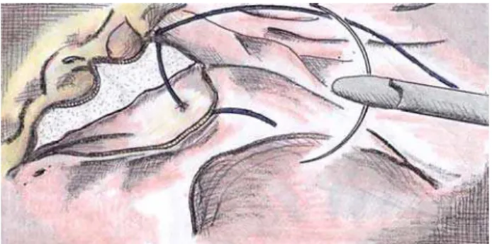
For diaphragmatic repair, no additional trocars were needed. In most cases, usual working port con-figuration for adrenalectomy and nephrectomy allowed intracorporeal suturing. However, if necessary an additional 5 mm port was placed on left or right flank respectively. The defect was repaired with separate 0-poliglactine sutures while pneumoperitoneum was decreased in 12 mmHg. Before the stitches were se-cured, the anesthesiologist administered a large inspira-tory breath (Figures-1 and 2).
Criteria used for chest tube placement was pneumothorax greater than 20% of lung volume or associated with hemodynamic or ventilatory changes.
RESULTS
A total of 6 cases of diaphragmatic injury were recorded during three adrenalectomies, one par-tial nephrectomy, one radical nephrectomy and one left retroperitoneal lymph node dissection respectively. In all cases, the cause of pleural lesion was iatrogenic injury to the diaphragm.
Case 1 - A 24-year-old woman (BMI 21) with a history of ulcerative colitis and an incidental 12 cm left adrenal cyst underwent laparoscopic adrenalec-tomy. During surgery an increase in end inspiratory pressure and end tidal carbon dioxide levels was noted. This prompted the inspection of the operative field. A 2 cm lesion was found on the left diaphragm, no pul-monary parenchyma was evident. The defect was repaired with 0-poliglactine sutures while pneumoperi-toneum was decreased in 12 mmHg with the anes-thesiologist administering a large inspiratory breath before securing the stitches. Adrenalectomy was
com-pleted with a total time of 135 minutes. A chest tube was placed and left for 12 hours. Follow-up chest x-ray showed no residual pneumothorax. The patient was discharged home after 48 hours.
Case 2 - A 40-year-old woman (BMI 24.5) underwent a right adrenalectomy for an incidental 7 cm right adrenal mass. During adrenal dissection a 3 cm iatrogenic injury was identified on the right dia-phragm with pulmonary parenchyma exposure. The pleural cavity was inspected with the laparoscope and a pleural mass was observed. A biopsy specimen was taken and the diaphragmatic defect was repaired us-ing the technique previously described. The proce-dure was finished in a total of 60 minutes. Follow-up x-rays showed no residual pneumothorax. Final histo-logic examination revealed an adrenal adenoma and a pleural teratoma. The patient was discharged home after 48 hours.
Case 3 - A 70-year-old female patient (BMI 48.9) with a 3 cm lateral right mid pole renal tumor underwent a partial nephrectomy. The partial nephrec-tomy was carried out under warm isquemia of 25
min-Figure 2 – Diaphragmatic repair completed.
325
Management of Diaphragmatic Injury during Laparoscopy
326
Management of Diaphragmatic Injury during Laparoscopy
utes with a total operative time of 120 minutes. While the upper pole of the right kidney was being dissected the “floppy diaphragm” sign was observed. After in-spection a 1 cm diaphragmatic lesion was evident. No pulmonary parenchyma was seen. The defect was re-paired with the technique described above. There was no need for tube thoracostomy and follow-up x-rays revealed complete resolution of the pneumothorax. The patient was discharged after 48 hours.
Case 4 - A 52-year-old man (BMI 32) that underwent a previous open left partial nephrectomy for renal carcinoma was submitted to a laparoscopic left radical nephrectomy for a 5 cm recurrent tumor. During surgery a 4 cm diaphragmatic injury was ob-served and repaired laparoscopically as described before. The procedure was finished in 150 minutes. There was no need for chest tube and the patient was discharged on postoperative day 5.
Case 5 - A 68-year-old male patient (BMI 20) with a history of laparoscopic left radical nephroureterectomy and previous chemotherapy un-derwent a left retroperitoneal lymph node dissection for a 7 cm left para-aortic mass. During surgery a 3 cm diaphragmatic defect was evidenced in associa-tion with the sudden inferior billowing of the diaphrag-matic wall. The defect was repaired as previously described. The procedure was completed in 120 min-utes. There was no need for tube thoracostomy and follow-up x-rays showed no residual pneumothorax. The patient was discharged on postoperative day 3.
Case 6 - A 65-year-old woman (BMI 22.5) underwent a left adrenalectomy for an incidental 5 cm left adrenal mass diagnosed during lung cancer staging. During the procedure two 1 cm diaphragmatic lesions were produced while the peritoneum was dis-sected over the colon. The “floppy diaphragm” sign was unmistakable in this case. The lesions were re-paired and the procedure was finished in a total of 60 minutes. No chest tube was put in place and follow-up x-rays showed no residual pneumothorax. The patient was discharged on postoperative day 3.
COMMENTS
Iatrogenic injury of the diaphragm during gen-eral laparoscopy is unusual. In laparoscopic renal and
adrenal surgery this complication does not exceed 0.6% in the largest series (2). The occasional occur-rence of this complication is due to the clear separa-tion that exists between the kidneys and the diaphragm (1). However, with the advances made in laparoscopic renal and adrenal surgery, more surgeons are expand-ing the limits for laparoscopy by attemptexpand-ing very de-manding procedures. This may sustain or even in-crease the incidence of iatrogenic diaphragmatic inju-ries. It is noteworthy that this series reflects the ex-perience of a single surgeon that has surpassed the learning curve of standardized techniques (OAC).
All of the patients that we report have addi-tional factors that may have lead to diaphragmatic jury. Morbid obesity, large tumors, inflammatory in-testinal pathologies, previous surgeries and chemo-therapy are some of the factors that can facilitate the occurrence of diaphragmatic lesions. However, adre-nal surgery by itself has an inherent risk for diaphrag-matic injury because the adrenal gland is juxtaposed against the diaphragm. Table-1 summarizes patient data, injury specifications and operative management. Diaphragmatic injury can originate from im-proper trocar placement or direct contact with monopolar electrocautery or harmonic scalpel (2). When the retroperitoneal approach is preferred for renal or adrenal surgery improper trocar placement can easily lead to diaphragm injury (2).
The anesthesiologist involvement is decisive in the diagnosis and timing of repair. Carbon dioxide pneumothorax may go undetected intraoperatively and close monitoring of cardiopulmonary status may alert of the injury.
In order to avoid diaphragmatic injury, care must be taken when large adrenal masses are dis-sected and during the mobilization of intra-abdominal structures for kidney exposure.
Our report is the largest series after the mul-ticentric work published by Del Pizzo et al. of the New York Presbyterian Hospital (2). Similar to what was described by Del Pizzo we also chose inter-rupted polyglactin sutures for the laparoscopic re-pair regardless of lesion size and location. Previous reports show that multiple techniques can be used to repair the diaphragm. In one specific case of a hand-assisted nephrectomy, the author chose to leave a dual layer mesh of polypropylene and polyglactin for a 1 cm lesion. The mesh was secured to the dia-phragmatic rent by aid of a laparoscopic stapler and the surgeon’s hand (5). Several reports confirm the feasibility of diaphragmatic repair by means of intracorporeal suturing (2,3,6). We believe that dia-phragm suturing must always be attempted due to the simplicity and reliability of this technique. Nev-ertheless there has been one successful report of diaphragmatic injury repair without the use of stitches (7). This was achieved by employing a matrix gel and a thrombin solution (Floseal) with interposition
of the omentum over a 1 cm diaphragmatic lesion. The authors refer to their technique as a suitable option for small lesions. To reach an effective repair of the diaphragm, air must be evacuated before the stitches are secured by means of either a suction device or the administration of a long forced inspira-tory breath. In addition, repair of diaphragmatic in-jury has to be timed according to patient parameters and feasibility of repair. When the patient is in stable condition surgery can continue and the injury may be addressed at the end of the procedure. In cases of large tumors that may obstruct the surgeon’s di-rect access to the lesion, surgical specimen should be removed first in order to ease repair. Neverthe-less we think that if possible, the diaphragm injury should be repaired without delay. This was the case
in all of our patients in which early recognition of diaphragmatic injury allowed for a prompt repair without the interference of the surgery. Pneumotho-rax greater than 20% of lung volume or associated with hemodynamic or ventilatory changes is man-aged with thoracostomy (6). Pleural lesions produced by trocar placement or important residual capnothorax may also warrant thoracostomy. Com-pared to air, carbon dioxide has higher solubility and increased diffusion coefficient, this allows a greater amount of molecules to diffuse across a membrane in a given time. This explains why capnothorax usu-ally resolves spontaneously and allows for expect-ant management in stable patients (6).
In case n° 1 ventilatory changes were evi-dent with carbon dioxide retention, but the patient re-mained hemodynamically stable, the lack of experi-ence in the management of capnothorax prompted a chest tube placement. Retrospectively we think this could have been avoided. Abreu et al., reported a higher incidence of gas collections associated with the retroperitoneal over the transperitoneal approach (6.6% vs. 0.7%) (8). However, they concluded that asymptomatic, subclinical, spontaneously resolving gas collections in the chest are more common with retroperitoneoscopy but the incidence of symptom-atic or serious thoracic complications is similar be-tween transperitoneal and retroperitoneal laparoscopy (9). We did not observe injuries from direct trocar entry in our series; this can be explained by the fact that we prefer the transperitoneal to the retroperito-neal approach for renal or adrenal surgery.
CONCLUSIONS
328
Management of Diaphragmatic Injury during Laparoscopy
CONFLICT OF INTEREST
None declared.
REFERENCES
1. Vallancien G, Cathelineau X, Baumert H, Doublet JD, Guillonneau B: Complications of transperitoneal laparoscopic surgery in urology: review of 1,311 pro-cedures at a single center. J Urol. 2002; 168: 23-6. 2. Del Pizzo JJ, Jacobs SC, Bishoff JT, Kavoussi LR, Jarrett
TW: Pleural injury during laparoscopic renal surgery: early recognition and management. J Urol. 2003; 169: 41-4.
3. Potter SR, Kavoussi LR, Jackman SV: Management of diaphragmatic injury during laparoscopic nephrectomy. J Urol. 2001; 165: 1203-4.
4. Voyles CR, Madden B: The “floppy diaphragm” sign with laparoscopic-associated pneumothorax. JSLS. 1998; 2: 71-3.
5. Gonzalez CM, Batler RA, Feldman M, Rubenstein JN, Nadler RB, Schoor RA: Repair of a diaphragmatic in-jury during hand assisted laparoscopic nephrectomy using an onlay patch of polypropylene and polyglactin mesh. J Urol. 2002; 167: 2512-3.
6. Venkatesh R, Kibel AS, Lee D, Rehman J, Landman J: Rapid resolution of carbon dioxide pneumothorax (capno-thorax) resulting from diaphragmatic injury during laparoscopic nephrectomy. J Urol. 2002; 167: 1387-8.
7. Bhayani SB, Grubb RL 3rd, Andriole GL: Use of gelatin matrix to rapidly repair diaphragmatic injury during laparoscopy. Urology. 2002; 60: 514.
8. Abreu SC, Sharp DS, Ramani AP, Steinberg AP, Ng CS, Desai MM, et al.: Thoracic complications during uro-logical laparoscopy. J Urol. 2004; 171: 1451-5. 9. Shanberg AM, Zagnoev M, Clougherty TP:.Tension
pneumothorax caused by the argon beam coagulator during laparoscopic partial nephrectomy. J Urol. 2002; 168: 2162.
Accepted after revision: January 31, 2007
Correspondence address:
Dr. Octavio Castillo Av Santa Maria 500
Providencia, Santiago, Chile Fax: 0056-2461-2875
E-mail: [email protected]
EDITORIAL COMMENT
Inadvertent diaphragmatic injury is a rare, but a serious matter for laparoscopic surgery of the up-per urinary tract. On the one hand, as stated by the authors, aggravating circumstances such as obesity, large tumors, or previous surgery may increase the risk of an inadvertent injury; on the other hand, lack of experience and technical errors can enforce this complication. Whereas major surgical difficulties are
best managed by extensive experience of the surgeon, technical errors can be avoided by paying attention to a few guidelines.
ac-cess technique the following trocars, in particular the upper ones, are placed under palpatory control, there-fore usually avoiding injuries to the diaphragm (1).
Due to the increasing numbers of laparoscopic centers worldwide, special care has to be taken to train the individual surgeon. In our opinion and as stated previously, every surgeon should run through a train-ing program prior to the clinical setttrain-ing, which imparts the basics of laparoscopic intracorporeal suturing and knotting techniques. Only thereafter is it possible to ensure that a quick and safe management of compli-cations can be provided (2,3).
REFERENCES
1. Rassweiler J, Seemann O, Frede T, Henkel T, Alken P: Retroperitoneoscopy: experience with 200 cases. J Urol. 1998; 160: 1265-9.
2. Teber D, Dekel Y, Frede T, Klein J, Rassweiler J: The Heilbronn laparoscopic training program for laparoscopic suturing: concept and validation. J Endourol. 2005; 19: 230-8.
3. Rassweiler J, Klein J, Teber D, Schulze M, Frede T: Mechanical simulators for training for laparoscopic surgery in urology. J Endourol. 2007; 21: 252-62.