Enzymatic activity analysis of MMP-2 and 9 collected by
swab from lower limb venous ulcers
Análise da atividade enzimática de MMP-2 e 9 coletadas por swab em
úlcera venosa de membro inferior
Flávio Santos da Silva1, Diego Neves Araujo1, João Paulo Matos Santos Lima2, Adriana Augusto de Rezende3,
Bento João da Graça Azevedo Abreu4, Fernando Augusto Lavezzo Dias1,5
Abstract
Metalloproteinases play a role in repair of venous ulcers of the lower limbs. he great majority of studies of metalloproteinase enzyme activity conducted to date have employed material from biopsies of ulcers. We evaluated the viability of using zymography to measure the enzyme activity of metalloproteinases 2 and 9 in samples of venous ulcer exudate collected on swabs. he method chosen for processing the samples proved viable in terms of its ability to provide adequate protein concentrations for analysis. Using zymography, we observed that the parameters that provided the best results for analysis of gelatinolytic activity were 0.125 to 0.5 mg of total protein content in the gels and enzymatic activation time of 19 hours (at 37 °C). Collection of venous ulcer luid using swabs proved to be a simple, rapid and efective method for obtaining samples for measurement of gelatinolytic activity with a minimum degree of invasivity.
Keywords: venous ulcer; metalloproteinases; gelatinases.
Resumo
As metaloproteinases participam do reparo das úlceras venosas de membros inferiores. Até o momento, estudos de atividade enzimática utilizaram, em sua maior parte, biópsia das úlceras. Objetivamos avaliar a viabilidade da mensuração da atividade enzimática de metaloproteinases 2 e 9, extraídas por swab, em amostras de exsudato de úlcera venosa, através de zimograia. O método de processamento da amostra coletada mostrou-se viável, visto que foi possível obter concentração proteica adequada para análise. Através de zimograia, observamos que as quantidades de proteína total das amostras carregadas nos géis entre 0,125 e 0,5 mg, além do tempo de ativação enzimática de 19 horas (a 37 °C), foram parâmetros adequados e de melhor resultado para a análise da atividade gelatinolítica. A coleta através de swab mostrou-se um método simples, rápido e eicaz para a coleta de luido de úlcera venosa com o objetivo de mensuração da atividade gelatinolítica com grau mínimo de invasividade.
Palavras-chave: úlcera venosa; metaloproteinases; gelatinases.
1Universidade Federal do Rio Grande do Norte – UFRN, Graduate Program in Physical herapy, Natal, RN, Brazil. 2Universidade Federal do Rio Grande do Norte – UFRN, Department of Biochemistry, Natal, RN, Brazil.
3Universidade Federal do Rio Grande do Norte – UFRN, Department of Clinical and Toxicological Analysis, Natal, RN, Brazil. 4Universidade Federal do Rio Grande do Norte – UFRN, Department of Morphology, Natal, RN, Brazil.
5Universidade Federal do Paraná – UFPR, Department of Physiology, Curitiba, PR, Brazil.
Financial support: his study was supported by Fundação de Amparo à Pesquisa do Estado do Rio Grande do Norte (FAPERN) and CT-INFRA, ailiated with MCT/ CNPq (PPP 2009), by the Brazilian Ministry of Science and Technology and Conselho Nacional de Pesquisa e Desenvolvimento (CNPq) (UNIVERSAL 2010), and by Coordenação de Aperfeiçoamento de Pessoal de Nível Superior (CAPES) (MSc scholarships received by FSS and DNA).
Conlicts of interest: No conlicts of interest declared concerning the publication of this article. Submitted: 03.22.14. Accepted: 05.27.14.
INTRODUCTION
Cutaneous venous ulcers (VUs) secondary to
chronic venous insuficiency are characteristically
recurrent and slow to heal and can cause edema, pain, discomfort, restricted mobility and impaired quality of life.1,2 Venous ulcers account for 70 to 80% of
ulcers of the lower limbs and the cost of treating them places a heavy burden on health service budgets.3,4
Both treatment of these ulcers, aiming for complete healing, and determination of their prognosis for
healing are dificult.
The metalloproteinases (MMPs) are involved in regulation of the process of tissue repair.5 They are a
family of zinc-dependent enzymes that break down several different molecules in the extracellular matrix (ECM). The equilibrium between production of ECM proteins and MMP activity is the primary determinant of tissue remodeling both in homeostasis and in the response to injury.6 However, when expression and
activity kinetics of MMPs are uncontrolled, due to high rates of synthesis or impaired regulatory or inhibitory mechanisms, they can have harmful effects. This dysregulation can contribute to destruction of connective tissue and the loss of its normal mechanical properties, with the result that wounds become chronic.7,8
Studies have detected widespread increase in both activity and expression of MMPs in VUs7,9
and MMPs 2 and 9, which are also known as the
gelatinases, have received special attention. Speciic tissue expression of MMP-9 has been identiied as a
prognostic marker of healing of chronic ulcers.10 A
previous study has demonstrated high levels of both MMP-2 and MMP-9 in chronic VUs of the lower limbs,9 suggesting that these two MMP isoforms
play a critical role in healing of venous ulcers. In view of this, these enzymes’ expression and activity could serve as markers of the effectiveness and mechanism of action of therapeutic interventions that are intended to accelerate the healing process of chronic ulcers.
The activity of MMPs 2 and 9 can be evaluated using zymography, which is a practical, reproducible and low-cost method. However, the method hitherto employed to collect biological material for MMP assays in studies involves tissue biopsies, which
makes serial testing more dificult and involves
discomfort and risk to patients, in addition to not being technically practical for all classes of health professionals. Other techniques, such as aspiration
of exudate, require use of speciic dressings and are
time consuming.11
The objective of the present study was to evaluate the viability of assaying MMP 2 and 9 enzyme
activity using a modiied zymography protocol to
test samples of lower limb VU exudate collected with swabs. We have conceived of a simple method that is rapid, minimally invasive and inexpensive, for serial collection (at different stages of healing) of this type of biological material and analysis of MMP activity.
METHOD
Participants
Samples were collected from six patients with chronic VU of lower limbs who were patients of the local University Hospital’s outpatients service. Procedures were approved by the Research Ethics Committee (Protocol 376/09). The results are preliminary data from registered clinical trial number RBR-44xmsx. Patients had had VU for at least 8 weeks, with no clinical signs of infection or peripheral arterial occlusive disease, tested using the ankle-brachial index.
Sample collection and processing
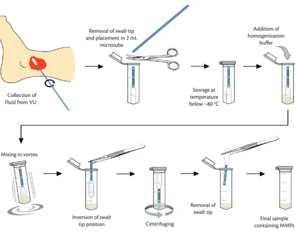
Samples of luid from VUs were taken using
sterile cotton buds (swabs), using a rolling motion, from border to border, after light irrigation with sterile saline solution. This procedure was conducted during primary dressing changes. Next, the tips of the swabs bearing the samples were removed and placed into 2 mL microtubes, fast-frozen in liquid nitrogen and stored at –80 °C until processing for protein extraction, from 2 to 3 weeks later.
To achieve homogenization, 250 mL of homogenization buffer was added to each tube containing a swab tip. The buffer comprised 50 mM Tris-HCl (pH 7.4); 3.1 mM Sucrose; and 0.1% Triton X-100, with no reduction agent, at a temperature of
4 °C. Samples were mixed in a vortex ive times for
3 s each time, with a 15 s interval at rest between each mixing. Swabs were then inverted in their microtubes and centrifuged (2 minutes, 10,000 rpm, 4 °C). The samples were kept in ice throughout these procedures. Figure 1 summarizes these stages.
Protein concentration was determined using the
Bradford method and quantiied in an ELISA reader
using standard controls made from bovine serum albumin. Readings were taken at 595 nm, 10 minutes after the assay, against a reference white.
Determination of enzyme activity of MMPs by zymography
Figure 1. Flow diagram of procedures involved in collecting and processing samples showing steps from collection of venous exudate with a swab to acquisition of the inal sample containing MMPs, ready for subsequent analyses.
pH of 8.8, containing 1 mg/mL of gelatin (from pig skin, type A, G2500 - Sigma-Aldrich). The stacking gel had a concentration of 4% and pH of 6.8 and contained 0.1% SDS. Proteins were separated under a constant voltage of 100 V in a buffer with the following concentrations: 192 mM Glycine; 25 mM Tris; 0.1% SDS.
After electrophoresis, the gels were immersed and agitated in Triton solution (2.5% Triton X-100; 50 mM Tris, pH 7.4; 5 mM CaCl2) to remove the SDS (three changes in 60 minutes).
Gels were then immersed at a temperature of 37 °C for 19 hours in an incubation solution consisting of 50 mM Tris (pH 7.4); 5 mM CaCl2; and 150 mM NaCl, in order to activate MMPs. After incubation, staining was performed with Coomassie Brilliant Blue G-250 (0.5% Comassie blue in 30% methanol and 10% acetic acid). Gels were submerged in the dye for 30 minutes, under agitation, before being destained (35% Methanol; 10% acetic acid) for 30 minutes and then placed in deionized water.
Gels were digitized on a bench scanner to enable densitometric analysis. Metalloproteinase enzyme activity was determined by the intensity of the pale bands against the stained background and MMPs were identified by their molecular weights, as described elsewhere,9,12 using ImageJ 1.46 software
(NIH, http://imagej.nih.gov/ij/). The program was
irst used to transform the image into 8-bits and then
a rectangular area was selected containing the bands (areas of enzyme digestion) and part of the stained gel background. The contrast between these two elements could then be demonstrated graphically, indicating the degree to which the gelatin in the gel had been broken down.13
RESULTS
Initially, we observed that an adequate quantity of
VU luid protein had been collected on the swabs,
using the Bradford method on ELISA plates (Table 1). We tested the polyacrylamide gel concentration in order to determine the best conditions for viewing and quantification of bands, defining the ideal concentration as 8%, the ideal incubation time for enzymatic activation as 19 hours and the ideal quantity of total protein loaded in each well on the gels as from 0.125 mg to 0.50 mg,.
Figure 2 illustrates the zymography results. Bands
representing the forms of pro-MMP-9 and MMP-9,
and pro-MMP-2 and MMP-2 were identiied with
the aid of previous descriptions9,12 and molecular
mass calculations. It was also possible to observe
gelatinolytic activity between 100 kDa and 150 kDa (~130 kDa), which has been described in the literature as an MMP complex that probably contains MMP-9.9,12 Bands corresponding to the latent and
active forms of each enzyme were selected together (model shown in Figure 2D) and quantified by densitometry (Figure 3) for illustration.
DISCUSSION
This article has described a viable and reproducible method for extraction of MMPs from VU exudate collected on swabs and for analysis of enzyme activity by zymography. As far as we know, this is
the irst description of this minimally invasive form of extraction of MMPs and the irst demonstration of
the viability of biochemical analysis of these small sample volumes.
There are descriptions of abnormal activation of MMPs and imbalances between MMP isoforms in chronic venous insufficiency patients, which contribute to formation and maintenance of VU.7-9,14
Among the different MMPs, MMP-9 appears to be a prognostic marker of healing of chronic ulcers.10
Furthermore, the course of the healing process in chronic wounds is associated with reduced expression of certain MMPs.14 In view of this, development of a
simple technique for collection of biological material for analysis of MMPs makes it possible to conduct serial evaluations of the process of ulcer healing and opens a wide range of applications for clinical research.
This study has demonstrated that collecting VU exudate on swabs is a viable and reproducible method for evaluation of the enzyme activity of MMP-2 and MMP-9 using zymography. Zymography is a relatively simple technique that is effective and
sensitive for identiication of MMPs by degradation
of their preferred substrates and by their molecular weights, making quantitative results possible.12
The technique described here is based on a minimally invasive procedure that is sterile, rapid and easy to conduct, making it an attractive alternative to
Table 1.Protein concentrations in samples.
Patient [protein] (mg/mL)
invasive methods used in earlier studies that involve biopsies and histopathological analysis.9,15 Although
biopsy can provide topographical information, for example, when tissue from the wound border and from the ulcer bed are compared, the technique requires local anesthesia and causes discomfort to patients, which makes serial analysis of wounds
dificult. Using exudate obtained by aspiration, when
possible, or by collection on swabs, as described
in this study, provides a more speciic relection of
the activity of MMPs in the ulcer bed. Although the results of these two collection methods have not been compared for venous ulcers, there is evidence that
they correlate in the case of diabetic ulcers.16 Future
studies could be designed to evaluate the agreement between the swab-based method and other methods, such as biopsy, with venous ulcers.
Finally, the technique described in this study also offers the advantage that it allows samples to be collected by many different classes of health professionals, since it does not demand local anesthesia, incisions or punctures, thereby facilitating analysis of the biological effectiveness of therapeutic interventions.
CONCLUSIONS
Collection of luid from VU using swabs proved
to be an effective, minimally invasive, sterile and rapid method offering great ease of use and one that is appropriate when the objective is to extract an adequate quantity of total protein from VU exudate for analysis of the enzyme activity of MMPs 2 and 9 using the zymography protocol proposed in this article.
REFERENCES
1. de Aguiar ET, Pinto LJ, Figueiredo MA, Savino Neto S. Úlcera de Insuficiência Venosa Crônica: Diretrizes sobre Diagnóstico, Prevenção e Tratamento da Sociedade Brasileira de Angiologia e Cirurgia Vascular (SBACV). J Vasc Bras. 2005;4(Supl.2):S195-200. 2. Lopes CR, Figueiredo M, Ávila AM, Soares LMBM, Dionisio
VC. Avaliação das limitações de úlcera venosa em membros inferiores. J Vasc Bras. 2013;12(1):5-9. http://dx.doi.org/10.1590/ S1677-54492013000100003.
3. de Araujo T, Valencia I, Federman DG, Kirsner RS. Managing the patient with venous ulcers. Ann Intern Med. 2003;138(4):326-34. http://dx.doi.org/10.7326/0003-4819-138-4-200302180-00012. PMid:12585831
4. Wipke-Tevis DD, Rantz MJ, Mehr DR, et al. Prevalence, incidence, management, and predictors of venous ulcers in the long-term-care population using the MDS. Adv Skin Wound Care. 2000;13(5):218-24. PMid:11075021.
5. Benjamin MM, Khalil RA. Matrix metalloproteinase inhibitors as investigative tools in the pathogenesis and management of vascular disease. EXS. 2012;103:209-79. http://dx.doi. org/10.1007/978-3-0348-0364-9_7. PMid:22642194
6. Dalton SJ, Mitchell DC, Whiting CV, Tarlton JF. Abnormal extracellular matrix metabolism in chronically ischemic skin: a mechanism for dermal failure in leg ulcers. J Invest Dermatol. 2005;125(2):373-9. PMid:16098049.
7. Meyer FJ, Burnand KG, Abisi S, Tekoppele JM, van Els B, Smith A. Effect of collagen turnover and matrix metalloproteinase activity on healing of venous leg ulcers. Br J Surg. 2008;95(3):319-25. http:// dx.doi.org/10.1002/bjs.5946. PMid:17854113
8. Murphy G, Nagase H. Progress in matrix metalloproteinase research. Mol Aspects Med. 2008;29(5):290-308. http://dx.doi. org/10.1016/j.mam.2008.05.002. PMid:18619669
9. Wysocki AB, Staiano-Coico L, Grinnell F. Wound fluid from chronic leg ulcers contains elevated levels of metalloproteinases MMP-2 and MMP-9. J Invest Dermatol. 1993;101(1):64-8. http://dx.doi. org/10.1111/1523-1747.ep12359590. PMid:8392530
10. Rayment EA, Upton Z, Shooter GK. Increased matrix metalloproteinase-9 (MMP-9) activity observed in chronic wound fluid is related to the clinical severity of the ulcer. Br J Dermatol. 2008;158(5):951-61. http://dx.doi.org/10.1111/j.1365-2133.2008.08462.x. PMid:18284390
11. Trengove NJ, Stacey MC, MacAuley S, et al. Analysis of the acute and chronic wound environments: the role of proteases and their inhibitors. Wound Repair Regen. 1999;7(6):442-52. http://dx.doi. org/10.1046/j.1524-475X.1999.00442.x. PMid:10633003 12. Snoek-van Beurden PA, Von den Hoff JW. Zymographic
techniques for the analysis of matrix metalloproteinases and their inhibitors. Biotechniques. 2005;38(1):73-83. http://dx.doi. org/10.2144/05381RV01. PMid:15679089
13. Hu X, Beeton C. Detection of functional matrix metalloproteinases by zymography. J Vis Exp. 2010;(45):e2445. PMid:21085107. 14. Beidler SK, Douillet CD, Berndt DF, Keagy BA, Rich PB, Marston
WA. Multiplexed analysis of matrix metalloproteinases in leg ulcer tissue of patients with chronic venous insufficiency before and after compression therapy. Wound Repair Regen. 2008;16(5):642-8. http://dx.doi.org/10.1111/j.1524-475X.2002008;16(5):642-8.00415.x. PMid:19128259
15. La Rocca G, Pucci-Minafra I, Marrazzo A, Taormina P, Minafra S. Zymographic detection and clinical correlations of MMP-2 and MMP-9 in breast cancer sera. Br J Cancer. 2004;90(7):1414-21. http://dx.doi.org/10.1038/sj.bjc.6601725. PMid:15054465 16. Schmohl M, Beckert S, Joos TO, Königsrainer A,
Schneiderhan-Marra N, Löffler MW. Superficial wound swabbing: a novel method of sampling and processing wound fluid for subsequent immunoassay analysis in diabetic foot ulcerations. Diabetes Care. 2012;35(11):2113-20. http://dx.doi.org/10.2337/dc11-2547. PMid:22837363
Correspondence Fernando Augusto Lavezzo Dias Universidade Federal do Paraná – UFPR Setor de Ciências Biológicas, Departamento de Fisiologia, Centro Politécnico Avenida Coronel Francisco H. dos Santos, 210 – Jardim das Américas CEP 81531-970 – Curitiba (PR), Brazil E-mail: [email protected]
Author information
FSS and DNA are MSc in Physical herapy from Universidade Federal do Rio Grande do Norte (UFRN), RN, Brazil. JPMSL is a PhD in Biochemistry from Universidade Federal do Ceará (UFC), and an adjunct professor at the Department of Biochemistry at Universidade Federal do Rio Grande do Norte (UFRN), RN, Brazil. AAR is a PhD in Biochemistry from Universidade de São Paulo (USP), and an adjunct professor at the Department of Clinical and Toxicological Analysis. BJGAA is a PhD in Cellular Biology from Universidade Federal de Minas Gerais (UFMG), and an adjunct professor at the Department of Morphology at Universidade Federal do Rio Grande do Norte (UFRN), RN, Brazil. FALD is a PhD in Cellular and Molecular Biology (Physiology) from Universidade Federal do Paraná (UFPR), Adjunct professor at the Department of Physiology at Universidade Federal do Paraná (UFPR), PR, Brazil.
Author contributions
Conception and design: FALD Analysis and interpretation: FSS, DNA JPMSL, AAR, BJGAA, FALD Data collection: FSS, DNA Writing the article: FSS, DNA, FALD Critical revision of the article: JPMSL, AAR, BJGAA, FALD Final approval of the article*: FSS, DNA, JPMSL, AAR, BJGAA, FALD Statistical analysis: N/A Overall responsibility: FALD