(1) Departmento de Radiologia Oral, Escola de Odontologia, Pontifícia Universidade Católica de Minas Gerais, Belo Horizonte, Brasil.
(2) Departmento de Dentística Restauradora, Escola de Odontologia, Pontifícia Universidade Católica de Minas Gerais, Belo Horizonte, Brasil. (3) Departmento de Anatomia, Escola de
Odontologia, Pontifícia Universidade Católica de Minas Gerais, Belo Horizonte, Brasil.
Conlict of interest: non-existent
Use of transcranial radiograph to detect morphological
changes in mandibular condyles
Uso da radiografia transcraniana para detectar alterações morfológicas
no côndilo mandibular
Laís Cristina Fonseca Pietra(1)
Mônica de Oliveira Santiago(2)
Claudia Scigliano Valerio(1)
Paulo Franco Taitson(3)
Flávio Ricardo Manzi(1)
Paulo Isaias Seraidarian(2)
Received on: July 28, 2016 Accepted on: November 09, 2016
Mailing address:
Flávio Ricardo Manzi
Departmento de Radiologia Oral, Escola de Odontologia, Pontifícia Universidade Católica de Minas Gerais
Av. Dom José Gaspar, 500 - Prédio 45, Belo Horizonte, Minas Gerais, Brasil CEP: 30535-901
E-mail: [email protected]
ABSTRACT
Purpose: to evaluate the accuracy of conventional transcranial radiographs (TRANS) to identify morpholo-gical changes in mandibular condyles.
Methods: the sample consisted of 36 mandibular condyles, obtained from 18, randomly selected, dried human skulls, without the identiication of age, gender, or ethnicity. Three experts in dental radiology examined the TRANS to identify possible changes in the condyles. The fourth examiner performed the macroscopic examination, which was considered the gold standard of the study. The condyles in both TRANS images and macroscopic examinations were classiied as mandibular condyles with change (1) or no change (0). Statistical analyses were performed using the X2 and the receiver operating characteris -tic (ROC) curve. Kappa intra- and interobserver tests were performed for examiners 1 to 3.
Results: the X2 test showed a statistically signiicant association between changes in the condyle in the TRANS images and the presence of macroscopic changes in the condyle (p < 0.05). The area under the curve was 0.83, with 96% sensitivity and 70% speciicity. The weighted kappa value for intraobserver agreement was 0.78, while the interobserver agreement was 0.71.
Conclusion: the use of TRANS proved to be an effective method to detect morphological changes in the mandibular condyle.
Keywords: Temporomandibular Joint; / radiography; Temporomandibular Joint Disorders; Diagnostic
Imaging
RESUMO
Objetivo: o objetivo deste estudo foi avaliar a acurácia das radiograias transcranianas (TRANS) conven-cionais na identiicação das alterações morfológicas nos côndilos mandibulares.
Métodos: a amostra consistiu em 36 côndilos mandibulares, obtidos a partir de 18 crânios secos huma-nos, aleatoriamente selecionados, sem identiicação de idade, gênero ou etnia. Três especialistas em radiologia oral examinaram as TRANS para identiicar possíveis alterações nos côndilos. Um quarto exa-minador realizou o exame macroscópico, que foi considerado o padrão ouro do estudo. As imagens das TRANS e os exames macroscópico foram classiicados como (1) côndilos com alteração ou (0) côndilos sem alteração. A análise estatística foi realizada através do teste X2 e da curva ROC (receiver operator
characteristic). O teste Kappa intra e interexaminadores foi realizado para os examinadores 1 a 3. Resultados: o teste X2 mostrou uma associação estatisticamente signiicativa entre as alterações no côn-dilo vistas nas imagens TRANS e a presença de alterações macroscópicas (p < 0,05). A área sob a curva ROC foi de 0,83, com 96% de sensibilidade e 70% de especiicidade. O valor Kappa para a concor-dância intraobservador foi de 0,78, enquanto que a concorconcor-dância interexaminador foi de 0,71.
Conclusão: o uso de radiograias transcranianas apresentou-se como método eicaz para a detecção de alterações morfológicas no côndilo mandibular.
Descritores: Articulação Temporomandibular; / radiograia; Transtornos da Articulação Temporomandibular;
INTRODUCTION
Temporomandibular disorders (TMDs) are classiied
as musculoskeletal degenerative disorders associated with morphological and functional deformities1-3.
These present a multifactorial etiology1,2,4-7 and
constitute a common complaint in many public and private dental services. TMDs are associated with one’s quality of life, presenting a positive correlation between the severity of the temporomandibular dysfunction and the negative impact upon one’s quality of life4-8.
Prior epidemiological studies, using diverse
populations, have examined the prevalence of TMDs9.
Some studies indicate a prevalence of 5-12%2,10,11, while
others have shown that nearly 40-60% of the general population can present the symptoms and signs of
TMD9, but only a small percentage of these affected
individuals actually seek treatment1,12, This diversity
of results concerning prevalence stems from the wide range of methodologies used in studies, especially as regards the sample selection criteria and the lack
of standardization related to TMD diagnoses12,13. As
regards gender, it is estimated that TMD is more prevalent in women than in men1,7,10.
Patients with TMD may also present other symptoms in addition to pain, such as clicking and crackling, changes or limitations in mandibular movements, and
headaches and/or muscular pain1,14. TMDs can also
affect the ears, eyes, and throat. Patients commonly report headaches that involve part or all of the head (frontal, temporal, parietal, occipital) and neck1,2,5-7,15.
TMD diagnosis is based on the patient’s medical history, in the clinical exam, and in the evaluation of the imaging exam1-3,14,16. The clinical diagnosis evaluates
the mandibular movements and the pain associated with these movements, joint noises, and symptoms found upon palpation of the region of the joints and muscles associated with temporomandibular joint
(TMJ)17. As regards the imaging exam, various
radio-graphic techniques have been proposed to evaluate TMJ, such as the transcranial radiograph (TRANS), computed tomography (CT), cone beam computed tomography (CBCT), arthrography, and magnetic resonance (MR)4,14,17-19.
Transcranial radiographs have been used worldwide
a way to reach a diagnosis speedily, easily, inexpen-sively, and with a low dose of radiation1,18,20-23.
Considering the importance of the evaluation of the morphological changes of mandibular condyles in the radiographic exams of TMJs in patients with TMD, this work seeks to evaluate the accuracy of transcranial radiographs in identifying osteophytes, worn or
lattened surfaces, erosion, among other changes in
mandibular condyles.
METHODS
Subjects
Research for this study began after having received approval from the local ethics committee (CAAE: 51173515.2.0000.5137 seems 1421150). The sample consisted of 36 mandibular condyles from 18 mandibles from dried human skulls selected from the collection of anatomical specimens at the Department of Dentistry of the University. The condyles were numbered from 1 to 36 for this study. The selection criterion to select
the skulls was random, with no identiication of age,
gender, or ethnicity.
Transcranial radiographs
To obtain the TRANS scans, a utility wax sheet
was ixed to the articular fossa of the skulls (Wilson,
Polidental, Cotia, SP, Brazil) to create space between the mandibular condyle and the mandibular fossa.
Next, the mandible was ixed to the maxilla using hot
melt adhesive (CIS, Sertic, São Paulo, SP, Brazil) with teeth in occlusion. The skulls were positioned using the
Accurad-200 head holder (Whip Mix Corporation, Fort
Collins, CO, USA). The Frankfort horizontal plane was used as a reference to position the dried human skull, while the midsagittal plane was aligned using vertical supporting bars, simulating the position of a patient. The head holder directs the central x-ray beam to the long axis of the condyles, causing the condyle to be radiographed so as to align itself perpendicularly to the
ilm and parallel to the x-ray path. The imagens were
taken on a traditional Oralix Gendex X-ray machine
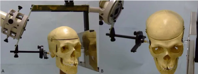
(Gendex Dental Systems, Hatield, PA, USA), which
Figure 1. Transcranial radiograph. (A, B) Stabilization of the dried human skull using the Accurad-200 head holder
Image analysis
All structural changes in the condyles shown both in the radiographs and in the anatomical specimens,
including osteophytes, worn and lattened surfaces,
and erosion, were grouped as being visible changes and received the code (1), i.e., “with change.” The condyles that did not present changes received code (0), i.e., “no change” (Fig. 2).
Figure 2. Condylar bone changes observed macroscopically. (A) Erosion with exposure to bone marrow. (B) Osteophyte. (C) Flattening of the condyle with the preservation of the cortical bone
radiologists drafted reports about the images, consid-ering the value (1) for the images in which the condyle presented change, and (0) when the condyle presented no change (Fig. 3).
(Instituto de Desenvolvimento Sustentável Mamirauá, Belém, Brazil). True negatives, true positives, false positives, and false negatives were calculated.
The intra and interobserver KAPPA test was performed for examiners 1 to 3. To interpret the KAPPA statistics, values of 0.81–1.00 indicated very good agreement, 0.61–0.80 indicated good agreement, 0.41–0.60 indicated moderate agreement, 0.21–0.40 indicated fair agreement, and 0.20 or less indicated poor agreement24.
RESULTS
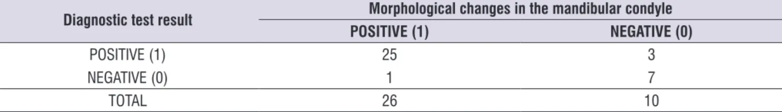
According to macroscopic examination, of the 36 mandibular condyles, 26 presented morphological changes, whereas 10 presented no morphological changes (Table 1).
A fourth oral radiologist (examiner 4) examined the dried human skulls macroscopically and drafted a report about the images, considering the same TRANS criteria, that is, value (1) for the images in which the condyle presented change, and (0) when the condyle presented no change. This result was considered the gold standard of the study.
Statistical analyses
The X2 test was used to determine the association
between the diagnosis of the change in the condyle in the TRANS and the morphological changes in the mandibular condyle observed in the anatomical
specimen. The signiicance level was p<0.05. The receiver operating characteristic (ROC) curve was used to measure the accuracy. The area under the ROC curve (AUC) was calculated using BioEstat Software, v. 5.0
Table 1. Frequencies of test results for 26 mandibular condyles with morphological changes and 10 mandibular condyles without
morphological changes
Diagnostic test result Morphological changes in the mandibular condyle
POSITIVE (1) NEGATIVE (0)
POSITIVE (1) 25 3
NEGATIVE (0) 1 7
TOTAL 26 10
Considering the evaluation of the TRANS images by examiners 1-3, the weighted KAPPA value for intraob-server agreement was more than 0.78. The weighted KAPPA value for interobserver agreement was 0.71. These results showed good intra and interobserver agreement24.
The X2 test showed a statistically signiicant
association between the change in the condyle in the TRANS and the presence of macroscopic change in the condyle (p < 0.05). The area under the curve was
0.83, with 96% sensitivity and 70% speciicity (Fig. 4).
The positive and the negative predictive values were determined to be 89% and 87%, respectively.
Table 2. Sensitivity, speciicity,positive predictive value, negative
predictive value, and accuracy of transcranial radiographs
Sensitivity 0.96
Speciicity 0.7
Negative predictive value 0.87
Positive predictive value 0.89
DISCUSSION
In this study, the accuracy of TRANS was assessed in its capacity to detect the morphological changes present in mandibular condyles through transcranial radiographs, using the macroscopic examination of anatomical specimens as the gold standard.
Some criteria are used to classify and guide the clinical approach of the TMDs2,14,16,25, associating the
history of the patient with the data that can be found in the patient’s clinical exam and radiograph2,3,14.
However, in the absence of an imaging exam, the
sensitivity should be of 55% and the speciicity of
61% for degenerative joint diseases of TMJ2. Thus, for
better accuracy in TMD diagnoses, imaging exams are essential.
Condylar bony morphological changes, such as bony surface erosion, concavity, spurring, and
lattening are bone alterations related to TMD12 and
that provide radiological signs. The imaging techniques used to evaluate such TMJs include: radiographic techniques (panoramic, lateral transcranial, transpha-ryngeal, and transmaxillary view), arthrography, as well
contradictory results. For some, there is a good
corre-lation between these exams28. For others, when a
condyle is not symmetrically positioned due to torsion within the joint cavity, the evaluation in the conventional radiograph is only partially relevant29. MR imaging uses
no radiation, has a high diagnostic accuracy, and is capable of assessing the disk position, disk shape, and bony changes; however, its cost is rather high4,9,17,30.
The correct three-dimensional positioning of the head is necessary to properly conduct a TRANS, given that the long axis of the condyle must be radiographed
perpendicularly to the ilm on the axial and coronal
planes. Due to the angle of 75° of the X-ray beams, the lateral third of the glenoid fossa and condyle are
projected upon the ilm. As a result, the central and
medial portions of the condyle are projected below the radiographic image of the condyle and cannot
be observed in the TRANS31. Thus, the TRANS
radio-graph possesses a limitation, as it is essentially a
proile or cross-sectional view of the lateral third of
the glenoid fossa and condyle31. By contrast, bony
changes occur on the articular surface32, and although
same31. Hence, the TRANS radiograph can be used as
a reference of condylar position within the fossa and as an aid in the diagnosis and treatment of pain caused by TMJ dysfunction27,31.
In the literature, there are few works that assess the accuracy of TRANS in the evaluation of the
degen-erative processes of TMJ. The work of Scarfe et al.21
show a sensitivity of 42.9%, a speciicity of 75.3%, and
an accuracy of 69.2% in the detection of osteophytes, with a digital sensor and a sensitivity of 52.4%, a
speci-icity of 68.7%, and an accuracy of 65.6% with conven
-tional ilm. As regards the CT exam, Scarfe et al.21 show
a sensitivity of 61.9%, a speciicity of 84.1%, and an
accuracy of 79.9% in the detection of osteophytes. In
a position of maximum mouth opening, Menezes et al.4
found a sensitivity of 84%, a speciicity of 92%, and an
accuracy of 91% for TRANS; however, the purpose of these authors was not to assess the accuracy of TRANS in the detection of the degenerative processes of the condyle, but rather in the positioning of the condyle within the TMJ. The present study obtained a sensitivity
of 96%, a speciicity of 70%, and an accuracy of 88%
in the detection of degenerative processes and
struc-tural changes, with the use of conventional ilms, values
which proved to be quite similar to those obtained by MR. According to Kumar et al.12, MR has a sensitivity
of 82.6% and a speciicity of 66.7% in symptomatic
patients.
MR is considered the gold standard to assess the position of the condyle and the mandibular joint disc in the glenoid fossa; however, its cost is quite high4,12.
The tomographic digital images are also of high quality
and precision1,2,4, however, they also have a high
cost33,34. By contrast, TRANS has been exclusively
used to assess the position of the condyle within the fossa as well as to evaluate bone changes, all at a low cost. Nevertheless, to date, no study has assessed the
accuracy of TRANS speciically regarding the detection
of the morphological changes of the condyles. This study demonstrated that TRANS presents an appro-priate accuracy for the evaluation of the morphological changes of the condyles.
TMD treatment does have an economic impact. The annual cost related to TMD treatment in the USA, not including imaging exams, has doubled over the past
decade, reaching nearly US$4 billion2,35. Thus, due
to the cost and high prevalence of TMD, it is crucial
that these problems be assessed in speciic primary
healthcare units, using a standardized clinical exam and a diagnostic protocol1,2,5,7,8. For this to occur,
complementary exams must have an accessible cost without a loss in the quality of data, so that they can be considered in the complex system of public funding.
The present study showed that TRANS is an eficient
and inexpensive exam that is capable of aiding in TMD diagnoses in cases in which the condyle presents morphological changes.
This study has some limitations. First, the imaging on dried skulls are more accurate than a live individual because the skulls can be accurately oriented for the radiographic examination, whereas, the live person
may be more dificult to get the correct alignment of
the radiograph beam with the long axis of the condyle. Second, the soft tissues in a live individual could
possibly blunt some of the deinition of bony changes
seen on dried skulls. Third, TRANS evaluates only the lateral one third of the condyle, so pathology that may exist in the middle or medial aspect of the joint may not
be identiied. Fourth, drying skulls could possible cause
some desiccation of the condylar surface resulting in greater arthritic bone changes than when the patient was alive. Finally, radiographic assessment was done by three dental radiology experts who have a far better expertise to identifying minor osseous changes as compared to the average clinician that may use this radiographic approach.
CONCLUSION
In conclusion, it was possible to compare the reports of the images obtained by transcranial radio-graphs on dried skulls with the macroscopic viewing of radiographed anatomical specimens. This present study performed on dried skulls proved that TRANS is
an eficient and inexpensive exam that can be used to
detect morphological changes in condyles.
ACKNOWLEDGEMENTS
This study was supported by the Permanent Program of Teachers Capacitation of PUC Minas (PPCD). Dr C.S. Valerio’s studies were supported by the Coordination for the Improvement of Higher Education Personnel (CAPES Foundation).
REFERENCES
1. Murphy MK, MacBarb RF, Wong ME, Athanasiou
2. Schiffman E, Ohrbach R, Truelove E, Look J, Anderson G, Goulet JP et al. Diagnostic Criteria for Temporomandibular Disorders (DC/ TMD) for Clinical and Research Applications: Recommendations of the International RDC/TMD Consortium Network* and Orofacial Pain Special Interest Group†. J Oral Facial Pain Headache. 2014;28(1):6-27.
3. Mohl ND. Reliability and validity of diagnostic modalities for temporomandibular disorders. Adv Dent Res. 1993;7(2):113-9.
4. Menezes AV, de Almeida SM, Bóscolo FN,
Haiter-Neto F, Ambrosano GMB, Manzi FR. Comparison of transcranial radiograph and magnetic resonance imaging in the evaluation of mandibular condyle position. Dentomaxillofac Radiol. 2008;37(5):293-9.
5. Barros VM, Seraidarian PI, Côrtes MI, de Paula L V. The impact of orofacial pain on the quality of life of patients with temporomandibular disorder. J Orofac Pain. 2009;23(1):28-37.
6. Conti PCR, Pinto-Fiamengui LMS, Cunha CO, Conti ACCF. Orofacial pain and temporomandibular disorders: the impact on oral health and quality of life. Braz Oral Res. 2012;26(Suppl 1):120-3.
7. Blanco-Aguilera A, Blanco-Hungría A,
Biedma-Velázquez L, Serrano-Del-Rosal R, González-López L, Blanco-Aguilera E, et al. Application of an oral health-related quality of life questionnaire in primary care patients with orofacial pain and temporomandibular disorders. Med Oral Patol Oral Cir Bucal. 2014;19(2):e127-35.
8. Oliveira BH, Nadanovsky P. Psychometric
properties of the Brazilian version of the Oral Health
Impact Proile-short form. Community Dent Oral
Epidemiol. 2005;33(4):307-14.
9. Okeson JP. Management of Temporomandibular Disorders and Occlusion. 6th ed. Elsevier Inc.; 2008.
10. National Institutes of Dental and Craniofacial
Research. Prevalence of TMJD and its signs and symptoms. 2014. [cited 2016 Jul 19]. Available from: http://www.nidcr.nih.gov/DataStatistics/
12. Kumar R, Pallagatti S, Sheikh S, Mittal A, Gupta D, Gupta S. Correlation Between Clinical Findings of Temporomandibular Disorders and MRI Characteristics of Disc Displacement. Open Dent J. 20151;9(Suppl 2: M4):273-81.
13. da Silva CG, Pachêco-Pereira C, Porporatti AL, Savi MG, Peres MA, Flores-Mir C et al. Prevalence of clinical signs of intra-articular temporomandibular disorders in children and adolescents: A systematic review and meta-analysis. J Am Dent Assoc. 2016;147(1):10-8.
14. Dworkin SF, LeResche L. Research diagnostic
criteria for temporomandibular disorders: review,
criteria, examinations and speciications, critique. J
Craniomandib Disord. 1992;6(4):301-55.
15. Carlsson GE. Epidemiology and treatment need
for temporomandibular disorders. J Orofac Pain. 1999;13(4):232-7.
16. Emshoff R, Brandlmaier I, Bösch R, Gerhard S, Rudisch A, Bertram S. Validation of the clinical diagnostic criteria for temporomandibular disorders for the diagnostic subgroup - Disc derangement with reduction. J Oral Rehabil. 2002;29(1996):1139-45.
17. Park JW, Song HH, Roh HS, Kim YK, Correlation JYL. TMJ disorders Correlation between clinical diagnosis based on RDC / TMD and MRI indings
of TMJ internal derangement. Int J Oral Maxillofac Surg. 2012;41(1):103-8.
18. Incesu L, Taşkaya-Yilmaz N, Oğütcen-Toller
M, Uzun E. Relationship of condylar position to disc position and morphology. Eur J Radiol. 2004;51(3):269-73.
19. Nah KS. Condylar bony changes in patients with temporomandibular disorders: A CBCT study. Imaging Sci Dent. 2012;42(4):249-53.
20. Kersey ML, Nebbe B, Major PW.
Temporomandibular joint morphology changes with mandibular advancement surgery and rigid
internal ixation: a systematic literature review.
Angle Orthod. 2003;73(1):79-85.
21. Scarfe WC, Farman AG, Silveira A, Fairbanks BW,
23. Schellhas KP, Piper MA, Omlie MR. Facial skeleton remodeling due to temporomandibular joint degeneration: an imaging study of 100 patients. Cranio. 1992;10(3):248-59.
24. Landis JR, Koch GG. The measurement of observer agreement for categorical data. Biometrics. 1977;33(1):159-74.
25. Brandlmaier I, Grüner S, Rudisch A, Bertram S,
Emshoff R. Validation of the clinical diagnostic criteria for temporomandibular disorders for the diagnostic subgroup of degenerative joint disease. J Oral Rehabil. 2003;30(4):401-6.
26. Hansson LG, Petersson A. Radiography of the
temporomandibular joint using the transpharyngeal projection. A comparison study of information obtained with different radiographic techniques. Dentomaxillofac Radiol. 1978;7(2):69-78.
27. Weinberg LA. Practical evaluation of the lateral
temporomandibular joint radiograph. J Prosthet Dent. 1984;51(5):676-85.
28. Van Sickels JE, Bianco HJ, Pifer RG. Transcranial radiographs in the evaluation of craniomandibular (TMJ) disorders. J Prosthet Dent. 1983;49(2):244-9.
29. Eckerdal O, Lundberg M. Temporomandibular joint relations as revealed by conventional radiographic techniques. A comparison with the morphology and tomographic images. Dentomaxillofac Radiol. 1979;8(2):65-70.
30. Helms CA, Kaplan P. Diagnostic imaging of the temporomandibular joint: recommendations for use of the various techniques. AJR Am J Roentgenol. 1990;154(2):319-22.
31. Weinberg LA. What we really see in a TMJ
radiograph. J Prosthet Dent. 1973;30(6):898-913.
32. Kurita H, Koike T, Narikawa J, Nakatsuka A,
Kobayashi H, Kurashina K. Relationship between alteration of horizontal size and bony morphological change in the mandibular condyle. Dentomaxillofac Radiol. 2003;32(6):355-8.
33. Scarfe WC, Farman AG, Sukovic P. Clinical
applications of cone-beam computed tomography in dental practice. J Can Dent Assoc. 2006;72(1):75-80.
34. Sezgin O, Kayipmaz S, Yasar D, Yilmaz A, Ozturk
M. Comparative dosimetry of dental cone beam computed tomography, panoramic radiography, and multislice computed tomography. Oral Radiol. 2012;28(1):32-7.
35. Gatchel RJ, Stowell AW, Wildenstein L, Riggs R, Ellis E. Eficacy of an early intervention for patients