300
Cutaneous metastasis in choroidal melanoma
Metástases cutâneas em melanoma de coróide
Fabricio Lopes da Fonseca1;Suzana Matayoshi2
A
BSTRACTMetastasis in choroidal melanoma is frequent on advanced diseases, involving mainly the liver, lungs and central nervous system. We report a case of cutaneous metastasis in choroidal melanoma because is an unusual condition, even in advanced disease.
Keywords: Melanoma; Choroid neoplasms; Neoplasm metastasis; Eye neoplasms; Skin neoplasms/secondary; Case reports
R
ESUMOA ocorrência de metástases do melanoma de coróide é frequente na doença avançada, acometendo principalmente fígado, pulmões e sistema nervoso central. Relatamos um caso de melanoma de coróide com metástases cutâneas, por se tratar de acometimento raro, mesmo em casos avançados da doença.
Descritores: Melanoma; Neoplasias da coróide; Metastase neoplásica; Neoplasias cutâneas/secundário; Relatos de casos
1MD, Fellow of Oculoplastic Service, Ophthalmology and Otorhinolaryngology Department,Hospital das Clínicas da Faculdade de
Medicina da Universidade de São Paulo - USP – São Paulo (SP), Brazil;
2Ph.D, Associate Professor and Chief of Oculoplastic Service, Ophthalmology and Otorhinolaryngology Department. Hospital das
Clínicas da Faculdade de Medicina da Universidade de São Paulo - USP – São Paulo (SP), Brazil.
Study carried out at
Oculoplastic Service - Ophthalmology and Otorhinolaryngology Department;Hospital das Clínicas da Faculdade de Medicina da Universidade de São Paulo- USP – São Paulo(SP),Brazil
Interest conflitct - None
R
ELATO DEC
ASORecebido para publicação em 27/8/2010 - Aceito para publicação em 29/3/2011
301
I
NTRODUCTIONM
elanomas are malignant neoplasms that develops from dendritic melanocytes in the skin, eyes, mucous epithelium and leptomeninges. Although the skin is the most common site of development, melanoma can occur in any tissue that contains melanocytes(1).The uveal melanoma is the more frequent intraocular malignancy in adults, affecting approximately 6 to 7 cases per million people in the United States of America2. Although rarer than in cutaneous melanoma,
metastasis is frequent and involves the liver, the lungs, the bones, the kidneys and the brain. Cutaneous metastasis is a rare manifestation of advanced disease(2,3).
Besides primary ocular melanoma, arising from eyelids, conjunctiva and uvea, the eye region may be affected by cutaneous melanoma, most often manifesting in the uvea densely vascularized, particularly the choroid, and orbit(3).
This paper aims to report a case of choroidal melanoma with cutaneous metastases, because it is an uncommon finding.
Case report
A 47-year-old female was referred to the HCFMUSP ophthalmology clinic with a history of insidious and painless decrease in visual acuity in the right eye for 8 years, proptosis which was initially noticed 2 years ago and the arising of large and pigmented lesions in the scalp are 3 months. She also reported some weight loss and anorexia for 3 months before diagnosis. She was diagnosed with neovascular glaucoma elsewhere, but lost follow-up.
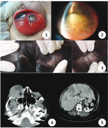
Ophthalmic examination revealed no light perception and total limitation of eye movements in all gaze positions, severe proptosis and palpable hard mass involving the entire orbital rim, with superior temporal hyperpigmentation (Figure 1). Slit-lamp examination showed intense neovascularization of the anterior segment, cataract and flat anterior chamber (Figure 1). Medical examination revealed hepatomegaly, with hardened liver surface and irregular edges, signs of asciites and three hyperpigmented and elevated lesions in the scalp (Figure 1).
She underwent computerized tomography (CT SCAN) of the skull, orbits, chest and abdomen and also a skin biopsy of the scalp lesions.
The CT scan of the orbit revealed a mass with heterogeneous pattern and irregular limits, affecting the posterior portion of right eye, with bone erosion and
invasion of anterior and middle ethmoidal, maxillary and sphenoid sinus (Figure 1). There was no significant intracranial extension. The abdominal CT scan revealed diffuse hepatic involvement, with heterogeneous areas compatible with necrosis and consistent with metastatic neoplasia (Figure 1).
The result of anatomo-pathological examination of the biopsy showed a lesion characterized by intense cellular pleomorphism, with bizarre nuclei and presence of melanin material (Figure 2). The presence of perilesional inflammatory infiltrate was also observed, as macrophages filled with material melanin (Figure 2). On the contrary seen in primary skin lesions, the epidermis remained intact, indicating metastatic involvement of an uveal primary lesion (Figure 2).
The patient was assessed jointly by the Clinical Oncology Service, and refered to palliative radiotherapy and chemotherapy.
Comments
The differentiation between primary uveal melanoma and primary cutaneous melanoma may be
Figure 1: 1) Hard mass involving the entire orbital rim, with supe-rior temporal hyperpigmentation ; 2) Intense neovascularization of the anterior segment, cataract and flat anterior chamber; 3) Hyperpigmented and elevated lesions in the scalp; 4) CT showing orbital mass, with spreading for paranasal sinuses and bone erosion; 5) CT showing diffuse hepatic involvement, consistent with metastatic neoplasia
Cutaneous metastasis in choroidal melanoma
302
difficult, as observed in this report. Clinical history and examination findings are useful, since they increase the likelihood of metastatic disease, as history of nonocular melanoma metastasis, faster growth, multiple and flat lesions, and pigmented cells in the vitreous4. In this report, the history of eye symptoms
for a long time, multiple skin lesions and the radiological and pathological features leads to the diagnosis of primary choroidal melanoma with skin metastasis.
Although less frequently, choroidal melanoma may course with metastasis in 75% of patients at time of death, opposite to 96% of cases of cutaneous melanoma2.
The liver is the main site of metastasis. Skin lesions is a rare manifestation which may can represent metastatic disease of choroidal melanoma, mainly in advanced ca-ses as we reported.
Figure 2: 10 Intense cellular pleomorphism, with bizarre nuclei and the presence of melanin material (Hematoxylin-Eosin 400x); 20 P e r i l e s i o n a l inflammatory infiltrate, with macrophages filled with material melanin (Hematoxylin-Eosin 50x); 30 Epidermis remained intact, indicating metastatic i n v o l v e m e n t (Hematoxylin-Eosin 400x)
R
EFERENCES1. Hurst EA, Harbour JW, Cornelius LA. Ocular melanoma: a review and the relationship to cutaneous melanoma. Arch Dermatol. 2003;139(8):1067-73. Comment in: Arch Dermatol. 2003;140(7):884-5; author reply 885.
2. Bell DJ, Wilson MW. Choroidal melanoma: natural history and management options. Cancer Control. 2004;11(5):296-303. 3. Shields CL, Shields JA. Ocular melanoma: relatively rare but
requiring respect. Clin Dermatol. 2009;27(1):122–33. 4. Rosenberg C, Finger PT. Cutaneous malignant melanoma
metastatic to the eye, lids, and orbit. Surv Ophthalmol. 2008;53(3):187-202.
Correspondence
A/C Fabricio Lopes da Fonseca
Rua General Bagnuolo, 616 apto 41 – CEP 03152130 Quinta da Paineira - São Paulo (SP) – Brazil
E-mail: [email protected]
Fonseca FL; Matayoshi S